Neurosurgical and Neuroradiological Critical Care


Neurosurgical and Neuroradiological Critical Care


Michael L. Mcmanus

Craig D. Mcclain

Robert C. Tasker


Critical care in neurosurgical and neuroradiological cases spans a continuum that begins with the operating theater or image neuroradiology suite and continues through the PICU. In addition, good patient care often requires the intensivist to provide expertise outside the PICU, in the emergency department, the radiology suite, and even into general or rehabilitative care settings. It is important, therefore, for a critical care physician to maintain a “systems” perspective and good working knowledge of the specialties and practices comprising this continuum of care. The focus of this chapter is upon close coordination of neurosurgery, neuroradiology, anesthesia, and critical care practices in this population of critically ill children. We begin with an overview of considerations important to our colleagues in anesthesiology, neurosurgery, and neuroradiology; continue with discussion of routine postoperative care; and then proceed with more detailed discussion of specific surgical entities. We conclude with discussion of a few specific postoperative challenges that are common in neurointensive care.


CLINICAL SCIENTIFIC FOUNDATIONS


Procedural and Operating Room Issues

The fundamental principles of postoperative and post-procedural neurocritical care include knowledge of the patient’s pre-procedure neurologic status, of the details of the procedure, of the patient’s post-procedure neurologic status, of the potential ongoing concerns, and whether there are interventions image that will optimize outcome.


General Anesthetic Considerations

The general goal of anesthetic management is to maintain stable hemodynamics and adequate gas exchange while providing analgesia and, when needed, sedation. In neuroanesthesia, this translates to maintenance of oxygenation, provision of appropriate carbon dioxide levels, preservation of adequate cerebral blood flow (CBF), and avoidance of exacerbations of intracranial hypertension. Hyponatremia, hypo-osmolarity, and hyperglycemia can contribute to cerebral swelling and neurological injury and are avoided. Anesthetic agents and depth are managed so as to permit a crisp wakeup and immediate neurological assessment once surgery is complete. Despite these seemingly straightforward goals, hypotension (1), hyperglycemia (2), and inadvertent hyperventilation (3) are persistent, but modifiable, risk factors in the perioperative period.

Induction of anesthesia usually involves premedication followed by titration of intravenous or inhaled agents at a rate that minimizes the hemodynamic consequences of transition from waking to anesthetic states. Premedication is administered with caution to avoid respiratory depression and is often avoided when significant intracranial hypertension is present. Induction agents are carefully titrated to avoid hypotension and decreased CBF. Thiopental (not available in United States), propofol, etomidate, and ketamine are common induction agents, and their effects on blood pressure (BP), intracranial pressure (ICP), CBF, and cerebral metabolic rate for oxygen (CMRO2) are considered prior to use. Recently, concerns about these deleterious effects of ketamine have been questioned (4,5). To minimize vasodilation that accompanies volatile agents, anesthetic maintenance classically involves a “balanced” technique using nitrous oxide, a narcotic, and lowdose volatile agent. The effect of volatile agents on CBF can vary with the dose of agent (increased vasodilation at higher dose), the region of brain studied (more pronounced on surface vessels), and the use of hyperventilation to cause vasoconstriction (often requested by neurosurgeons, to shrink brain tissue, even when ICP is normal). All volatile agents cause dose-dependent increases in CBF that can be attenuated with controlled ventilation. There is longstanding debate around the use of nitrous oxide since it can cause some degree of cerebral vasodilation, it may contribute to postoperative vomiting,
as well other limitations (6). In addition, nitrous oxide is relatively contraindicated when air collections are present (such as after recent craniotomy) since its diffusion may cause their expansion. Increasingly, there is addition or substitution of ultrashortacting agents such as remifentanil or dexmedetomidine for anesthetic maintenance. The general effects of commonly used anesthetics on CMRO2, CBF, pressure autoregulation, and ICP are summarized in Table 60.1.


Pre- and Intraoperative Fluid Management

Euvolemia is preferred before induction of anesthesia because the associated loss of sympathetic tone produces hypotension. The primary intraoperative fluids are exclusively isotonic as vasodilation and acute blood loss can necessitate sudden infusion of large volumes. Of the available solutions, normal saline is often the fluid of choice since it is slightly hypertonic to plasma and is compatible with all medications (including phenytoin). Fluid replacement is best guided by heart rate, BP, and perfusion since urine output can be profoundly influenced by stress-related changes in antidiuretic hormone (ADH) secretion. In addition, general anesthesia and prone positioning can interfere with urine production and urinary catheter drainage. Overall, urine output is an unreliable marker of volume status since oliguria can arise from many unrelated sources. Nonetheless, maintenance of euvolemia is critically important both to support CBF and to avoid venous air embolism via skull dural sinuses. A special situation arises in cerebrovascular cases that receive 1.5 times maintenance fluid requirements as preoperative preparation (see section on Cerebrovascular Surgery); in these cases intrinsic homeostasis will lead to excretion of the excessive salt load in the postoperative period, which may be mistakenly interpreted as postoperative “cerebral salt wasting” (CSW; see section on Dysnatremia).

In regard to the use of intraoperative glucose-containing fluids, the stress response should maintain normal serum glucose levels without exogenous glucose administration. However, in postoperative infants and small children, particularly if there has been an effective fast of 6-12 hours, there is the risk of perioperative hypoglycemia. Therefore, glucose-containing fluids can be used to meet baseline demands for neonates and susceptible infants. In general, older children and adolescents can tolerate 18-24 hours of fasting before requiring glucose-containing fluid administration. One risk of giving exogenous glucose is that along with the stress of critical illness (and resulting insulin resistance) hyperglycemia may be induced and, in turn, may be associated with neurologic injury and poor outcome. Concern persists that hyperglycemia can worsen injury due to ischemia, but it remains unclear if tight glycemic control offers significant benefits to children.


TABLE 60.1 EFFECTS OF ANESTHETICS, BENZODIAZEPINES, AND OPIOIDS ON CEREBRAL METABOLISM, CIRCULATION, AND ICP


AGENT


CMRO2


CBF


PRESSURE AUTOREGULATION


ICP


Inhaled anesthetic


▪ Sevoflurane


▪ Isoflurane


▪ Desflurane


□□


Absent


Inhaled nitrous oxide


□ or no change


Present


Intravenous anesthetic


▪ Propofol


▪ Thiopental


□□


□□


Present


□□


Dissociative anesthesia


▪ Ketamine


No change


□□


?


□□


Sedative benzodiazepines


□□


Present


Analgesic opioids


No change


No change


Present


No change


See Chapter 58 for pressure autoregulation. The direction of arrows indicates increased or decreased, and the number of arrows indicates the strength of the derangement above “no change” qualitatively.


Hemodynamic Management

As a general rule, intravenous anesthetics agents preserve CBF and autoregulation while volatile agents cause some degree of vasodilation and autoregulatory impairment (Table 60.1). As depth of anesthesia increases, so too does the impact on the circulation and CBF. To closely monitor and direct the control of mean BP and cerebral perfusion during anesthetic administration, an intra-arterial catheter is used. Especially in cerebrovascular surgery, vasoactive medications are always kept immediately available if needed to manipulate the circulation. Although a -2 agonists have direct constrictive effects on cerebral vasculature, all agents that increase mean BP will increase CBF when the limits of autoregulation are exceeded.

There are interactions between anesthetic agents and vasopressors. In short time frames, BP and cerebral autoregulation vary with anesthetic depth (i.e., as depth of anesthesia increases, BP and the ability to autoregulate CBF decrease). Over longer time frames, vasopressors can impact their effectiveness. In animal models, catecholamine infusion produces circulatory changes that increase the distribution of propofol and reduce its anesthetic effects (7,8). A similar phenomenon can be demonstrated with remifentanyl (9). The practical implication of this phenomenon is that hemodynamics and depth of anesthesia must be continuously balanced both in the operating room and throughout the transition to the PICU.


Surgical Considerations

Cranial surgeries require rigid skull fixation and pins may be placed prior to positioning. Complications of the use of pins include sudden increases in heart rate, BP, and ICP, as well as pin-related skull fractures and intracranial hemorrhages in infants and small children. The patient position carries risks and the sitting position is associated with an increased risk of venous air embolism. In pediatric neurosurgery, the sitting position is infrequently necessary and many adult neurosurgeons often choose to avoid it. Perioperative antibiotics are routinely administered. Blood loss varies widely among procedures and
can be particularly challenging in hemispherectomies and craniofacial reconstruction. In addition, chronic and even short-term anticonvulsant use may predispose to platelet dysfunction, thrombocytopenia, and factor deficiencies that can lead to increased blood loss.


Cranial Imaging Considerations

Imaging for neurosurgical patients presents many challenges. Cranial computed tomographic (CT) and magnetic resonance (MR) imaging may require transport and sedation of unstable patients. Complex imaging and interventional procedures may require prolonged immobility, controlled ventilation, and manipulation of the circulation. The neuroradiology and imaging suites can be “unfriendly” environments, in the sense that many of the supports routinely available in the operating room and PICU are either unavailable or difficult to secure. Full monitoring, safe positioning, access to the patient, and help during emergencies may all present challenges to anesthesia and PICU staff.

In some centers, portable CT devices have been introduced to mitigate transport hazards and decrease barriers related to the imaging of critically ill patients. Although the resolution and versatility of portable scanners are below those of fixed machines, their diagnostic utility is similar (10). The radiation doses attendant to portable machines is approximately 15% higher than fixed machines, so extra precautions are necessary to limit patient exposure and assure radiation safety of staff.

MR imaging is an indispensable tool in neurosurgery and neurocritical care, but MR safety is an important concern since the behavior of ferromagnetic materials in the MR suite can be dangerous. The terms “MR conditional,” “MR safe,” and “MR unsafe” have been proposed to clarify discussion of these hazards (11), and the MR suite itself has been divided into four zones of varying risk (12). MR unsafe materials must be excluded from the suite. MR safe equipment presents no threat when its operating instructions are followed. MR conditional refers to an object that has been demonstrated to pose no known problems under specified MR conditions (i.e., the magnetic field strength, its spatial gradient, its time rate of change, the radiofrequency field strength, and its specific absorption rate). For critically ill patients, special monitors, infusion pumps, and MR safe or MR conditional anesthesia machines may all be required. Historically, cardiac pacemakers have been considered a contraindication to MR imaging, but compatible devices are now available. Programmable cerebrospinal fluid (CSF) shunts may be altered by magnetic fields, so those devices should always be interrogated following an MR examination. To prevent malfunctions or thermal injury, manufacturers of nerve stimulators offer strict guidelines regarding radiofrequency and gradient fields.


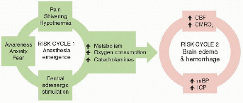
FIGURE 60.1. Potential stress-induced pathophysiology during emergency from anesthesia. mBP, mean blood pressure.

Intraoperative MR imaging has been developed to improve surgical navigation and resections. First-generation facilities employed an “open” configuration wherein both patient and magnet are stationary and surgery is conducted with limited access but real-time imaging capabilities. In later configurations, either the patient or magnet is mobile, permitting better access but requiring repositioning between scan sequences. In open systems, all anesthesia and surgical equipment must be MR safe. Unfortunately, some important equipment found in conventional operating rooms (e.g., precordial Doppler ultrasonography, core temperature probes, fluid warmers) do not yet have MR safe or MR conditional counterparts. Nevertheless, numerous neurosurgical procedures have been safely performed in children in suites with combined MR and operating room capability, and the equipment for these procedures is rapidly evolving (13).


Emergence

Regardless of the anesthetic technique used during the procedure, quick postoperative emergence is a basic requirement by neuroanesthesia. Awakening, extubation, and a screening neurological examination are important before transfer to the PICU in order to ensure that the patient has experienced no occult injury and is following the expected postoperative trajectory. When extubation is not possible, as in the patient with multiple trauma, respiratory impairment, or other comorbidities, it is prudent to confirm some level of responsiveness upon arrival in the PICU.

There are, potentially, two interlocking viscous cycles in pathophysiology that may lead to hemodynamic instability, cerebral edema, and intracranial hemorrhage in the immediate postoperative period (Fig. 60.1). Hence, in some high-risk children, there might be a plan for postoperative mechanical ventilation or deep sedation (Table 60.2). Emergence agitation may be due to pain, a full bladder, dysnatremia (see later), drug reaction (e.g., paradoxical reaction to midazolam or diphenhydramine), or emergence delirium (e.g., reaction to sevoflurane). Treatable causes will have been identified and dealt with, or even anticipated, by the anesthesiologist. For example, pain and agitation can be anticipated and treated with bolus doses of opioids, propofol, clonidine, or dexmedetomidine. The stress response with emergence hypertension can be treated with low-dose fentanyl infusion (1-1.5 mcg/kg/h) and an antihypertensive. In regard to the antihypertensive, the choice is between a β-blocker (labetalol or esmolol) and a calcium channel antagonist (nicardipine). There is no ideal vasodilator: β-blockers have the risk of bradycardia and cardiac conduction delays; the calcium channel antagonists cause cerebral vasodilatation, impaired autoregulation, and risk of
hypotension. Dexmedetomidine infusion is being used increasingly in pediatric neurosurgery—there are no data, but its use is gaining popularity since it acts as a sedative, sympatholytic, and analgesic.


TABLE 60.2 LIKELY CANDIDATES FOR DELAYED EMERGENCE AND DELAYED ENDOTRACHEAL EXTUBATION AFTER NEUROSURGERY


CAUSES FOR CONSIDERATION OF DELAYED EXTUBATION AND EMERGENCE FROM ANESTHESIA


▪ Preoperative altered level of consciousness


▪ Surgery lasting >6 h


▪ Large tumor resection with preoperative midline shift


▪ Injury to cranial nerves IX, X, XI


▪ Complications during surgery


▪ Intraoperative brain swelling


▪ Hypothermia


▪ Coagulopathy


▪ Acid-base abnormality


▪ Dysnatremia (see text for details)


TABLE 60.3 FACTORS TO CONSIDER IN THE PATIENT FAILING TO WAKE UP AT THE END OF NEUROSURGERY


FACTOR


INTERVENTION


Neuromuscular blockade


Impaired hepatic and/or renal metabolism may prolong the effect


Altered drug metabolism


Consider drug dosing and drug clearance of continuous infusions used (also see hypothyroid)


Seizure


Assess and treat accordingly


Intracranial bleeding


Imaging required and surgery


Intracranial pressure/ischemia


Imaging required


Hypothermia


Warm patient


Hypoglycemia


Check blood glucose and treat accordingly


Hypercapnia


Check blood gas and support ventilation as needed


Hypo-osmolality


Check serum electrolytes and treat accordingly


Hypothyroidism


Thyroid hormone required for benzodiazepine metabolism; support patient until full emergence and thyroid function tests available


Occasionally, a patient may unexpectedly fail to awaken at the end of surgery. A number of factors will need to be considered and corrected (Table 60.3) and, if not improved, the patient may need emergency imaging.


POSTOPERATIVE NEUROSURGICAL CARE IN THE PICU

The overall goal of postoperative neurosurgical care is to manage the patient with optimal management of physiology and pharmacology for the best expected outcome for the child’s underlying condition. In order to achieve these objectives, there must be clear documentation in the patient’s medical record and good communication between all team members. All hospitals have their own Policy and Procedures Manual that covers the detail of what is expected of medical practitioners, and what is considered best care practices in preparing a child for a neurosurgical procedure. Clinical assessment, coma scale scores, neuromonitoring, mechanical ventilation, and head and spinal cord injury are covered elsewhere in this textbook.


Initial PICU Care


Patient Hand-Off

Table 60.4 outlines a checklist for the general neurosurgical admission to the PICU. After the operation, those caring for the child will also need information in a systematic manner. We use the Formula One hand-off technique (14). This idea addresses the limited time to transfer information and the necessary details need to be conveyed to the next team caring for the child. Table 60.5 outlines a checklist for the postoperative transition of care. The neurosurgeon and anesthesiologist need to convey succinctly what happened in the operating room and what they want to happen in the immediate postoperative period. The postoperative attendants need to be cognizant of any concerns about bleeding, compromised tissue, derangements in homeostasis, and requirements for postoperative antibiotics, steroids, etc. We find that this process is best served by attending-to-attending sign-out. The critical care team examines every patient on arrival, documenting physiological stability, level of consciousness, and the presence or absence of focal neurological findings.


Mechanical Ventilation

Most cases requiring PICU care will not require mechanical ventilation (15). However, when prolonged mechanical ventilation is necessary, the aim is to support gas exchange while permitting ongoing neurosurgical assessment. End-tidal carbon dioxide monitoring helps prevent inadvertent hyper- or hypoventilation and positive end-expiratory pressure is delivered as necessary to support oxygenation (16,17). When inflation pressures are high, care is taken to avoid falls in mean BP and cerebral perfusion pressures (CPP, the difference between mean BP and mean ICP). High-frequency oscillatory ventilation may be used without significant impact on ICP (18). ICP monitoring may be necessary in cases in which the sedation needed to tolerate intubation and ventilation interferes with the ability to follow neurologic examination or in which the administration of PEEP may contribute to increasing ICP.


Hemodynamic and Fluid Management

In general, hemodynamic management targets BP within 20% of preoperative values or normal for age. When increased ICP is a consideration, CPP targets are those used in trauma (see Chapter 61).

Isotonic fluids are always used intraoperatively and should be continued in the immediate postoperative period. Since the neurosurgical population has increased risk of postoperative hyponatremia, as high as 12% (19), the dysnatremias will be discussed in some detail subsequently. Because of the risk of hyponatremia in the perioperative period, many clinicians choose to avoid using hypotonic solutions altogether. It should be noted that Ringer’s Lactate sodium (130 mmol/L) might also result in a fall in serum sodium. This fluid is sometimes used intraoperatively as it is a balanced solution with a physiologic amount of base, calcium and potassium, and will limit the hyperchloremic acidosis that occurs with large volumes of normal saline.


TABLE 60.4 INFORMATION REQUIRED AT TIME OF NEUROSURGICAL ADMISSION TO THE PICU


ACTIVITY


DETAILS


ACTION


History


▪ Presenting complaint


▪ Previous medical history


▪ Developmental level


▪ Previous anesthetics


▪ Medications


▪ Allergies


▪ Seizures


▪ Documented


Examination


▪ Central and peripheral nervous systems


▪ Presence of raised ICP


▪ Cardiopulmonary


▪ General level of hydration


▪ Documentation of neurologic deficits


▪ May need preoperative resuscitation and treatment


Comorbidities


▪ Intercurrent illness


▪ Any cardiopulmonary disease


▪ Any gastroesophageal reflux


▪ Endocrinopathy


▪ Discuss with anesthesiologist


Blood testing


▪ Hemoglobin


▪ Urea and electrolytes


▪ Blood cross-matched


Fasting perioda

Stay updated, free articles. Join our Telegram channel

Jun 4, 2016 | Posted by in CRITICAL CARE | Comments Off on Neurosurgical and Neuroradiological Critical Care

Full access? Get Clinical Tree

Get Clinical Tree app for offline access