CHAPTER 28 NEUROPATHIC PAIN
1. What is neuropathic pain?
The term neuropathic pain is applied to any acute or chronic pain syndrome in which the mechanism that sustains the pain is inferred to involve aberrant somatosensory processing in the peripheral or central nervous system (PNS/CNS). Neuropathic pain is commonly distinguished from two other inferred pathophysiologies, nociceptive pain and psychogenic pain. The sustaining mechanisms of nociceptive pain are inferred to involve ongoing activation of pain-sensitive afferent peripheral nerves. This activation may be caused by injury to either somatic (known as somatic pain) or visceral (known as visceral pain) structures. Psychogenic pain is a generic term used to refer to those pains that have sustaining mechanisms related to psychological processes. Other descriptive terms, such as those codified in the American Psychiatric Association’s Diagnostic and Statistical Manual (DSM-IV-R), can be used to classify the latter pain syndromes.
2. Why do the definitions of neuropathic pain, nociceptive pain, and psychogenic pain refer to these disorders as “inferred” pathophysiologies?
This classification by pathophysiology is inferred because there is no way to prove or disprove that any particular mechanism is operating in the clinical setting to maintain a chronic pain syndrome. The type of pathophysiology that may be involved is conjectured on the basis of the pain description and associated findings on examination and ancillary tests. Because the diagnosis is inferred, there is the potential for imprecision and oversimplification when labeling patients. Indeed, it is likely that patients often have more than one set of pathophysiology and that each type of pathophysiology actually refers to multiple specific mechanisms. Nonetheless, a pathophysiologic classification has become widely accepted by clinicians, who have observed that it may be useful in defining the type of evaluation that may be needed, selecting appropriate therapies, and determining the prognosis for improvement.
3. What findings on clinical evaluation suggest that a pain is neuropathic?
Neuropathic pain is suggested when patients use terms to describe their pain that are consistent with a dysesthesia, which is defined as an abnormal pain complaint. Pain may be described as burning, electric-like, or shooting. Patients often say that the pain is unfamiliar, unlike any pain experienced before. The examination may reveal allodynia (pain created by a normally non-painful stimulus), hypalgesia or hyperalgesia (relatively decreased or increased perception of a noxious stimulus, respectively), hypoesthesia or hyperesthesia (relatively decreased or increased perception of a nonnoxious stimulus, respectively), or hyperpathia (exaggerated pain response).
There may be other focal neurologic deficits, such as weakness or focal autonomic changes. Focal autonomic phenomena may include swelling and vasomotor instability (observed as color changes, livedo reticularis, and focal temperature changes). There may also be trophic changes, including alterations of the skin and subcutaneous tissues or the hair and nails.
Ancillary tests, such as electrodiagnostic studies (electromyogram and nerve conduction velocities), can sometimes be helpful in confirming the existence of a neurologic lesion. Tests such as thermography are occasionally useful to confirm autonomic dysregulation. Quantitative sensory testing (QST) may also be used to aid in the diagnosis.
4. What are some of the challenges inherent in diagnosing neuropathic pain?
When a dysesthesia occurs in the setting of an overt neurologic lesion, the diagnosis of neuropathic pain may be straightforward. Even in this setting, however, it may be difficult to exclude a contribution to the pain of coexisting processes, such as damage to somatic structures sufficient to produce nociceptive pain or psychological processes that exacerbate the pain. Furthermore, neuropathic pain can occur without an overt neurologic deficit (for example, complex regional pain syndrome type 1 can follow minor soft-tissue injury, and pain may be the first and only manifestation of a small fiber polyneuropathy). Finally, neuropathic pain can be nondysesthetic, such as the deep aching that commonly occurs from nerve or nerve root compression. All of these factors can complicate the diagnosis of neuropathic pain.
5. The clinical diversity of neuropathic pain suggests that the mechanisms responsible are both numerous and complex, presumably involving interactions between the PNS and CNS. Is there a useful model for conceptualizing these interacting mechanisms?
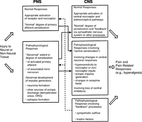
Three decades of basic research have shown that neuropathic pain may result from any of a variety of mechanisms that interact in complex ways. The normal response of the PNS and the CNS following exposure to a noxious stimulus can become disturbed at multiple levels concurrently (Fig. 28-1). This process may involve altered peripheral input (e.g., from sensitization of primary afferent neurons) or central processing (e.g., any of the mechanisms involved in so-called central sensitization; see Question 9), changes in efferent activity in the sympathetic nervous system, and shifts in pain modulatory processes in both the PNS and CNS. Further research is needed to determine the specific processes that result in these pathologic distortions of normal nociception.
6. From the clinical perspective, what is a useful classification of the heterogeneous population with chronic neuropathic pain?
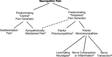
Although patients with neuropathic pain are traditionally categorized on the basis of diagnosis (e.g., painful diabetic polyneuropathy) or site of the precipitating lesion (e.g., peripheral nerve), it may be most useful to extend the classification based on inferred pathophysiology, and to suggest that some patients with neuropathic pain have disorders that are primarily sustained by processes in the CNS, whereas others have disorders sustained by processes in the PNS. This distinction is suggested by both clinical and experimental data (Fig. 28-2). For example, a predominant peripheral pathophysiology is suggested by the observation that some patients with neuropathic pain precipitated by nerve injury are cured by a local intervention, such as resection of a neuroma. A predominant central pathophysiology is obvious in those patients whose neuropathic pain is precipitated by stroke.

Figure 28-2 Classification of neuropathic pains by putative predominating mechanism. 1. Response to either peripheral or central nervous system injury. 2. Associated with focal autonomic dysregulation (e.g., edema, vasomotor disturbances), involuntary motor responses, and/or trophic changes that may improve with sympathetic nerve block. 3. Multiple mechanisms probably involved. 4. The patterns of peripheral activity or peripheral and central interaction that yield the lancinating quality of these pains are unknown. 5. Nociceptive nervi nervorum (small afferents that innervate larger nerves) may account for neuropathic pain accompanying nerve compression or inflammation. 6. Injury to axons may be followed by neuroma formation, a source of aberrant activity likely to be involved in pain.
7. What neuropathic pain syndromes are presumably sustained by aberrant somatosensory processing in the CNS?
Neuropathic pains that are inferred to have sustaining mechanisms in the CNS can be broadly divided into two groups: (1) disorders known as the deafferentation pains, and (2) disorders collectively known as complex regional pain syndromes (previously known as reflex sympathetic dystrophy and causalgia; see Question 50). The deafferentation pains include a large number of specific syndromes, such as central pain (pain following injury to the CNS), pain resulting from avulsion of a plexus, pain resulting from spinal cord injury, postherpetic neuralgia, phantom pain, and others. Complex regional pain syndrome is presumably sustained by a number of different mechanisms. One such mechanism is believed to involve efferent activity in the sympathetic nervous system and produces a pain known as sympathetically maintained pain.
8. Which pain syndromes are presumably sustained by aberrant somatosensory processing in the peripheral nervous system?
Neuropathic pains that are inferred to have sustaining mechanisms in the PNS can be divided into a group of painful polyneuropathies and a group of painful mononeuropathies. Each of these groups, in turn, subsumes many specific syndromes.
9. Describe the specific CNS mechanisms likely involved in the various types of deafferentation pain
Studies in experimental models and humans suggest that a state of “central sensitization” may be relevant to all deafferentation syndromes. Although peripheral input may be important in some syndromes (as suggested by the transitory relief of deafferentation pain that is commonly observed following interruption of proximal somatosensory pathways), the sustaining pathophysiology presumably relates to changes in the response characteristics of central neurons that are at least partly independent of this input.
Central sensitization may involve functional and structural changes in CNS pathways involved in nociception (see Fig. 28-1 and Table 28-1). Each of these changes presumably occurs as a consequence of specific mechanisms, which have only begun to be elucidated. Recent studies, for example, have indicated the importance of an interaction between excitatory amino acids (specifically glutamate) and the N-methyl-D-aspartate receptor in producing sensitization of nociceptive neurons in the dorsal horn of the spinal cord. Although the relationship of these functional and structural changes to chronic pain in humans is conjectural, the range of phenomena underscores the plasticity of central connections and suggests a focus for future research targeted at the prevention or treatment of neuropathic pain.
TABLE 28-1. Changes that may be Involved in Neuropathic Pains Sustained by Abberrant Processes in the Central Nervous System
Functional Changes | Structural Changes |
---|---|
Lowered threshold for activation | Transsynaptic degeneration |
Exaggerated activation | Transganglionic degeneration |
Ectopic discharges | Collateral sprouting |
Enlarging receptive fields | |
Loss of normal inhibition |
10. Phantom pain is commonly considered to be a type of deafferentation pain. What is phantom pain?
Although the prototype phantom pain follows limb amputation, the term is applied to pain following amputation of any body part. For example, surveys have described phantom pain following mastectomy and tooth extraction. Some authors also use the term to describe pain in regions of the body that are completely denervated (rendered anesthetic) but not amputated, such as the area below a transected spinal cord or the area supplied by a severely injured peripheral nerve. This usage may be confusing, however, and it would be preferable to use the term “central pain,” or one of its subtypes (see Question 35), to describe a pain that occurs in an area denervated as a result of a CNS lesion, and to use either the generic term “deafferentation pain” or the older term “anesthesia dolorosa” to describe a pain that is inferred to have a central mechanism induced by a severe peripheral nerve injury.
11. What is known about the specific mechanisms that may result in phantom pain?
The specific mechanisms that cause phantom pain are not known. One conceptualization suggests that phantom pain is a somatosensory “memory” that does not reside in a specific region of the CNS, but instead involves complex interactions of neural networks in the brain. Recent studies have suggested that the “shrinkage” of the somatosensory cortical representation of an amputated limb correlates with the development of pain. It has also been suggested that pain management with opioids results in reexpansion to its original representation and that this change correlates with pain reduction. The precise pathophysiology underlying these associations is not known.
12. What is the epidemiology of phantom pain?
Epidemiologic surveys of phantom pain must distinguish this phenomenon from both nonpainful phantom sensations and stump pain. Failure to be precise may be the cause of variation in older surveys, which have reported transitory or occasional discomfort in the phantoms of 25% to 98% of amputees. Although some surveys suggest that about half the patients with phantom pain continue to experience pain for a period of at least 1 to 2 years, others indicate that a large majority experience resolution of pain (though not necessary all phantom sensation) within 1 year of amputation.
Several studies have attempted to define predisposing factors for the development of phantom pain. Phantom limb pain is rare in congenital amputees or children who lose a limb before the age of 6. This observation suggests that some degree of CNS maturation is required before phantom pain can occur. The experience of pain in the limb prior to amputation has been noted to predispose to the development of phantom pain in some, but not all, surveys. A recent study observed that 57% of patients who experienced pain immediately before amputation developed phantom pain that resembled the preexisting pain in quality and location. A strong association between stump pain and phantom pain has also been reported. Other surveys have suggested that older age, proximal amputations, upper limb lesions, sudden amputations, and preexisting psychological disturbances may increase the likelihood of phantom pain, but these factors have not been confirmed in more recent studies.
13. What is the natural history of phantom pain?
Although most patients develop phantom sensations and phantom pain soon after the nerve injury, symptoms may develop at any time after denervation. Most surveys observe that the pain substantially declines over time.
14. How is phantom pain different from phantom sensation?
Phantom pain is one element among many phantom sensations. Pain has been termed an exteroceptive sensation, a description that has also been applied to the perception of touch, temperature, pressure, itch, and other sensations. Kinesthetic sensation, which involves the perception of posture, length, and volume, and kinetic sensations, including the perceptions of willed movements and spontaneous movements, also occur. Among the more common kinesthetic sensations are unusual or bizarre postures, foreshortening of a limb (“telescoping”), or distortions in the size of body parts (usually reduction in proximal regions and expansion of distal regions). All these sensations are usually most vivid immediately after amputation. Over time, the size of the phantom often shrinks, and the intensity of all sensation gradually fades.
15. How is phantom pain different from stump pain?
In contrast to phantom pain, in which the inferred “generator” of the pain is in the CNS, stump pain is a peripheral neuropathic pain presumably related to the development of a neuroma at the end of a severed nerve. Patients usually report some combination of aching, squeezing, throbbing, stabbing, and electrical discomfort localized to the distal stump. The onset of the pain is usually delayed for months, and the incidence is lower than that in phantom pain. Following limb amputation, many patients have both stump pain and phantom pain.
16. Can phantom pain be prevented?
Phantom pain may be difficult to treat, and prevention would clearly be ideal. The possibility of prevention has been highlighted by a small trial that demonstrated the efficacy of a 72-hour preoperative epidural infusion of local anesthetic and/or morphine in reducing postoperative phantom pain among patients with preamputation limb pain. Although the data are too limited to recommend regional anesthesia prior to all amputations, this approach should be considered in a selected group of patients who have intense preexisting pain.
17. What initial management strategies are used for phantom pain?
Patients with established phantom pain should be evaluated for the existence of potentially treatable factors that may be exacerbating the pain, such as stump neuroma or depression. Management of these factors may improve outcome. Although there is no compelling evidence that the use of a prosthesis or physical therapy yields analgesic effects in patients with phantom pain, such rehabilitative therapies can have salutary effects on function and should be considered on this basis alone. For the phantom pain itself, a large number of potentially analgesic treatments can be tried in an effort to improve comfort and facilitate functional gains.
18. Which analgesic agents are most effective in treating phantom pain?
There have been very few analgesic clinical trials in patients with phantom pain, and trials of adjuvant analgesic drugs are generally offered in a manner identical to that in other types of neuropathic pain (see Chapter 37, Adjuvant Analgesics). A placebo-controlled trial suggested that calcitonin (200 IU via brief intravenous infusion) may be effective, at least in patients with relatively short-lived phantom pain, and a trial of this drug by intranasal or subcutaneous administration should be considered early. The long-term use of opioids can sometimes be effective in treating phantom pain.
19. Are nerve blocks helpful to the patient experiencing phantom pain?
Although sympathetic nerve blocks usually produce minimal or transitory benefit, rare patients appear to do well, and this small potential for long-term favorable effects warrants a trial of sympathetic blockade in selected patients with refractory pain. Prolonged relief from sensory nerve blocks appears to be even more rare than benefit from sympathetic blocks, and cases have been described in which sensory blockade paradoxically increased the pain.
Chemical or surgical neurolysis of proximal somatosensory pathways has more risk than temporary nerve blocks, including the potential to worsen pain, and these procedures are not used to manage phantom pain. The dorsal root entry zone lesion has had promising results in a specific type of deafferentation pain syndrome, plexus avulsion (see Questions 47-49), but results have not been favorable in phantom pain. Local injection into the stump, which may be useful for stump pain, also has very limited efficacy in the management of phantom pain.
20. Describe the role of neurostimulatory approaches in phantom pain
Neurostimulatory approaches are safer than neurodestructive procedures and have been used in the management of phantom pain. Although the results are usually disappointing with transcutaneous electrical nerve stimulation (TENS), its inherent safety warrants a trial in most patients. A large experience with invasive neurostimulatory procedures, including dorsal column stimulation and deep brain stimulation, has yielded mixed results. These approaches should not be considered until conservative treatments have failed, and the patient has undergone a comprehensive evaluation by experienced clinicians.
21. True or false: Psychological approaches are unlikely to be successful in the treatment of phantom pain
False. The value of psychological interventions as part of a multimodality approach to phantom pain deserves emphasis. In the case of phantom pain, disfigurement and physical impairments may compound the distress related to pain, making psychological intervention all the more important.
22. Postherpetic neuralgia (PHN) is another common deafferentation pain syndrome. What is the definition of PHN?
Although it is axiomatic that postherpetic neuralgia (PHN) is defined solely by the experience of prolonged pain following acute herpes zoster infection, the specific time criterion used to diagnose this condition is a matter of debate. In the medical literature, various reports have defined PHN as pain that persists beyond the crusting of lesions, or pain that continues beyond 1, 1.5, 8, or 24 weeks following resolution of the rash. For research purposes, it is probably most reasonable to require a criterion of 4 months from onset of the lesion (1 month for healing of the lesion followed by 3 months of pain). This criterion is used to define an early period, during which therapies for acute herpes zoster are appropriate, and an open-ended period that follows, during which treatments appropriate for PHN should be implemented.
23. What is known about the mechanisms that result in postherpetic neuralgia?
Following resolution of a systemic varicella infection, which usually occurs in childhood, the virus maintains a dormant phase in dorsal root ganglia. Herpes zoster is the segmental recrudescence of the varicella virus, the appearance of which presumably involves some type of breakdown in immune surveillance.
Herpes zoster produces diffuse inflammation of peripheral nerve, dorsal root ganglion, and, in some cases, the spinal cord. Long after the acute infection resolves, the pathology reveals chronic inflammatory changes in the periphery, neuronal loss in the dorsal root ganglion, and a reduction of both axons and myelin in affected nerve. Quantitative sensory nerve testing has suggested that this injury produces different types of damage, which in turn, results in pathophysiologic subtypes of PHN. In one type, destruction of peripheral nerve leads to deafferentation, and in another, small peripheral nerve fibers are present and involved in the pathophysiology of the pain. Some studies have suggested the relatively selective loss of large peripheral nerve fibers in several patients, which could underlie reduced peripheral inhibitory processes mediated by these fibers.
24. What is the epidemiology of herpes zoster and postherpetic neuralgia?
The incidence of herpes zoster, which overall is approximately 1.3 to 4.8 cases per 1000 person-years, increases in the older person and the immunocompromised (e.g., patients with cancer or AIDS). Some reports have suggested that the incidence may also be influenced by various systemic insults, such as surgery, toxic exposures, and infections, and focal pathologic processes affecting the spine or nerve roots.
Although only 10% of all those with acute herpes experience pain for more than 1 month, the incidence rises steeply with age. In one survey, the prevalence of pain 1 year after the eruption was 4.2% in patients 20 years old and younger and 47% in those older than 70. PHN also is relatively more likely among those with severe zoster eruptions and those with a high level of distress.
25. What is the natural history of postherpetic neuralgia?
PHN gradually improves in most patients, and clinical experience suggests that the best predictor of future improvement is the course during the recent past. Given this natural history, the interpretation of uncontrolled therapeutic trials must be very cautious, particularly if the treatment was administered to patients relatively soon after the acute herpes zoster infection. Many ineffective treatments for PHN have been introduced on the basis of “favorable” effects observed during uncontrolled trials. Clinical studies of treatments for PHN should be controlled, if possible, and include a stringent time criterion, such as 4 months from onset of the lesion.
26. What are the important clinical features of PHN?
Herpes zoster erupts in the thoracic dermatomes in more than 50% of patients. The trigeminal distribution (usually V1) is next most common. Lumbar and cervical zosters each occur in 10% to 20% of patients. Regardless of location, the pain of PHN is usually complex, described as some combination of deep aching, superficial burning, and paroxysmal pain. Itch is also commonly reported. Allodynia or hyperpathia is variable; in some patients, the sensitivity to touch is the most distressing component. About 10% of patients with herpes zoster infection experience pain without the concomitant presence of skin lesions. The course of this syndrome—zoster sine herpetum—and the incidence of PHN thereafter is believed to be comparable to the more typical herpes zoster presentation. PHN in the ear can follow the so-called Ramsay-Hunt syndrome, a frequently misdiagnosed disorder in which varicella spreads from the geniculate ganglion and produces facial paralysis, hearing loss, vertigo, and pain in the ear. Vesicles in the tympanic membrane may be the only observable sign of the latter disorder.
27. Can postherpetic neuralgia be prevented?
With the advent of the varicella vaccine, primary prevention of PHN is now feasible. A reduction in the incidence of this lesion presumably will be observed many years after its use becomes widespread. A recent large trial also has determined that repeat vaccination during late adulthood reduces the incidence of PHN. This treatment may soon become widespread. Studies of antiviral treatment, such as famcyclovir and valacyclovir, have shown that early treatment shortens the time of pain associated with the acute attack, in essence reducing the incidence of PHN (see Question 29).
28. What about studies of other medications?
There is no good evidence that any of the other currently available therapies for acute herpes zoster prevent PHN. Corticosteroids and early sympathetic nerve block can have valuable analgesic effects during acute zoster, but do not prevent PHN. Finally, one study suggested that the use of low-dose amitriptyline during acute zoster reduces PHN, but this finding requires confirmation.
29. What is an appropriate management strategy for acute zoster?
Antiviral therapy is the first issue to consider when developing a strategy for the treatment of acute herpes zoster. The threshold for using antiviral therapy in the hope of reducing the likelihood of prolonged pain has been lowered as the result of recent studies. Antiviral therapy certainly should be used if patients are at significant risk for tissue injury from the virus itself or if patients are immunocompromised. Those who have overt or imminent injury to the cornea or evidence of damage to motor nerves or nerves supplying viscera (such as the bladder) are in this category. Patients at relatively higher risk of PHN—including older patients (≥60 years), those with intense cutaneous eruptions, and those with very severe pain—are also candidates for antiviral therapy. In the nonimmunocompromised patient, the use of a systemic corticosteroid, such as prednisone, can reduce the acute pain. The same response is likely in those who undergo sympathetic nerve block. The latter procedure is believed by some to reduce the incidence of PHN, but this has not been adequately confirmed. Similarly, the use of low-dose amitriptyline during the acute phase was suggested to reduce the incidence of PHN in one trial and requires confirmation. In all patients, the pain should be aggressively managed with some combination of local measures and analgesic drugs (nonsteroidal antiinflammatory drugs [NSAIDs] or opioids).
30. Both topical and systemic analgesic drugs are commonly used in the treatment of PHN. What are the topical therapies for this condition?
Studies have established that a 5% lidocaine patch (Lidoderm) can be an effective therapy. Some patients benefit from application of the eutectic mixture of lidocaine and prilocaine (EMLA) or from a 5% or 10% lidocaine gel or cream. Although EMLA and high-concentration lidocaine can produce dense cutaneous anesthesia if applied thickly under an occlusive dressing, it may not be necessary to do so, and most patients initiate this therapy using a thin application several times per day. If this is not helpful, and the area of pain is small enough, cutaneous anesthesia can be produced to determine the effect.
Some data support the use of topical antiinflammatory drugs, such as aspirin in chloroform. Clinical experience with this approach has not been very favorable, and it is seldom used.
Another topical therapy, capsaicin cream, has been advocated. Capsaicin is a naturally occurring compound that selectively depletes peptide neurotransmitters (such as substance P) from small-diameter primary afferent neurons. Current experience suggests that an adequate trial of this drug, which is generally believed to require three to four applications daily for approximately 4 weeks, will identify a small proportion of patients who report substantial pain relief. Some patients develop local burning and are unable to proceed with a trial.
31. What systemic analgesic therapies have been used for PHN?
Systemic drug therapy for PHN follows the same general approach recommended for other types of neuropathic pain (see Chapter 37, Adjuvant Analgesics). Although the usual first-line approach comprises one or more adjuvant analgesics, consider a trial of an opioid if the pain is severe. Controlled trials have recently been completed and have demonstrated the potential efficacy of opioid therapy in this disorder.
Full access? Get Clinical Tree