Neuromuscular Disorders: Muscular Dystrophies and Congenital Myopathies


Neuromuscular Disorders: Muscular Dystrophies and Congenital Myopathies


Neil R. Friedman


Childhood myopathies are a heterogeneous group of disorders primarily affecting skeletal muscles. They must be differentiated from primary neuropathic processes affecting other elements of the motor unit (anterior horn cell or peripheral nerve). Both typically present with weakness, but a number of distinguishing features may be identified (Table 36.1).


CLASSIFICATION

The childhood myopathies can be broadly classified into six major groups. The myasthenic syndromes, which involve disorders of the neuromuscular junction, are included in this classification scheme because their presentation is often similar to that of myopathies and they are frequently considered in the differential diagnosis:


  • Muscular dystrophies


  • Congenital myopathies


  • Inflammatory myopathies


  • Metabolic myopathies


  • Channelopathies


  • Myasthenic syndromes

This chapter deals with the two most common groups of pediatric neuromuscular diseases, muscular dystrophies and congenital myopathies.


MUSCULAR DYSTROPHIES

Muscular dystrophies are a group of genetically determined primary degenerative myopathies. Their distinguishing feature is the muscle pathology that shows dystrophic changes; these include variation in muscle fiber size, increased number of internal nuclei, increased amount of fat and connective tissue, and evidence of degeneration/regeneration (e.g., whorled fibers, split fibers, small and angulated fibers). The pattern of genetic inheritance and, more recently, the identification of specific proteins associated with the cytoskeletal framework have improved our understanding of the pathophysiology of these disorders and have made possible a better and more precise classification of the muscular dystrophies as a whole. The major proteins associated with this framework (Fig. 36.1) include dystrophin (a subsarcolemmal protein discovered in the late 1980s), dystrophin-associated glycoproteins (α-, β-, γ-, and σ-sarcoglycans and the dystroglycan complex), and merosin/laminin 2 (extracellular matrix protein). An absence or reduced amount of these proteins is associated with a number of different muscular dystrophies. A fifth sarcoglycan (δ-sarcoglycan) has been mapped to chromosome 7q21 and appears homologous to ε-sarcoglycan. More recently, mutations in this gene has been associated with myoclonus-dystonia syndrome, but affected individuals have no signs or symptoms of muscle disease.


TABLE 36.1 DIFFERENTIATING A MYOPATHY FROM A NEUROPATHY


Myopathy


Neuropathy


Weakness


Usually proximal > distal (e.g., iliopsoas, deltoid); with chronicity, distal musculature becomes involved


Tends to be distal predominantly, although some notable exceptions (e.g., SMA in anterior horn cell disease, congenital hypomyelinating neuropathy)


Reflexes


Present or minimally diminished early on; with chronicity, become reduced/absent


Absent or markedly diminished


Muscle bulk


Either disuse atrophy without fasciculations or pseudohypertrophy from fatty replacement of muscle


Atrophy associated with fasciculations


EMG/NCV


EMG is myopathic (i.e., decreased amplitude and duration, polyphasic motor unit potentials with increased recruitment).


EMG is neuropathic (i.e., increased amplitude and duration, polyphasic motor unit potentials with fibrillations, fasciculations, or sharp waves).


NCV normal


NCV reduced/slowed (demyelinating > axonal)


Sensory involvement


Absent


Usually present


Creatine kinase


Variable: normal, mildly, or markedly elevated


Usually normal or only mildly elevated


EMG, electromyography; NCV, nerve conduction velocity; SMA, spinal muscular atrophy.


Muscular dystrophies can be classified according to their mode of inheritance. An incomplete list of the more common muscular dystrophies appears in Table 36.2.


X-Linked Dystrophies


Duchenne Muscular Dystrophy and Becker Muscular Dystrophy

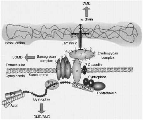
Duchenne muscular dystrophy (DMD) and Becker muscular dystrophy (BMD) are the prototype muscular dystrophies affecting 1 in 3500 and 1 in 30,000 liveborn male infants, respectively. They are also known as the Xp21 dystrophies because of the locus of the involved gene. The defective gene product is dystrophin (see Fig. 36.1). In DMD, dystrophin is essentially absent and its absence is associated with a severe phenotype; in BMD, the gene deletion or point mutation results in a reduction in the amount of dystrophin, or in the formation of a truncated protein, and a milder phenotype. Because the inheritance is X-linked-recessive, males are affected and
females are carriers. Female carriers may in fact manifest the disease to varying degrees because of skewed inactivation of the X chromosome (Lyon hypothesis).


Figure 36.1 Diagrammatic association of the dystrophin-associated protein complex. BMD, Becker muscular dystrophy; CMD, congenital muscular dystrophy; CYS, cysteine; DMD, Duchenne muscular dystrophy; LGMD, limb-girdle muscular dystrophy; NOS, nitric oxide synthetase. (From Bonnemann CG, McNally EM, Kunkel LM. Beyond dystrophin: current progress in the muscular dystrophies. Curr Opin Pediatr 1996;8:569-582, with permission.)


TABLE 36.2 CLASSIFICATION OF MUSCULAR DYSTROPHIES


X-linked-recessive


Duchenne muscular dystrophy (dystrophin) (Xp21)


Becker muscular dystrophy (dystrophin) (Xp21)


Emery-Dreifuss muscular dystrophy (emerin) (Xq28)


Autosomal dominant


Limb-girdle muscular dystrophy (LGMD) (type 1)


LGMD 1A (5q31)


LGMD 1B (1q11-21)


LGMD 1C (6q23)


LGMD 1D (caveolin) (3p25)


LGMD 1E (7q)


Emery-Dreifuss muscular dystrophy (Lamin A/C) (1q21.3)


Myotonic dystrophy (19q13)


Facioscapulohumeral muscular dystrophy (4q35)


Oculopharyngeal muscular dystrophy (14q11.2-q13)


Bethlem myopathy (21q22.3 and 2q37)


Autosomal recessive


Congenital muscular dystrophy (CMD)


Classic or pure CMD (without CNS involvement)


Merosin-positive


Merosin-negative (6q2)


CMD with CNS involvement


Fukuyama CMD (fukutin) (9q31-q33)


Walker-Warburg syndrome (9q34)


Muscle-eye-brain disease (1p3)


Limb-girdle muscular dystrophy (type 2)


LGMD 2A (calpain) (15q15)


LGMD 2B (dysferlin) (2p13)


LGMD 2C (γ-sarcoglycan) (13q12)


LGMD 2D (α-sarcoglycan) (17q21)


LGMD 2E (β-sarcoglycan) (4q12)


LGMD 2F (δ-sarcoglycan) (5q33-34)


LGMD 2G (telethonin) (17q11-12)


LGMD 2H (e3-ubiquitin ligase) (9q31-34)


LGMD 2I (fukutin related protein) (19q13)


LGMD 2J (titin) (2q31)


Gene loci and gene products are within parentheses.


CNS, central nervous system.


Children with DMD typically present after the age of 2 years with a history of excessive falling, apparent clumsiness, and toe walking. Evidence of proximal weakness may be elicited while taking the history (difficulty in negotiating stairs or getting up from the floor, Gower maneuver). Examination reveals calf hypertrophy (pseudohypertrophy), proximal muscle weakness, hyporeflexia, and the presence of a Gower sign. The creatine kinase level is typically markedly elevated (in the thousands). Electromyography (EMG) is usually unnecessary for the diagnosis and shows a myopathic pattern. Muscle biopsy shows dystrophic changes (see preceding text), and special immunohistochemical stains show an absence of dystrophin. DNA studies reveal a large deletion or duplication in approximately 60% of cases. Full gene sequencing, however, will pick up smaller deletions and/or point mutations in the remainder. Children are nonambulatory by age 13, and premature death typically occurs in the early twenties.

Although no cure is available, treatment is aimed at prolonging independent ambulation (orthoses or steroids), avoiding contractures, and providing early surgical stabilization of scoliosis to preserve respiratory function and facilitate seating in a wheelchair. Cardiac and respiratory failures develop during the teenage years. Nocturnal hypoventilation resulting in early morning headache, nausea, and lethargy secondary to hypercarbia can be effectively managed with nasal bilevel positive airway pressure (Bi-PAP) ventilation. Children with DMD often have an intelligence quotient that is lower than normal and may require special educational services.

In BMD, the preservation of a reduced or altered form of dystrophin is associated with a milder phenotype. Children typically present at a slightly later age and are usually independently ambulant beyond their 16th birthday. The motor problems and the clinical, biochemical, and pathologic features are essentially the same as in DMD. Staining of the
muscle for dystrophin, however, shows some preservation of the protein, and gene deletion studies, when results are positive, may be able to predict the phenotype. If the mutation is associated with preservation of the reading frame sequence, BMD is typically predicted (with a few exceptions), whereas if the mutation involves the promoter region or disrupts the translational reading frame (out of frame), a Duchenne phenotype is typical. In BMD, prolonged ambulation with orthoses may be possible, and life expectancy is longer. However, cardiomyopathy often develops earlier, and must be closely monitored.

Female siblings and mothers of affected children should be screened by determining the creatine kinase level to look for possible carrier status. However, if a deletion is found in the index case, this is a more sensitive and specific marker for screening family members. Approximately two thirds of mothers of children with DMD or BMD carry the deletion. The reasons why not all mothers carry the deletion or point mutation are the high frequency of spontaneous mutation of the gene (1 in 10,000 gametes) because of its large size (2.5 million base pairs, or approximately 1% of the X chromosome) and germline mosaicism. The presence of a deletion allows for prenatal diagnosis.


Emery-Dreifuss Muscular Dystrophy

Emery-Dreifuss muscular dystrophy (EDMD) is characterized by a triad of:

Jul 5, 2016 | Posted by in CRITICAL CARE | Comments Off on Neuromuscular Disorders: Muscular Dystrophies and Congenital Myopathies

Full access? Get Clinical Tree

Get Clinical Tree app for offline access