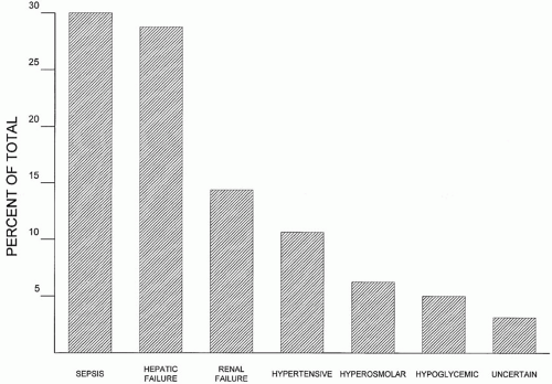
Neurological Emergencies


Neurological Emergencies


Alterations of consciousness and cognition are both significant daily problems in the intensive care unit (ICU). It is not shocking that depressed consciousness, especially that sufficient to be called coma, has profound prognostic importance but many clinicians are surprised to learn that development of confusion or delirium also has powerful predictive value.


▪ DELIRIUM

Delirium is a rapidly developing, often unrecognized, syndrome of confusion and inattention that affects more than 80% of all ICU patients. Two distinct subtypes of the condition are described. In the hyperactive form, patients are agitated and combative, in the hypoactive form, they are calm and withdrawn. Because the patients with agitated delirium are bigger management problems, not surprisingly it is recognized and reported more frequently. The elderly are more likely to manifest the hypoactive variety of the disorder. The most frequent conditions associated with delirium are shown in Figure 34-1.


FIGURE 34-1 Relative frequency of (nonsedative) causes of delirium in nontrauma ICU patients. Sepsis, hepatic failure, and renal failure rank as the most common causes.

The mechanism(s) of delirium is not known but strong associations have been recognized. Delirium is much more common in the elderly, in patients with baseline intellectual or cognitive impairment, and those with impaired hearing or vision. Alcoholism and drug abuse are also risk factors but there does not appear to be any particular acute medical or surgical illness that carries a disproportionate risk of causing the disease. However, there are contributing environmental factors. For example, sleep deprivation, invasive catheter and tube use, and application of physical restraints all appear to be associated with the condition. With regard to drugs, sedatives, analgesics, and medications that have anticholinergic properties are commonly linked with delirium.

Diagnosing delirium is not difficult and many ICUs have now incorporated one of the three most popular scoring schemes into routine care. Of available methods, the Confusion Assessment Method for ICU (CAM-ICU) is perhaps the simplest validated system. (http://www.icudelirium. org/delirium/CAM-ICUTraining.html) When combined with a standardized scoring system for consciousness, like the Richmond Agitation Sedation Scale (RASS) (see Chapter 17), both wakefulness and delirium can be accurately quantitated in seconds.

Development of delirium is an independent predictor of morbidity and mortality. The odds of reintubation are increased; the duration of mechanical ventilation and ICU and hospital length of stay are prolonged by delirium. The 6-month mortality rate is 20% higher among patients experiencing ICU delirium, and among survivors, residual cognitive impairment is common. Unfortunately, little is known about the treatment of delirium. Retrospective studies suggest the use of haloperidol may be associated with higher survival. In one of the few randomized trials conducted, olanzapine appears to have comparable safety and effect to haloperidol. Although benzodiazepines can prevent the development of delirium in the setting of alcohol or sedative withdrawal, as a practical matter they often exacerbate the condition once it is manifest. The effectiveness of preventative measures is unknown. Nevertheless, making sure that patients have their glasses, dentures, and hearing aids, and that they are frequently “reoriented” to time, place, location, and situation may serve as a deterrent. Likewise, providing a quiet environment with periods of sleep that coincide with normal solar cycles makes sense.
Removal of unnecessary tubes and monitoring devices and beginning a program of physical activity as soon as feasible may also serve to prevent or reverse delirium. Even if none of these measures are beneficial for delirium, they will not be harmful and may offer noncognitive therapeutic benefits.


▪ COMA

Coma is a sleeplike state of unconsciousness from which patients cannot be awakened. Although some involuntary movements (spinal reflexes and “posturing”) may occur, there is no speech or purposeful eye or limb movement. Less-profound stages of suppressed consciousness are often termed obtundation, stupor, and lethargy. Imprecise terms such as semicoma or light coma may confuse families and subsequent examiners and should be abandoned in favor of a simple unambiguous description of the highest level of function.


Pathophysiology

Consciousness has two components: arousal and awareness. Failure of arousal results from reticular activating system (RAS) or diffuse bilateral hemispheric dysfunction. Continuous stimulation by the RAS is required for the appearance of wakefulness. Conversely, awareness, a cognitive function, requires coordinated function of both cerebral cortices. Arousal may occur without awareness, but not the converse. From its origin in the midpons, the RAS radiates diffusely outward to the cerebral cortex. It is this protected origin and wide distribution that prevents coma, unless a diffuse process impairs both the cerebral cortices or the RAS is interrupted near its pontine root. Although this simple schema generally explains arousal and awareness, the dominant cortical hemisphere plays a disproportionate role in maintaining consciousness, and selective damage to both frontal lobes can also result in coma.


TABLE 34-1 CLINICAL CHARACTERISTICS OF CAUSES OF COMA


CAUSE OF COMA


CHARACTERISTIC


TRAUMATIC OR VASCULAR


TOXIC OR METABOLIC


History


Injury


Uncontrolled hypertension


Seizure


Toxin, drug exposure history History of liver or kidney failure, COPD or diabetes Recent hypoxic event


Onset


Abrupt


Gradual


Rate of progression


Rapid (minutes) deterioration


Slower (hours) progression common


Pattern of progression


May be stuttering or gradual Rostrocaudal loss of function


Global impairment from the start No rostrocaudal pattern


Focality


Focal lesions common


Focal lesions rare


Coma may arise from a wide variety of diffuse or focal conditions affecting the central nervous system (CNS); however, all coma results from four basic pathophysiologic mechanisms: (a) metabolic or toxic encephalopathy, (b) generalized seizures, (c) compression of the midbrain or cerebral cortices by structural lesions or increased intracranial pressure (ICP), and (d) inadequate cerebral perfusion. The extent of neurological impairment of any potential cause of coma is modified by the patient’s age and underlying neurological and vascular status. Metabolic insults that do not change cognition in a healthy young person can result in profound coma in an elderly patient with impaired circulation.


Etiology


Metabolic Disorders

Outside the neurosurgical setting (e.g., head trauma, subarachnoid hemorrhage (SAH), brain tumor), metabolic encephalopathy is the most common cause of coma. A comparison of the usual clinical features of structural and metabolic coma is presented in Table 34-1. Because metabolic encephalopathy affects the cortex and brainstem diffusely, abrupt focal deficits and progressive rostrocaudal losses seen with supratentorial mass lesions do not usually occur. Rather, patients exhibit slowly evolving symmetric deficits, often preceded by somnolence or confusion. A variety of common disorders, including drug overdose, hypoxia, hypotension,
hypoglycemia, dehydration, sepsis, hepatic encephalopathy, and uremia can all contribute to impaired consciousness. The common reversible causes of coma are summarized in Table 34-2.


TABLE 34-2 COMMON REVERSIBLE CAUSES OF ALTERED MENTAL STATUS


MEDICAL CONDITIONS


SURGERY AND TRAUMA


Hypoperfusion


Drug overdose


Hypoperfusion


Hypoxemia


Carbon monoxide


Hypoxemia


Hypercapnia


Hypercalcemia


Cholesterol embolism


Hypoglycemia


Wernicke encephalopathy


Fat embolism


Status epilepticus


Temperature disorders


Air embolism


Myxedema


CNS hemorrhage


Traumatic subdural hematoma


Hypertension


CNS infections (e.g., brain abscess, meningitis, encephalitis)


Diffuse neuronal shear injury


It is difficult to provide absolute guidelines as to the magnitude of a single abnormality necessary to cause coma, because multiple derangements often coexist, and the age of the patient, presence of underlying diseases, and the rapidity of development also determine impact. Yet, as a general rule, it is uncommon for glucose greater than 50 or less than 500 mg/dL, Na+ greater than 120 or less than 155 mEq/L, Ca2+ less than 12 mg/dL, or Mg2+ greater than 0.8 or less than 10 mg/dL to produce coma. Only very rarely do disorders of other electrolytes alter consciousness. The precise cause of unconsciousness in patients with renal failure is unknown; however, uremia alone rarely causes coma until the blood urea nitrogen (BUN) exceeds 100 mg/dL. (Renal insufficiency probably alters consciousness through multiple mechanisms, including metabolic acidosis, electrolyte abnormalities, metabolic toxin or drug accumulation, and increased permeability of the blood-brain barrier.) Similarly, the exact cause of coma in patients with liver failure is unknown, but accumulation of endogenous (e.g., glutamine) and exogenous toxins (e.g., drug metabolites) and cerebral edema all play some role in the process (see Chapter 31). Cerebral edema is a much bigger problem in the setting of acute hepatic failure. There is such a poor correlation between serum ammonia levels and mental status that no rule can be offered as to the level of ammonia associated with coma.

More firm guidelines can be offered with respect to the impact of hypoxemia and hypercapnia on consciousness. Tolerance of hypoxemia depends not only on the extent of desaturation but also on compensatory mechanisms available. The major mechanisms of acute compensation are increased cardiac output, O2 extraction, and anaerobic metabolism, and improved unloading of O2 resulting from tissue acidosis. Most individuals without cardiac disease or anemia remain asymptomatic until PaO2 falls below 50 mm Hg. At that level, malaise, lightheadedness, nausea, vertigo, impaired judgment, and incoordination are noted. Confusion develops as PaO2 falls into the 35 to 50 mm Hg range. As PaO2 declines below 35 mm Hg, urine output slows, bradycardia and conduction system blockade develop, lactic acidosis appears, and the patient becomes lethargic or obtunded. At levels near 25 mm Hg, the normal unadapted individual loses consciousness, and minute ventilation falls because of respiratory center depression. As for all other metabolic toxins, the effects of carbon dioxide (CO2) depend not only on the absolute level of CO2 but also on its rate of accumulation. A PaCO2 less than 60 mm Hg is often asymptomatic, but as levels rise above this threshold, headache and lethargy are commonly observed. Asterixis and myoclonus occur as levels mount even higher, but surprisingly massive elevations in CO2 (often double or triple normal values) are usually necessary to render a patient unconscious.

Generally, a cerebral perfusion pressure (CPP) (mean arterial pressure [MAP]—intracranial pressure [ICP]) must exceed 50 mm Hg to perfuse the brain adequately; however, otherwise young healthy patients may maintain consciousness despite severe reductions in MAP. By contrast, patients with chronic hypertension, cerebrovascular disease, or elevated ICP may be underperfused at much higher pressures.

Severe sepsis frequently alters consciousness; however, mental status changes usually fall short of true coma. The mechanisms are multifactorial. Obviously, direct infection of the CNS can render a patient unconscious. In addition, severe sepsis is usually associated with some degree of hypoxemia and almost 75% of septic patients will develop hypotension—both factors which contribute to mental status changes. Possibly most interesting,
is the finding that inflammatory cytokines (e.g., interleukin-1) may directly impair consciousness. Sepsis-induced coma is particularly common in the febrile elderly patient, where altered mental status presents a difficult management decision. Frequently deliberated diagnoses are severe sepsis alone, or sepsis complicated by dehydration, electrolyte abnormalities, meningitis, and intracranial structural lesions such as a brain abscess or hemorrhage. Timely antibiotic therapy is desirable in all infectious situations, but confirming the diagnosis and nailing down a specific organism so as to best choose antimicrobial therapy usually requires a lumbar puncture (LP). Unfortunately, LP incurs some risk in the presence of coagulopathy, anticoagulation, or intracranial mass lesion. Therefore, deciding the order in which to perform a head computed tomography (CT) scan, do an LP, and administer antibiotic therapy is often debated. In febrile nonimmunocompromised patients without evidence of a focal neurologic defect, papilledema, or history of trauma, an LP is almost certainly safe and can be performed before or without a head CT scan. (Not that long ago, the decision to perform an LP was always made only based upon history and examination.) If head trauma, papilledema, new onset seizures, or focal neurologic deficit complicates the evaluation of a potentially infected patient, probably the best course is to obtain blood cultures, administer empiric antibiotics, and proceed to the CT scanner before LP. In any case, if logistical limitations preclude prompt CT scanning and intracranial infection is a real possibility, it is best to begin antibiotic treatment, even if doing so precludes a definitive microbiological diagnosis. Although it is rational to administer antibiotics as quickly as practical in patients with suspected meningitis, there is little if any data to support the often-quoted need to dose antibiotics within 1 h of suspecting the diagnosis.

Medication ingestion or toxin exposure is the most frequent cause of nontraumatic coma. Opiates, benzodiazepines, ethanol, and antidepressants are the most common drug classes implicated. Chemical-induced mental status changes are typically multifactorial; commonly several consciousness-depressing drugs are simultaneously ingested, and the drugs or toxins often lead to changes in oxygenation or perfusion that can cause coma in and of themselves. For example, narcotic overdose is often combined with benzodiazepine and alcohol ingestion. Although direct effects of these drugs alone can cause coma, they can also impair consciousness by producing hypoventilation or hypotension (see Chapter 33).


Seizures

Generalized seizures may induce unconsciousness during the ictal phase or as a postictal phenomenon. Distinguishing a seizure from other causes of coma is easy if convulsive activity is observed or consciousness returns rapidly after a suspected seizure in an otherwise healthy patient. However, in the ICU many seizures do not cause visible convulsions. In addition, patients with previous stroke, metabolic encephalopathy, or prolonged seizures (status epilepticus) may suffer a long postictal period. In turn, status epilepticus often has an underlying structural or metabolic cause. Thus, delayed return of consciousness following a seizure should prompt consideration of a structural lesion (e.g., tumor, stroke, SAH, subdural hematoma) or underlying metabolic disorder (e.g., hypoglycemia, toxin ingestion, or electrolyte disturbance). As with metabolic encephalopathy, seizures that cause loss of consciousness generally tend to produce symmetric neurological defects unless there is an underlying structural abnormality. In contrast to metabolic encephalopathy, the loss of consciousness associated with seizures is usually instantaneous rather than gradual.


Structural Lesions

Although supratentorial mass lesions are generally confined to one hemisphere, they cause coma by increasing ICP or by impairing RAS function through pressure on the brainstem. Unless located exactly in the midline, supratentorial mass lesions usually produce hemispheric (unilateral) findings that precede loss of consciousness. Because cerebral function is progressively lost in a rostrocaudal manner, there is a relatively predictable progression. If unchecked, the process culminates in central or uncal herniation. In central herniation, the midbrain is pressed straight down into the foramen magnum. Clinical manifestations include miosis, decerebrate posturing, and a lateral gaze palsy as the sixth cranial (abducens) nerve is stretched. Uncal herniation occurs as asymmetric pressure forces the medial portion of the temporal lobe to cross the tentorium, compressing the midbrain. Clinical manifestations include a fixed dilated pupil, third cranial (oculomotor) nerve palsy, and hemiparesis. By contrast, when infratentorial lesions (e.g., brainstem strokes, cerebellar hemorrhage)
cause coma, very rapid loss of consciousness without rostrocaudal progression is typical.


Approach to the Comatose Patient


History

Because pathophysiology and treatment differ radically, metabolic and structural causes of coma must be distinguished as quickly as possible. The history is helpful in making this separation if it reveals trauma or drug ingestion. Sudden onset of coma suggests a seizure or a vascular event (e.g., SAH, brainstem stroke). The onset of coma after minutes to hours of a focal deficit suggests supratentorial intracerebral hemorrhage with progressively increasing ICP. A slowly evolving focal deficit occurring over days to weeks before loss of consciousness suggests abscess, tumor, or subdural hematoma, whereas progression to coma over minutes to hours without a focal deficit favors a metabolic cause. The setting in which coma develops may suggest trauma or hyperthermia or hypothermia from environmental or toxic exposure (e.g., organophosphates, carbon monoxide). Medications found near the comatose patient are particularly helpful. For example, not only may the offending compound(s) be discovered, but medication containers bearing the name of the prescribing physician may allow further history to be obtained. At the very least, knowledge of a patient’s medications provides a forensic picture of underlying diseases. In patients with alcoholism, intoxication with ethanol, ethylene glycol, methanol, or isopropyl alcohol should be suspected. In patients with diabetes, hypoglycemia and diabetic ketoacidosis (DKA) are common causes of coma. Underlying medical problems such as hypothyroidism, renal failure, cirrhosis, or psychiatric illness also increase the likelihood of a metabolic etiology, whereas a history of falling, previous stroke, brain tumor, extracranial neoplasm, or atrial fibrillation favors structural or vascular problems. Patients known to have malignant tumors are subject to both structural lesions (e.g., metastases and hemorrhage) and metabolic causes (e.g., hyponatremia, hypercalcemia). Similarly, uncontrolled hypertension can induce metabolic (e.g., hypertensive encephalopathy) or structural (e.g., intracerebral hemorrhage) coma. Immunocompromised patients, especially those with human immunodeficiency virus (HIV) infection are at higher risk for structural causes of coma including, CNS infections and lymphoid malignancies.


Physical Examination

Physical examination is most helpful in differentiating structural from metabolic causes of coma when it reveals evidence of focal or lateralizing signs. In such patients, a metabolic etiology is uncommon. (Exceptions to this rule occur in patients with hepatic failure, hypoglycemia, prior stroke, and postictal patients.) Comatose patients should always be fully disrobed and examined for evidence of occult trauma. Although physical evidence of head trauma suggests a structural cause, up to 50% of trauma patients also suffer from intoxication, oftentimes sufficient to produce coma. Boggy areas of the skull suggest depressed skull fracture, while the Battle sign (postauricular hematoma), “raccoon eyes,” and bloody nasal or aural discharge suggest basilar skull fracture. In traumatized patients, it is critical to exclude cervical spine instability before the neck is manipulated because serious spinal injuries commonly accompany cranial lesions severe enough to cause coma. Echymoses, mucosal bleeding, or petechiae may implicate coagulopathy as the cause of intracranial bleeding. Incontinence of stool or urine or tongue laceration strongly suggests recent seizure. Atrial fibrillation, a large heart, and history of recent myocardial infarction are all associated with cerebral embolic disease. (In general, embolic disease is an uncommon cause of coma because of the limited area of cerebral infarction caused by emboli.) Cardiac murmurs should raise suspicion of endocarditis-induced septic embolism or brain abscess. Arrhythmias only result in coma when they cause hypotension or cerebral emboli. In this setting, isolated carotid bruits are of little significance because occlusive carotid disease is a rare cause of coma. Nuchal rigidity suggests meningitis, encephalitis, or SAH.

Vital signs provide additional clues to diagnosis. Although hypertension may accompany any cause of increased ICP, severe hypertension suggests intracerebral or SAH, particularly if accompanied by nuchal rigidity or focal neurological signs, respectively. Hypertensive encephalopathy alone is less likely to produce coma and is usually characterized by a nonfocal examination and blood pressures greater than 240/130 mm Hg. Stimulant intoxication with amphetamines, cocaine, phencyclidine, or phenylpropanolamine, should also be
considered in patients with altered mental status and hypertension. Cocaine-induced hypertension alone may cause delirium but if coma is present, intracranial hemorrhage is likely. Although young patients without underlying vascular or cerebral disease may remain awake with very low (40 to 50 mm Hg) MAPs, the elderly, patients with chronic hypertension, and those with concurrent metabolic encephalopathy or structural lesions tolerate hypotension less well.

Heatstroke, serotonin syndrome, neuroleptic malignant syndrome, malignant hyperthermia, stimulant intoxication, and any number of infections may cause fever sufficient to impair consciousness. Patients with hyperthermia or hypothermia frequently have an infection accompanying the primary temperature disorder that may itself disturb consciousness (e.g., pneumonia, brain abscess, or meningitis). Although primary aberrations of body temperature can directly cause coma, they rarely do so until core temperatures exceed 105°F or fall below 80°F (see Chapter 28). Bradypnea and hypoventilation most often result from the sedative effects of drugs or alcohol, accumulated hepatic or renal toxins, hypothyroidism, or far advanced brainstem compression. Tachypnea is a nonspecific finding but usually arises from one of four basic causes: (a) inappropriate ventilatory control, (b) hypoxemia, (c) compensation for metabolic acidosis, or (d) reduced lung or chest wall compliance. Contrary to popular teaching, specific “pathognomonic” respiratory patterns have little localizing or prognostic value, with one notable exception— uncoordinated, irregular (ataxic) respiration usually indicates severe medullary impairment and impending respiratory collapse.


Expanded Neurological Examination

After the vital signs are obtained, ventilation and perfusion are stabilized, and initial history and physical examination are performed, neurological function should be examined in a stepwise fashion. The five key features of the initial neurological examination may be remembered by the mnemonic “SPERM”: (a) State of consciousness, (b) Pupillary response, (c) Eye movements, (d) Respiratory rate and pattern, and (e) Motor function.

One of four terms should be used to describe the state of consciousness: (a) alert, (b) lethargic (aroused with simple commands), (c) stuporous (aroused only with vigorous stimulation—usually pain), and (d) comatose (unarousable).

Pupil size, congruency, and response to light and accommodation should be described. Pupillary function is controlled by the midbrain. Therefore, if the pupils function normally, the cause of coma either is a structural lesion located above the midbrain or is metabolic. Small “pinpoint” pupils usually result from pontine hemorrhage or from ingestion of narcotics or organophosphates. (Meperidine frequently fails to produce the miotic pupils typical of other narcotics.) Pupillary responses almost always remain intact in metabolic causes of coma. (Exceptions to this rule include atropine and atropine-like substances and the now rare glutethimide intoxication in which “fixed and dilated” pupils may occur.) With uncal herniation from increased supratentorial pressure, the third cranial nerve is compressed on the tentorial edge resulting in unilateral pupillary dilation and fixation. If increased pressure goes unrelieved, complete diencephalic herniation can occur resulting in fixed midposition (3 to 5 mm) pupils.

Normal movement of the eyes requires an intact ponto-medullary-midbrain connection. The resting position of the gaze, the presence of nystagmus (horizontal, vertical, or rotatory), and the response to head movements (oculocephalic testing) or to cold tympanic membrane stimulation (oculovestibular testing) should be recorded. A normal response to oculocephalic testing is conjugate eye movement away from the direction of head rotation. A normal response to cold oculovestibular testing is conjugate eye movement toward the side of stimulation. Cervical spine stability must be ensured before oculocephalic maneuvers are performed. Likewise, tympanic membrane integrity should be confirmed before oculovestibular testing to prevent introduction of water into the cerebrospinal fluid (CSF) through a basilar skull fracture. Although endogenous toxins accumulated from hepatic or renal failure usually do not impair coordinated eye movements, exogenous toxins (drugs) frequently impair eye movements. In addition, depletion of thiamine (Wernicke syndrome) can result in horizontal and vertical nystagmus. In pontine disorders, the medial longitudinal fasciculus is often dysfunctional but the sixth cranial nerve function is preserved. Therefore, ipsilateral eye abduction is intact, but contralateral adduction is impaired. Quite simply, if rotation of the head (oculocephalic) and vestibular stimulation (calorics) produce no change in eye position, the pons is
nonfunctional. If only the eye ipsilateral to caloric stimulus abducts, a lesion of the medial longitudinal fasciculus (encapsulated by the pons) should be suspected.

Description of the respiratory pattern is less helpful than has been previously suggested; however, ataxic breathing is a marker of severe brainstem dysfunction. Despite the nonspecificity of most breathing patterns, the respiratory rate can provide valuable clues to the etiology of coma (see “Physical Examination” above).

The highest observed level of motor function should be noted (e.g., “Spontaneously moves all extremities,” “Withdraws only right arm and leg from noxious stimulus,” “No response to pain”). Motor function in pontine compression is often limited to extensor (decerebrate) posturing, whereas lesions above the pons can produce flexor (decorticate) posturing. If a structural lesion compresses the centers for respiration and heart rate control on the dorsal medullary surface, the patient also will be flaccid, without eye movements, and will have midposition, unreactive pupils. Although “posturing” is an ominous finding in most settings, rarely it is the result of metabolic encephalopathy, (especially acute hepatic failure) and in such cases, is completely reversible.

The results of the neurological examination are often reported using a standardized scale. Currently, the Glasgow Coma Score (GCS) is the most commonly used composite score. In this system, best motor, verbal, and eye opening responses are tallied in a score ranging from 3 to 15 (Table 34-3).


TABLE 34-3 THE GLASGOW COMA SCALE


Eyes


Open


Spontaneously


4


To verbal command


3


To pain


2


No response


1


Best motor response


To verbal command


Obeys


6


To painful stimulus


Localizes pain


5


Flexion—withdrawal


4


Flexion—decorticate


3


Extension—decerebrate


2


No response


1


Best verbal response


Oriented, converses


5


Disoriented, converses


4


Inappropriate words


3


Incomprehensible sounds


2


No response


1


Total


3-15


Localizing the Level of Dysfunction

If history or examination reveals a sequential, rostrocaudal loss of function, either a supratentorial mass lesion or diffusely increased ICP is the most likely etiology of coma. Fundascopic examination that demonstrates papilledema is virtually diagnostic of increased ICP or hypertensive encephalopathy.

Although the thalamus-diencephalon cannot be directly examined, injury to this area usually depresses consciousness but spares motor function. (Because pupillary and ocular movements are controlled by the midbrain and pons, respectively, they typically remain unaffected.) The respiratory pattern in thalamic dysfunction is unpredictable. Injury extending lower to the midbrain level usually results in loss of motor function and decorticate, or flexor, posturing. Although pupillary diameter is generally midposition (approx. 3 mm), midbrain injury tends to spare pupil reactivity and eye movements. When damage extends further to the pontine level, pupillary function is routinely impaired. Motor responses are often limited to extensor (decerebrate) posturing. If compression progresses to the medullary level, all motor function is usually lost, as are pupillary response and eye movements. It is only with medullary compression that respiratory rhythm is predictably affected, becoming ataxic.


Laboratory Evaluation

Body fluids should be collected to evaluate potential metabolic and toxic causes of coma. Situationally appropriate testing is indicated, even in patients
with obvious head trauma, because of the possibility that a metabolic cause may coexist or may have precipitated the trauma (e.g., alcohol, carbon monoxide). Laboratory determinations should include indices of renal and hepatic function, serum glucose and electrolyte determinations, hemoglobin and arterial blood gases, and when appropriate, carboxyhemoglobin determinations. If the history, physical examination, or initial laboratory testing suggests drug overdose or poisoning, a toxicology profile, and if indicated specific levels of compounds not included in a typical toxicology screen (e.g., aspirin, acetaminophen, ethylene glycol, methanol), should be obtained (see Chapter 33).


Treatment

The major diagnostic differential is to separate structural from metabolic causes of coma, but initially in all cases similar supportive treatment should be undertaken. The airway should be secured, perfusion stabilized, and oxygen administered. The cervical spine should be immobilized if there is any suspicion of trauma. An IV line should be established to obtain appropriate laboratory specimens and administer fluids and medications. Because of its time-sensitive importance, glucose levels should be tested at the bedside. If testing is not immediately available or if the measured glucose value is low, 50% dextrose in water (D50 W) and thiamine (approx. 1 mg/kg) should be given accompanied by thiamine.

Naloxone, a narcotic antagonist, and flumazenil, a benzodiazepine antagonist, can temporarily reverse narcotic and benzodiazepine-induced coma, respectively, and thus serve as useful diagnostic tools. The duration of action of both antagonists is less than that of their agonist counterparts; thus neither compound is a reliable substitute for intubation and mechanical ventilation in patients with sedative-induced respiratory failure. Recurrent coma often follows a single dose of either reversing agent in patients who are not closely monitored.

After initial stabilization and primary evaluation, a more detailed neurological examination and specific diagnostic testing can be undertaken. In most cases, a head CT scan will be performed. In patients admitted to the hospital with coma, the CT scan is much more likely to reveal a structural cause of coma than for patients who have been in the ICU for a period of time. (The important exception to this rule is the ICU patient who has a new focal deficit or first seizure.) Magnetic resonance imaging (MRI) scanning usually adds little to CT results in patients with coma, although occasionally subtle cerebral edema, venous sinus thrombosis, and acute ischemic strokes can be seen on MRI of patients with nondiagnostic CT scans. In most febrile comatose patients, blood cultures, LP, and institution of antibiotics are indicated. However, if lateralizing neurological signs are present or if there is a history of seizure, trauma, or immunocompromise, CT of the head should usually precede LP but not antibiotic administration. For patients with a clear metabolic cause of coma (e.g., hepatic or renal failure), organ specific treatment should be undertaken.


Prognosis

Surprisingly, the history and clinical examination over time are better predictors of outcome than sophisticated ancillary tests. Approximately 90% of comatose patients who will completely recover show significant neurological improvement within 3 days. As a general rule, the faster the motor activity returns the better the prospects for full recovery. (Most patients with successful recoveries have purposeful motor activity within 24 h.) With regard to etiology, drug-induced coma has an excellent prognosis. By contrast, the prognosis of coma persisting greater than 72 h is terrible if the etiology is ischemia or trauma. More than half of such patients with ischemic injury are dead and an additional 40% are in a persistent vegetative state or have severe impairment at one year. (Less than 5% have good recovery.) Outcomes from traumatic coma are better but still poor; about 30% of patients are dead and another 45% are in a persistent vegetative state or have severe cognitive deficits at one year. Fewer than 10% have good functional recovery.


▪ SEIZURES


Pathophysiology

Seizures result from paroxysmal neuronal discharges that cause generalized or focal neurological signs. Most generalized seizures begin as a focal cortical discharge. Knowledge of the cell biology of seizures has advanced considerably. For example, we now know seizures are associated with excessive activity of N-methyl D-aspartate (NMDA) receptors and inadequate stimulation of gamma amino butyric acid (GABA) receptors. Understanding the role these receptors helps explain the effectiveness of GABA stimulants as anticonvulsants.


Although seizures have been classified in many ways, it is probably most useful to think of them in terms of their duration (brief vs. continuous) and scope (generalized vs. focal). Duration is important because prolonged seizures become more refractory to treatment, irreversibly injure neuronal tissue, and cause systemic metabolic problems (e.g., acidosis, hypoxia, hyperthermia, and rhabdomyolysis). Although there is no standard definition of the commonly used term “status epilepticus,” it is generally agreed that a seizure lasting greater than 10 min or a series of recurring seizures without an intervening period of consciousness qualifies. Similarly, although not standardized, the term “refractory status epilepticus” has been applied to the highly lethal situation in which seizures last greater than 2 h or cannot be controlled with two or more anticonvulsants. Focality is also noteworthy because it suggests a discrete structural abnormality. Although seizures usually present as localized or generalized phasic muscle spasms, in the ICU, seizures occasionally masquerade as unexplained coma or puzzling sensory or psychiatric disturbances.


Etiology

One of five basic mechanisms is responsible for convulsions. Seizures arise from intrinsic electrical instability (epilepsy), toxic or metabolic disturbances (e.g., electrolyte imbalances, alcohol, drug effect), or structural lesions (e.g., trauma or tumor), infectious causes (e.g., meningitis, cerebritis, brain abscess), or abnormalities of brain perfusion (global hypoxia). In general, these etiologic factors segregate into two prognostic groups. Patients with idiopathic epilepsy, subtherapeutic drug levels, and alcohol-related seizures tend to have an excellent prognosis, whereas victims of stroke, trauma, tumor, encephalitis, or direct CNS poisons tend to have a poor prognosis.

For the most part, seizures occur at the extremes of age and the most frequent causes of seizures also vary by age. For example, children most commonly have seizures as the result of fever, infection, or a change in anticonvulsant medications used to treat idiopathic epilepsy. By contrast, young adults are much more likely to seize from SAH, trauma, noncompliance with anticonvulsants, or drug use or withdrawal (e.g., tricyclic antidepressants, cocaine, alcohol). In the older adult, stroke, subdural hematoma, and tumor become more common. Hypoglycemia and CNS infections (e.g., meningitis, encephalitis) effect patients of all ages.

High fever (especially in children), drug withdrawal (particularly anticonvulsants, ethanol, barbiturates, benzodiazepines, baclofen), and iatrogenic overdoses of isoniazid, penicillin, imipenem, tricyclic antidepressants, theophylline, or lidocaine are common metabolic causes. Electrolyte disturbances may also induce seizures, especially when such changes occur abruptly (e.g., acute hyponatremia, disequilibrium following dialysis).

Although most seizure disorders that occur in outpatients are idiopathic, this is true less often in the ICU, where such treatable conditions as drug or alcohol withdrawal, metabolic imbalances, drug toxicity, and acute structural lesions are more common. Most important among the metabolic precipitants are uremia, hypoglycemia, hypocalcemia, hypomagnesemia, and hyponatremia. CNS infections (meningitis, encephalitis, and brain abscess) are frequent causes of ictus; about one third of adults with bacterial meningitis will experience a seizure. HIV infection represents a particular hazard for CNS infections, including toxoplasmosis and viral encephalitis.


Diagnosis

A seizure diagnosis is usually made from the history and observation of an attack. Occasionally, historical features and/or the clinical appearance are so atypical as to require confirmation by electroencephalography (EEG). In such cases, an intra-ictal EEG may be diagnostic and the pattern of discharge may help determine etiology. For example, EEG localization of seizure discharge to the base of the temporal lobes (or appropriate MRI findings) suggests herpes encephalitis. EEG recording may also reveal unsuspected seizure activity in a patient with unexplained coma. In fact, up to 20% of unresponsive ICU patients may have occult seizures and an even higher percentage (50%) of patients who remain unresponsive after having a convulsion have been found to have ongoing nonconvulsive seizures. A head CT or MRI scan is indicated for new onset seizures, those accompanied by a preceding or persistent focal neurological deficit, and in those refractory to simple medical therapy. In such patients, a CT often reveals a structural cause (e.g., vascular malformation, primary or metastatic tumor, or subdural subarachnoid or parenchymal hemorrhage). In patients with a known seizure disorder, a CT scan is not necessary to evaluate each
recurrence; however, it should be remembered that even patients with epilepsy develop strokes, tumors, and CNS infections. Hence, recurrent seizures should not be reflexly ascribed to a singular cause in perpetuity.


Systemic Effects of Seizures

Brief ictal episodes are of little consequence provided they do not occur while the patient is involved in a dangerous activity, and the airway, oxygenation, and ventilation are preserved. However, continuous electrical firing during prolonged seizures depletes cellular reserves of oxygen and adenosine triphosphate (ATP) and allows intracellular accumulation of calcium, all processes that culminate in neuronal death. By damaging the cortex, recurrent or prolonged seizures are associated with long-term cognitive impairment. In humans, seizures ≥2 h in duration reliably result in permanent brain injury, but lasting injury may begin as early as 30 min after seizure onset. Seizure-induced neuronal damage does not require loss of consciousness nor convulsive muscular contraction.

Massive catecholamine release during convulsions may induce arterial and intracranial hypertension and cause pulmonary edema. Adrenergic stimulation initially produces hyperglycemia, but during prolonged seizures glucose consumption can cause hypoglycemia. Fever from central thermostatic reset and/or sustained muscular activity may rise to concerning levels (>105°F) and tends to respond poorly to antipyretics. Furthermore, thermoregulation may be disturbed for days after seizure cessation. Because fever and leukemoid reactions (peripheral leukocyte counts often exceed 20,000 cells/mm3) are common, infection is often suspected. Differentiating infectious fever from convulsive fever is further confounded by the common occurrence of cerebral fluid pleocytosis with total leukocyte counts up to 80 cells/mm3 and a predominance of neutrophils.

Profound and rapid onset acidosis often accompanies seizure activity. Half of postictal acidemic patients exhibit a lactic acidosis alone, whereas the other half have a mixed respiratory and metabolic acidosis. Although the seizure-associated acidosis may be severe (pH < 6.5), no evidence links pH with outcome, and most patients resolve the acidosis within 1 h. The same vigorous muscular contractions causing acidosis can result in rhabdomyolysis with hyperkalemia. Increased free water losses from sweating and hyperventilation may increase serum osmolarity and Na+ concentration. Hypotension and (rarely) seizure-induced cardiovascular collapse can further aggravate neurological damage, but unlike the setting of ischemic brain injury, cerebral blood flow is typically increased in seizing patients. The combination of direct neuronal damage and metabolic pandemonium of status epilepticus results in mortality rates of 30% to 35%.


Treatment

The most important factors determining the outcome of status epilepticus are the etiology of the episode and the time to terminate the seizure. The longer that time, the more difficult it is to control the seizure and the worse the ultimate outcome. Protection of the airway, oxygenation, and maintenance of perfusion are primary considerations. Aspiration risk can be reduced by proper patient positioning (lateral decubitus) and endotracheal intubation when clinical judgment dictates. If pharmacologic paralysis is necessary for intubation, EEG monitoring assumes greater importance since muscular activity will be halted, but neuronal discharges can continue unrecognized. As with all causes of altered consciousness, electrolytes and glucose should be tested and normalized. Thiamine (100 mg) should be administered in most cases to prevent Wernicke encephalopathy.

In patients who experience a solitary seizure or several brief seizures with known precipitant, longterm anticonvulsants are not always necessary; however, there is universal agreement that status epilepticus should be pharmacologically ended as rapidly as possible. Drugs that bind GABA receptors are the most effective seizure quashing drugs. One strategy for acute anticonvulsant administration is suggested in Table 34-4. There is no single ideal drug regimen for terminating seizures; however, benzodiazepines, specifically lorazepam, represent excellent initial choices because of their effectiveness, rapid action, and wide therapeutic margin. Benzodiazepines cannot be expected to provide long-term seizure control by themselves, but can “break” seizures long enough to accomplish intubation if necessary and to initiate therapy with phenytoin or another longer acting drug.

Initial intravenous doses of lorazepam (0.1 mg/kg) are very effective (>80%) in terminating seizure activity within minutes. Lorazepam’s 2- to 3-h half-life and avid GABA receptor binding provide seizure protection for up to 24 h. In a randomized
trial comparing lorazepam to phenytoin, diazepam plus phenytoin, or phenobarbital, lorazepam alone was the most effective initial therapy. A minor disadvantage is the need to refrigerate lorazepam. Diazepam (0.15 mg/kg) is an acceptable alternative that does not require refrigeration. Diazepam is also available as a rectal gel that can be used when IV dosing is not possible. The major disadvantages of diazepam are its shorter duration of action and potent scleroscent effect on peripheral veins. Midazolam (0.2 mg/kg bolus) can also be used in place of lorazepam, but because it has the shortest duration of action of all the benzodiazepines, a continuous infusion (0.2 to 1 μg/kg/min) is usually required. Hypotension and hypopnea are complications of all benzodiazepines but occur rarely (<5%) unless other anticonvulsants (especially, phenobarbital) have been administered.


TABLE 34-4 THERAPY OF STATUS EPILEPTICUS


Step 1 Stabilize vital signs


Establish an airway, administer oxygen


Ensure circulation with adequate blood pressure


Establish intravenous access


Collect blood for electrolytes, glucose, hemoglobin, creatinine, liver function tests, acid-base status, and possibly toxicologic analysis


Administer D50W (1 mg/kg) and thiamine (1 mg/kg) unless patient known to be normoglycemic or hyperglycemic


Step 2 Rapidly achieve seizure control


Lorazepam (0.1 mg/kg) IV


or


Diazepam (0.15) mg/kg mg IV


(initial doses of either may be repeated if necessary)


Less-desirable alternative: Midazolam


(0.2 mg/kg) bolus then 0.2-1.0 mg/kg/min infusion


Step 3 Achieve/maintain seizure control


Phenytoin 20 mg/kg unfused at <50 mg/min


or


Fosphenytoin 20 mg/kg phenytoin equivalents infused at 100-150 mg/min


Step 4 Salvage therapy for resistant status epilepticus


Propofol (2-10 mg/kg/h)


or


Phenobarbital 20 mg/kg given by slow (30-50 mg/min) infusion


Step 5 Advanced treatment for refractory disease


Pentobarbital 10 mg/kg load at 0.2-2.0 mg/kg/h, followed by 1-4 mg/kg/h


or


Thiopental 3-7 mg/kg load, followed by 50-100 mg/min IV


or


Midazolam infusion (0.2 mg/kg bolus followed by 0.05 to 0.5 mg/kg/h)


or


General anesthesia (e.g., isoflurane)


Step 6 Diagnostic evaluation


Consider CT, MRI, LP, toxicologic evaluation

Only gold members can continue reading. Log In or Register to continue

Stay updated, free articles. Join our Telegram channel

Jul 17, 2016 | Posted by in CRITICAL CARE | Comments Off on Neurological Emergencies

Full access? Get Clinical Tree

Get Clinical Tree app for offline access