Neurohormonal Control in the Immune System


Neurohormonal Control in the Immune System


Kathryn A. Felmet


Through most of the 20th century, scientists considered the immune system to be autonomous and self-regulating despite knowing that well-characterized neural circuits maintain homeostasis in other organ systems. The idea that states of mind, in particular, the stress of urbanization and industrialization, were capable of causing illness gained popularity among physicians and layperson in during the 18th century. During the same era, the power of placebo to mimic subjective outcomes, such as analgesia, was discovered. It was not until the 1960s that the placebo-controlled trial became the standard for testing new medical intervention. Today, we understand that the central nervous system (CNS) guides the mostly autonomous functions of the immune system through both humoral and neural pathways, much as it oversees functions of the heart, digestive, reproductive, and other systems. Although these pathways are not under our conscious control, they exist at the periphery of our experience, are influenced by pain and stress, are subject to suggestion and classical conditioning, and they influence our behavior and subjective experience.

A comprehensive understanding of the feedback loops that allow the CNS to fine-tune the immune response eludes us. Studies in neuroendocrine immune (NEI) interactions have focused on interactions between NEI mediators and individual cell types. While we still cannot hear the whole symphony, what follows is a description of the few notes and melodies that scientists have taught us to recognize.


THE NEUROHORMONAL RESPONSE TO ACUTE STRESS

Outgoing signals from the brain to the immune system were first understood under the paradigm of the stress response. Acute stress causes activation of the sympathetic nervous system (SNS), which leads to epinephrine release; activation of the hypothalamic-pituitary-adrenal (HPA) axis, which leads to cortisol release; and activation of endogenous opioids. Together these systems prepare a body for action by putting growth and housekeeping functions on hold, making fuel substrates available, and supporting blood pressure and intravascular volume. In addition to the immunosuppressive effects of adrenal steroids, endogenous epinephrine and opioids have also been found to have immunosuppressive effects.

The proinflammatory and immune-supportive influence of pituitary peptide hormones on immune function was first recognized in the 1970s. These proinflammatory peptides, such as vasopressin and prolactin, are also released as a part of the stress response. Dozens of molecules produced by the brain or released from nerve terminals have been discovered to have immunomodulatory properties.


Shared Chemical Language of Neuroendocrine and Immune Systems

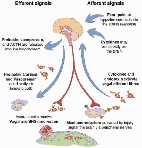
The neuroendocrine and immune systems share a chemical language. Immune cells express receptors for neurotransmitters and hormones. Endocrine cells, peripheral nerves, and cells of the CNS express receptors for immune-derived cytokines. Immune cells also produce hormones and signaling molecules classically thought of as neurotransmitters. Brain cells produce a wide variety of cytokines (1). Although the complex interplay of all of these signaling molecules remains poorly understood, the profusion of NEI mediators tells us that the brain provides guidance and fine-tuning for the immune response as it does for other organ systems (Fig. 82.1). To do this, the CNS monitors immune function with high acuity, sensing low levels of inflammatory cytokines and very early mediators of inflammation.


FIGURE 82.1. Bidirectional NEI communication. Stimulation of the acute stress response generates immunosuppressive signals (activation of the HPA axis, the SNS, and endogenous opioids) and immune-supportive signals (release of proinflammatory peptides, such as vasopressin and prolactin). A threat to the homeostatic milieu, in the form of trauma or infection, activates the immune system. Activated immune cells produce cytokines (e.g., TNF-α, IL-1, IL-6), which signal the CNS by stimulating afferent nerves or by circulating directly to the brain. Autonomic nerve activity delivers anti-inflammatory signals in response to immune activation.


The Immune System as a Sensory Organ

This cross talk between cells of the nervous, endocrine, and immune systems is not a static pattern of feedback loops, but a complex, integrated system sensitive to environmental signals perceived by both the brain and the immune system. The immune system can be thought of as a sensory organ that perceives microscopic threats. Day-to-day activities result in wear and tear on epithelial barriers, leading to microbial and other nonself invasion. These peripheral stresses activate immune cells, send signals to the CNS via peripheral or autonomic afferent nerves, and initiate an immunoregulatory response. Centrally mediated immune modulation can be initiated by other sensory input, the simplest example of this being classical conditioning. When an immunosuppressive stimulus (e.g., antilymphocyte serum) is paired with a sensory input (e.g., a flavored drink), the resulting immune suppression can be later reproduced with the flavored drink alone (2).


RECENT NEUROENDOCRINE IMMUNE MEDIATORS APPLIED TO CRITICAL ILLNESS

image Although our understanding of NEI interactions is limited, it is still clear that perturbations to any single pathway in this intricate system may have distant reverberations. Because therapies used in critical care interfere with or mimic many of these pathways, it is incumbent on the intensivist to pay attention to developments in the field of NEI interactions. In the past few years, corticosteroids, growth hormone, and vasopressin—all NEI mediators with immunomodulatory effects—have been proposed as therapeutic agents in the treatment of ICU patients.


Adrenal Replacement Therapy May Have Immunomodulatory Affects

Current recommendations for the use of adrenal replacement therapy in children with sepsis are conservative, calling for their use in patients with fluid refractory, catecholamineresistant shock and suspected or proven absolute adrenal insufficiency, such as that resulting from chronic steroid exposure or adrenal hemorrhage (3). Corticosteroids are known to have immunosuppressive effects; however, it is unclear whether the small doses used for adrenal replacement will increase patients’ susceptibility to nosocomial infection. In the setting of adrenal insufficiency resulting from chronic steroid exposure, the modest doses prescribed to replace adrenal function are unlikely to cause significant additional immune suppression. Recovery from severe sepsis requires a balance between control of inflammation and activation of the specific or adaptive immune response. Studies of adrenal replacement therapy published to date have yet to consider the impact of these drugs on immune function.


Treatment with Growth Hormone Increases Mortality due to Sepsis

The hypercatabolic state seen in critically ill patients has been attributed in part to growth-hormone dysfunction. Trauma, sepsis, and surgery are thought to induce a state of growthhormone resistance (4,5). The hyperglycemic, catabolic state induced by growth-hormone depletion and resistance is compounded by the normal stress response, the effects of immunederived cytokines, and inadequate calorie delivery in the ICU (4). Accordingly, exogenous growth hormone was proposed to preserve muscle and improve healing. In small studies, exogenous growth hormone given to postoperative patients and burn patients improved nitrogen balance and protein synthesis, increased muscle strength and lean body mass, and increased the rate of healing (4,6).

Unfortunately, use of exogenous growth hormone in critically ill patients is associated with increased mortality. In two large European studies of growth hormone use in critically ill adults, patients treated with growth hormone had a 1.9- to 2.4-fold relative risk of mortality, mostly because of multiorgan dysfunction syndrome, shock, or uncontrolled infection (7). The reasons behind this unexpected outcome may relate to growth hormone’s effect in immune function. At physiologic levels, growth hormone is immunostimulatory, but it has immunosuppressive effects at supraphysiologic levels in vitro (8).


Vasopressin Infusion in Vasodilatory Shock

A large randomized controlled trial comparing norepinephrine to low-dose vasopressin found that vasopressin did not offer a mortality advantage over norepinephrine in adults with septic shock. In a prospectively identified stratum of patients with less severe septic shock, vasopressin was associated with improved survival (9). Because patients treated with vasopressin had greater decreases in a broad array of cytokines, chemokines, and growth factors compared with those treated with norepinephrine, downregulation of the innate immune response has been proposed as a mechanism for this effect (10). As vasopressin has immunosuppressive effects when present in the CNS and immune-supportive effects when present in peripheral tissues (11), it is difficult to predict which effect would predominate during vasopressin infusion in the ICU.


PATHWAYS OF COMMUNICATION

This section summarizes the available data that describe the efferent pathways by which the brain communicates with the immune system, the afferent pathways by which the immune system informs the brain, and the impact of this communication on the function of immune cells. The vast majority of these data have been generated in vitro and in animal models that experimentally impair isolated NEI pathways. From these data, a sense of the overall impact of NEI pathways on the immune system and the critically ill patient as a whole may be synthesized.


Efferent Signals: How the Central Nervous System Communicates with the Immune System

Signals can travel between the brain and immune system on peripheral nerves or in the form of circulating chemical signals, such as hormones or cytokines. Due to the relative ease of experimental modeling, more is known about the humoral control of immune responses than about the signals that travel along nerves. The paucity of data belies the importance of direct innervation in control of immune responses. These pathways are evolutionarily conserved, suggesting a central role to the fine-tuning of the inflammatory response. Neural control has several advantages over humoral control. Humoral control requires molecules to diffuse to and from the site of action. By contrast, direct innervation can carry precise signals that are rapid in onset, discrete in location, and brief in duration.


Autonomic Control of Inflammation: The Cholinergic Anti-inflammatory Pathway

The importance of neural pathways in NEI communication is highlighted by the elegant work of Dr. Kevin Tracey and colleagues, who demonstrated that the vagus nerve is capable of sensing and regulating inflammation using simple and rapid feedback loops. Vagal efferent fibers, distributed throughout the reticuloendothelial system, modulate the immune response to endotoxin through cholinergic signaling (12).

In the presence of endotoxin, macrophages release cytokines that promote inflammation and potentiate activation of the specific immune response (13). Macrophages express nicotinic acetylcholine receptors. The binding of these receptors by an appropriate ligand decreases the release of tumor necrosis factor (TNF)-α and other endotoxin-inducible proinflammatory cytokines (IL-1β, IL-6, and IL-18) without altering release of the anti-inflammatory cytokine IL-10 (12,14) (Table 82.1). This acetylcholine-dependent downregulation of inflammation is reproduced by stimulation of the vagus nerve (12,14).

The vagally mediated anti-inflammatory signal seems to impact both local and systemic inflammation. Vagotomized animals release more TNF-α, IL-1, and IL-6 into the circulation during endotoxemia (12). Relative to sham-operated controls, these animals develop more severe local inflammation and are more sensitive to the lethal effects of endotoxin. Stimulation of the vagus nerve or administration of nicotinic acetylcholine receptor agonists reduces inflammatory markers, organ injury, and mortality from sepsis in murine models. Evidence suggests that vagally mediated inhibition of inflammatory responses to antigens routinely encountered in the intestine may protect from autoimmune disease and that some autoimmune disease may be ameliorated by nicotinic acetylcholine receptor agonists (12).


Autonomic Control of Inflammation: The Noradrenergic Pathway

The SNS also innervates the immune system. Postganglionic sympathetic neurons follow vasculature to innervate all primary and secondary lymphoid organs (15). The concentration of noradrenergic fibers in the spleen is concentrated in the white pulp around a central artery. These fibers play a role in controlling blood flow and thus may regulate lymphocyte traffic (16). Noradrenergic fibers also continue into lymphoid tissue without blood vessels. Norepinephrine release from peripheral nerves may impact immune function nonsynaptically by diffusion to the nearby cells of interest (16). The microenvironment around the synapse-like structures at their termination may be bathed in concentrations of norepinephrine as high as 0.3-3 mM on antigenic challenge (15,17). Cells of the bone marrow, thymic epithelial and dendritic cells, monocytes, and macrophages express both α– and β-adrenergic receptors, whereas B and T cells express the β2-adrenergic receptors exclusively (15).


TABLE 82.1 PROINFLAMMATORY AND ANTI-INFLAMMATORY CYTOKINES


PRODUCED BY


ACTIONS ON IMMUNE CELLS


RELEVANCE IN CRITICAL ILLNESS


Proinflammatory Cytokines


TNF-α


APCs, NK cells, T cells


Local inflammation, endothelial activation


An early mediator of inflammation and shock


IL-1β


APCs, epithelial cells


T-cell activation, macrophage activation


Causes fever and acute-phase protein production


IL-12


B cells, macrophages


Activates NK cells, induces CD4 T-cell differentiation into Th-1-like cells


Suppresses Th-2


IFN-γ


Th-1 cells, NK cells


Potent macrophage activation, suppresses Th-2 responses


Low levels associated with increased risk of infection


GM-CSF


Macrophages, T cells


Stimulates growth and differentiation of granulocytes and monocytes


Increases HLA-DR expression, may be clinically useful as an immune stimulant


Anti-inflammatory Cytokines


IL-4


Th-2 cells, mast cells


B-cell activation, suppresses Th-1 cells, IGE switch


Suppresses Th-1 response


IL-10


Th-2 cells, APCs


Potent suppressor of macrophage functions


Suppresses Th-1 response


Other Important Cytokines


IL-2


T cells


T-cell proliferation, supports Th-1 cells


Most important proliferative factor for T cells


IL-6


T cells, macrophages, endothelial cells


T- and B-cell growth and differentiation


Causes fever, and acute-phase protein production levels are related to severity of systemic inflammation


IL-8


Monocytes, endothelial cells


Chemotactic for neutrophils


Levels are related to severity of systemic inflammation


GCSF


Fibroblasts and monocytes


Stimulates neutrophil growth and differentiation


Exogenous GCSF safely increases neutrophil counts but does not alter mortality


APCs, antigen-presenting cells; HLA-DR, human leukocyte antigen-DR.


In the bone marrow, β-adrenoceptor activation suppresses cell proliferation and differentiation, whereas stimulation of the α-adrenoceptor suppresses myelopoiesis but enhances lymphopoiesis (15). In dendritic cells, monocytes, and macrophages, activation of β-adrenoceptors inhibits the endotoxininduced release of the proinflammatory cytokines IL-1β, TNF-α, IL-12, and interferon (IFN)-γ and increases the release of the anti-inflammatory cytokines IL-10 and IL-6 in response to endotoxin (15,18). IgG transcription is enhanced by binding to β-adrenergic receptors on B cells (18). In general, catecholamines are believed to favor T helper cell (Th)-2 cytokines and inhibit cellular immunity by suppressing Th-1 cytokine production (16,18). In murine models, activation of the SNS by Lipopolysachharide (LPS) engages an anti-inflammatory reflex that rapidly and dramatically decreases the TNF-α response. It is interesting to note that, in the NEI system, the sympathetic and parasympathetic nervous systems may act synergistically rather than in opposition.


Other Neurotransmitters

Researchers are still discovering new neurotransmitters, hormones, and cytokines, and continue to recognize new NEI roles for well-known molecules. The molecules described here are examples of this interface and add layers of complexity to the NEI system. These substances may function as neurotransmitters in neural pathways or as circulating messengers in humoral pathways.

Opioids. Pain can stimulate the immune response, and both exogenous opioids and endogenous endorphins are known to have immunosuppressive effects. Opioids are thought to induce immune suppression at analgesic doses by binding to classical, naloxone-sensitive opioid receptors in the brain. Immune cells have both classical and nonclassical opioid receptors, but it is unclear to what extent morphine interacts directly with these cells to cause immune suppression in vivo (19,20). Acutely, centrally acting morphine activates the SNS, and most of the observed immunosuppressive effects of morphine may occur via this pathway (21,22). Morphine also activates the HPA axis, which may lead to cortisol-mediated immune suppression, particularly during chronic administration (23).

In vitro, morphine has clear immunosuppressive effects. It decreases B-cell antibody production, reduces the T-cell proliferative response to mitogen, suppresses IL-2 gene expression, increases T-cell apoptosis, and decreases IL-6 levels. It is uncertain to what extent this mechanism is important in vivo (19,24). Acute and chronic exposure to morphine decreases splenic and peripheral natural-killer (NK)-cell activity in animals (20). Treatment with morphine for as little
as 36-72 hours impairs macrophage response to monocyte colony-stimulating factor in mice and affects phagocytosis and superoxide production ex vivo (20). In animal models, drugs that potentiate narcotic analgesia, such as clonidine and dexmedetomidine, also have immune suppressive effects (25).

Somatostatin and Somatostatin Receptor Ligands. Somatostatin analogs (e.g., octreotide) are used in the treatment of gastrointestinal hemorrhage. In addition to decreasing splanchnic blood flow, somatostatin inhibits the release of insulin and decreases secretion and absorption in the gastrointestinal tract (8). Somatostatin has direct immunosuppressive effects in vitro. Somatostatin receptors are expressed on peripheral T and B lymphocytes, on activated monocytes, and in hematopoietic precursors (26

Only gold members can continue reading. Log In or Register to continue

Stay updated, free articles. Join our Telegram channel

Jun 4, 2016 | Posted by in CRITICAL CARE | Comments Off on Neurohormonal Control in the Immune System

Full access? Get Clinical Tree

Get Clinical Tree app for offline access