NECROTIZING SOFT TISSUE INFECTION
CASE SCENARIO
A 53-year-old obese man presents to the emergency department complaining of one day of progressive pain and swelling of his scrotum, with a rash over his inner thighs and buttocks. His history is significant for diabetes, obesity, hypertension, and hyperlipidemia. He has a low-grade fever, tachycardia, and hypotension. He has erythema and swelling over his scrotum, penile shaft, perineum, and inner thighs, with tenderness extending 4 inches beyond the margin of erythema.
EPIDEMIOLOGY
Necrotizing soft tissue infection (NSTI) is a severe, life-threatening, rapidly progressive infection that fortunately is also rare. The Centers for Disease Control and Prevention (CDC) reported 4.3 cases of invasive Group A Streptococcus (GAS) infections per 100,000 people in the United States for 2011. Although this number likely underestimates the true frequency, as it does not account for polymicrobial infections (NSTI is classically monomicrobial), it serves as an approximate measure of the incidence.1
Risk factors for NSTI include diabetes, trauma, age >65, and immune compromise (including human immuno-deficiency virus [HIV], chemotherapy-related immune suppression, chronic alcoholism, and IV drug abuse). An association with nonsteroidal anti-inflammatory drug (NSAID) use has been proposed, but is not supported by evidence. Approximately 20% of patients have no apparent underlying risk factors.
PATHOPHYSIOLOGY
NSTI rapidly propagates through production of bacterial toxins that cause tissue necrosis, intra-vascular coagulation, weakening of the adhesion between the muscle and fascial layers, and decrease in pus viscosity—all changes that allow for quick spread of infection. The factors that allow an infectious inoculum to proceed to this aggressive necrotizing phenotype, instead of the more common fates of bacterial soft tissue infections (i.e., clearance, abscess, or cellulitis), are areas of active research, and no clinically modifiable factors have been discovered.2 Whatever the inciting factors, in NSTI, bacterial infection of the soft tissues progresses to the fascia, where toxins are transcribed and result in enzymatic degradation of the fascia (early changes), thrombosis of the microvasculature (late changes), and possibly gas formation and necrosis of the overlying skin (very late changes).
Traditionally NSTIs are divided into monomicrobial disease or polymicrobial disease, but this classification is of little interest to the surgeon or emergency physician, as it does not affect initial diagnosis or treatment. Recent retrospective series show that roughly two thirds of cases are polymicrobial and one third are monomicrobial.3 Common pathogens include GAS, Staphylococcus aureus, Klebsiella species, Escherichia coli, and anaerobic bacteria. Clostridium perfringens is of historical interest, but is uncommonly isolated in clinical disease.
NSTIs can also be differentiated on the basis of depth of invasion, spanning the spectrum from adipositis, to fasciitis, to myonecrosis. Similar to microbial status, these classifications are of little practical use. Finally, the eponym “Fournier’s gangrene” has been applied to NSTI of the perineum and is another artificial distinction with no impact on clinical management.
CLINICAL PRESENTATION
Sarani and colleagues provided a very useful review of NSTIs to characterize the most common symptoms.3 They report the most frequent findings on presentation to be erythema (between 66% and 100% of patients), pain or tenderness beyond the borders of erythema (73%–98% of patients), and swelling (75%–92% of patients). Other common findings include crepitus, induration, presence of bullae, fever (32%–53%) and hypotension (11%–18%).
These symptoms do not occur simultaneously. Patients first experience tenderness, swelling, and mild cellulitis as early signs of NSTI. As the toxins are produced and allow rapid spread of the infection, the tenderness spreads beyond the erythema along fascial planes. As the toxins take longer to produce microvascular thrombosis, the erythema and bullae overlying the infected tissue are late manifestations of the infection, as are systemic signs, i.e., tachycardia, hypotension, and oliguria. Gas production (if gas-forming bacteria are involved) causing crepitus is also a late development.
Typically patients present with low-grade fever, malaise, and leukocytosis, and complain of a tender erythematous lesion. There may be an associated recent trauma. Unfortunately, many of the conditions that place patients at risk for NSTI include conditions that make reporting of symptoms difficult and include chronic alcoholism, IV drug abuse, paraplegia, and diabetic neuropathy.
DIFFERENTIAL DIAGNOSIS
The list of conditions associated with tender erythema and swelling is limited (Table 24–1). The clinical challenge is not differentiating NSTI from a long list of potential disease states, but rather rapidly distinguishing NSTI from other soft tissue infections that will respond to antibiotics alone (i.e., cellulitis).
WORKUP AND CHOICE OF IMAGING
The most important factor to consider in NSTI is that while rarely encountered, it can be a rapidly progressive infection with both high morbidity and mortality. Early recognition and debridement confers significant improvements in outcome.
Initial history and exam should focus on risk factors and a dermatologic exam, noting the extent of any tenderness and/or erythema. Crepitus or bullae, systemic sepsis, and tenderness extending beyond the margin of erythema are worrisome signs for NSTI, as is pain out of proportion to exam.
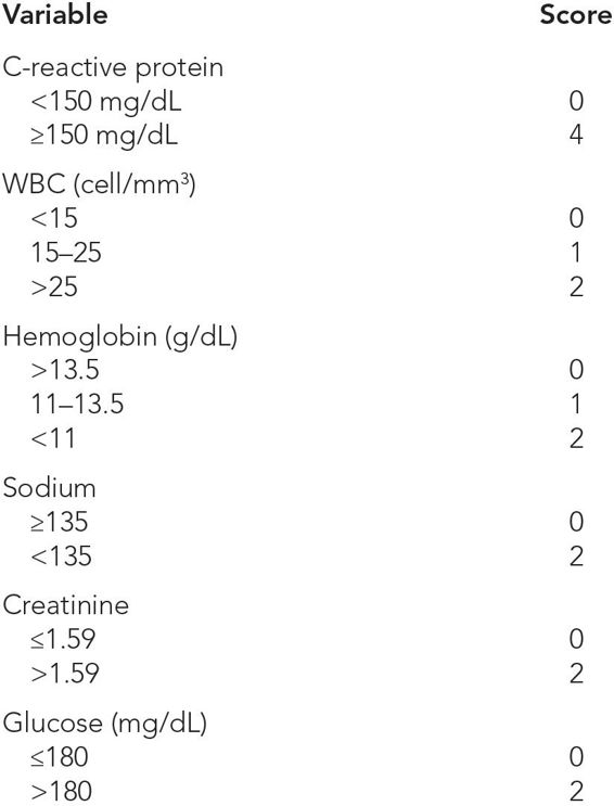
Initial laboratory investigation should include complete blood count with differential, coagulation studies in case operative debridement is necessary, a basic serum chemistry panel to investigate any metabolic derangements, and C-reactive protein as a marker of inflammation. There is no single test that can effectively rule in NSTI, but a number of variables can increase the positive predictive value. Wong and colleagues combined data from these basic laboratory studies to create the Laboratory Risk Indicator for Necrotizing Fasciitis (LRINEC).4 This index is calculable from data readily available on presentation, with good positive and negative predictive values (Table 24–2).
An LRINEC of ≤5 corresponds to 50% chance of NSTI. An LRINEC of ≥8 represents a >75% chance of NSTI.
Abbreviation: WBC, white blood cell.
The most important factor when considering potential imaging modalities in a patient with suspected NSTI is to remember that any delay in treatment will result in increased morbidity and mortality and so must be avoided. Thus, the role of imaging is to rapidly assist the clinician in distinguishing between NSTI and non-necrotizing pathology in cases when the diagnosis is unclear. In straightforward cases, no radiologic studies are necessary, and attaining imaging in these scenarios can create dangerous and unnecessary delays in care.
There have been no randomized, prospective, controlled trials comparing any imaging modalities. Plain film x-rays have no role; although historically they were used to detect subcutaneous emphysema, the increasing number of non-gas-forming necrotizing infections, as well as the prevalence of computed tomography (CT) scanning, has rendered this modality obsolete. Where ultrasound is concerned, some groups have reported success in the diagnosis of NSTI,5 but this remains a highly operator-dependent modality.
In contrast, CT scanning has the advantages of being commonly available, fast, and capable of providing detailed images in multiple views. In addition to assisting in making the diagnosis, CT scanning can elucidate the extent of disease. For these reasons, it is a preferred method in challenging cases, when the additional data assists in formulating a risk/benefit analysis regarding operative management.
The disadvantages of CT scan are ionizing radiation, nephrotoxic contrast material, and possible delay in definitive treatment. In the time required to transport the patient from the CT scanner to the operating room, this rapidly spreading infection may progress. For this reason, the decision to debride should be based on clinical, not radiographic, extent of the disease.
Some clinicians prefer the use of magnetic resonance imaging (MRI) in evaluating potential NSTI. MRI does provide superior imaging of soft tissue, and in locations with ready availability and expertise in rapid image acquisition, it is a viable imaging choice. The drawback with MRI is the significant time required for study completion at most institutions.
IMAGING FINDINGS
 Ultrasound
“Cobblestoning” of the subcutaneous tissue6 signifies soft tissue edema of the subcutaneous fat. Although a hallmark of NSTI, the finding is nonspecific, and is also seen in cellulitis, heart failure, venous thrombosis, hypoalbuminemia, lymphedema, and any other cause of tissue edema.
Increased echogenicity of the subcutaneous tissue is the ultrasound correlation of subcutaneous emphysema and results from sound waves reflecting off objects with different sound propagation characteristics. It is analogous to the “shadowing” that is seen with gallstones. Pockets of subcutaneous gas will disperse sound waves and result in “scattering” and a “dirty” appearance.7 Potential confounding factors include foreign bodies (e.g., “road rash”) and other sources of subcutaneous gas (e.g., stab/gunshot wounds); these are not likely to be confused with NSTI.
Thickened fascia with peri-fascial fluid collections can be seen by experienced ultrasound operators, are very concerning for NSTI, and should prompt operative exploration (Table 24–3 and Figure 24–1).


