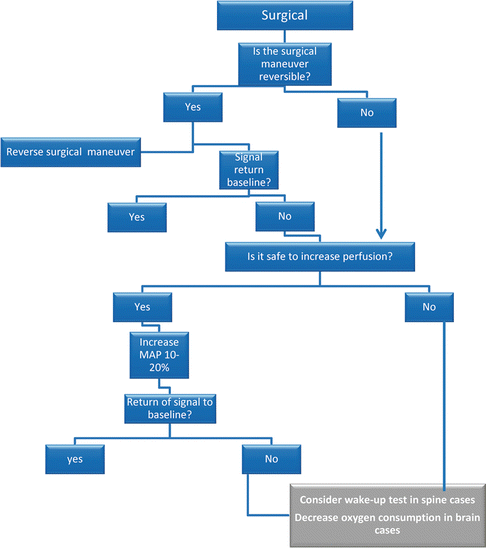
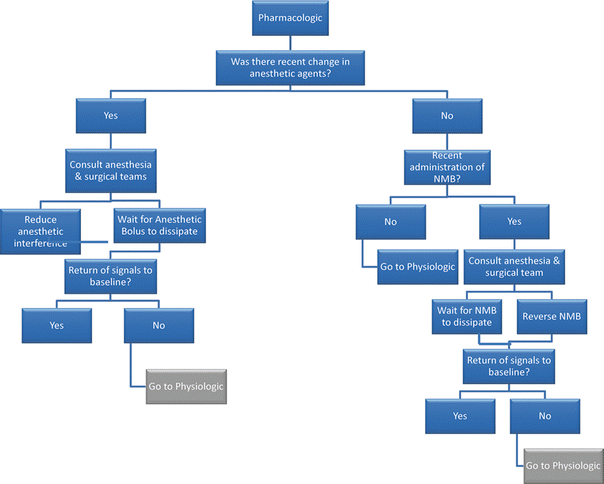
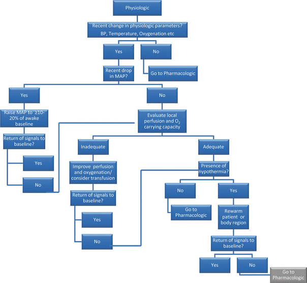
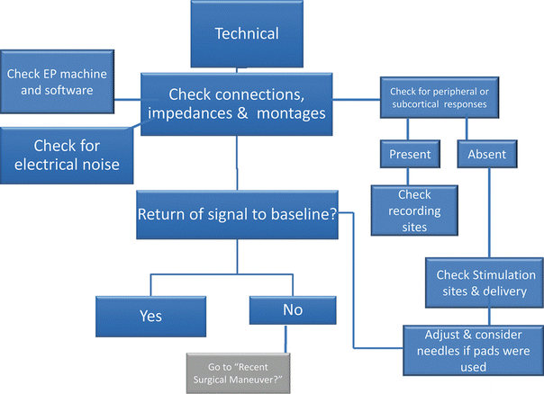
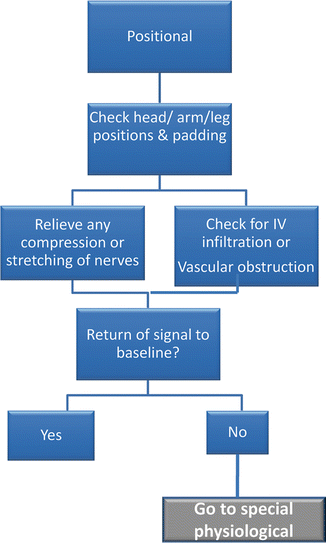
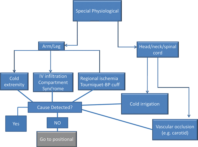
Fig. 20.1
(a–g) A basic algorithm for the anesthesiologist and other members of the monitoring team to use in evaluating the possible contributions to response deteriorations
The anesthesiologist is in a key position to evaluate the impact of anesthesia, physiology, and positioning, and therefore plays an important role in the search for an etiology . Often the specifics of the change can help identify possible causes. For example, anesthetic causes tend to be global and not to be focal in nature. Although certain IOM modalities are more sensitive to anesthetic agents (e.g., cortical somatosensory evoked potential [SSEPs] or muscle motor evoked potentials [MEPs] ) (see Chap. 19, “General Anesthesia for Monitoring”), it would be very unusual for anesthetics to alter only one hemisphere or one extremity more than the other. In addition, some modalities are rarely affected by anesthetic agents (e.g., short latency ABRs , subcortical SSEPs, EMG responses [aside from muscle relaxant effects], and spinal recordings of SSEPs and MEPs). Therefore, if the change in the IOM is related to an anesthetic effect, then a quick search for changes in the management becomes essential. Since a steady-state anesthetic protocol is least likely to produce changes, any abrupt change in drug delivery that may lead to an alteration in drug levels should be considered as a possible etiology. One exception is a slow change in IOM amplitude referred to as “fade ,” which is often ascribed to anesthesia but is not well understood [1]. In addition to being of global nature, anesthesia-related changes tend to occur within a short time (1–15 min) after alteration of the anesthetic levels.
A second category where the anesthesiologist must search for etiologies is that of physiology. Some of these effects are noted in several chapters. Like anesthetic effects, physiological effects may be global in their nature (i.e., they would usually expect to affect both sides of the body). Also like anesthesia, adverse physiology generally has more impact on cortical responses than subcortical responses, and within the spinal cord it has more of an effect on grey matter than white matter (see Chap. 40, “Surgery on Thoracoabdominal Aortic Aneurysms”). Hence, a quick scan of the physiological monitoring may help identify a change in blood pressure, heart rate, ventilation/oxygenation, etc. The effects of physiology can also be focal. For example, blood flow to one part of the brain could be reduced by carotid occlusion, hypothermia could occur primarily in one arm due to rapid infusion of cold fluids, and so on. In these cases, the question that must be asked is, what is the anatomic region that is affected based on the IOM responses, and what are the possible physiological circumstances in that region that could be unfavorable for the neural paths involved?
Stay updated, free articles. Join our Telegram channel

Full access? Get Clinical Tree


