Key points
- •
Noncontrast CT is the first-line imaging of acute stroke symptoms to assess for intracranial hemorrhage and evidence of edema related to ischemia.
- •
Head and neck CTA is useful in evaluation of acute stroke symptoms to detect LVO, dissection, or significant intracranial arterial stenoses.
- •
Diffusion-weighted brain MRI is the most sensitive method to confirm acute ischemia; however, patients may be unable to have MRI.
- •
CT and MR perfusion parameters have been established through clinical trials to stratify risk for revascularization in acute ICA and proximal MCA occlusion.
- •
Multiple vendors provide AI software to help expedite stroke imaging review. All are an aid to the physician rather than stand-alone diagnostic tool.
Neuroimaging should be obtained for all patients suspected of having acute ischemic stroke or transient ischemic attack. Noncontrast head computed tomography (CT) scans are used to exclude hemorrhage, evaluate for early brain injury, and exclude stroke mimics. CT angiography assists in identifying proximal vessel occlusions, dissection, or high-grade arterial stenoses. Additional imaging techniques have emerged to improve selection of patients likely to benefit from therapies. Artificial intelligence applications assist in acute stroke imaging assessment, identifying acute hemorrhage, and predicting risk of endovascular intervention in acute large vessel occlusion. Each should be considered an aid rather than stand-alone diagnostic tool.
Introduction
Neuroimaging should be obtained for all patients suspected of having acute ischemic stroke (AIS) or transient ischemic attack (TIA). Noncontrast head computed tomography (CT) scans are used to exclude hemorrhage, to evaluate for early brain injury, and to exclude stroke mimics, such as tumor. CT angiography (CTA) can assist in identifying proximal vessel occlusions, dissection, or high-grade arterial stenoses, which may be responsible for the ischemic deficit. Additional CT and magnetic resonance imaging (MRI) techniques have emerged to improve selection of patients likely to benefit from therapies, such as intravenous thrombolysis or mechanical thrombectomy.
American Heart Association (AHA) guidelines and the American College of Radiology (ACR) Appropriateness Criteria emphasize access to emergent brain imaging before initiating stroke therapy. It is crucial to remember that brain and neurovascular imaging studies represent a single moment in time within a dynamic process and must be interpreted in conjunction with clinical changes.
There are an increasing number of artificial intelligence (AI) applications that may assist in the acute stroke imaging assessment, to identify acute hemorrhage, and to predict risk of endovascular intervention in acute large vessel occlusion (LVO). These applications are marketed as a resource when there is a lack of timely, available expertise for CT scan interpretation. Each should be considered an aid, however, rather than stand-alone diagnostic tool. Medicolegal concerns regarding liability for AI misdiagnosis, a physician not acting on an AI finding, remain to be resolved. ,
Discussion
Noncontrast Head Computed Tomography
Rapid and accurate detection of stroke by emergency clinicians at the time of first contact is crucial for timely initiation of appropriate treatment of AIS patients. Noncontrast head CT is the preferred imaging study for evaluation of acute stroke symptoms because of widespread availability, rapid scan times, and ease of detecting intracranial hemorrhage. There is level 1 evidence that all patients with suspected acute stroke should receive emergent brain imaging on arrival, before initiating acute stroke therapy. Effective protocols and communication have confirmed that a median door–to–imaging time of 20 minutes or less can be accomplished across different hospital settings.
Imaging findings
An advantage of noncontrast head CT is the conspicuity of acute hemorrhage as bright signal relative to the gray of the brain parenchyma ( Fig. 1 ). Acute hemorrhage represents only 8% to 15% acute strokes, associated most commonly with hypertension; however, it also may be due to underlying arterial abnormalities, venous occlusion, cavernoma, and cerebral amyloid.

CT has poor sensitivity (20%–75%) and better specificity (56%–100%) in determining early ischemic changes within 6 hours to 8 hours; therefore, the diagnosis of acute stroke remains a clinical decision. Early infarct signs on noncontrast head CT are subtle and include loss of gray–white matter, cortical sulcal effacement, focal parenchymal hypoattenuation, and the insular ribbon or obscuration of the sylvian fissure ( Fig. 2 ). Both underestimation and overestimation of early infarct signs are common even in a controlled setting. ,

Evaluation for the subtle changes in gray–white matter definition in acute ischemia is difficult on CT, and preset options for stroke windows (40 WW:40 WL) displays ( Fig. 3 ) or alternate settings have been used for many years to enhance visual perception. Small studies suggest review may be adequate using a smartphone or laptop to identify features that influence intervention ; however, these do not have wider validation. The ACR maintains technical standards for image quality and review, which do not include mobile devices. AHA guidelines include that sites without in-house imaging interpretation expertise should have an approved teleradiology system for timely review of brain imaging. Several vendors have received Food and Drug Administration (FDA) clearance for AI applications to aid in detection of intracranial hemorrhage and work list/provider alert systems to help reduce time to decision making.

In the setting of acute stroke symptoms, identification of a focally dense (bright) artery on noncontrast CT should prompt emergent vascular imaging correlation ( Fig. 4 ). Vascular hyperdensity can indicate the presence of red-cell thrombus; however, it has been shown to be only 52% sensitive detecting LVO in a meta-analysis, including Third International Stroke trial patients. Smaller single-site series have reported increased sensitivity to 75% for basilar thromboses and 76% for M1 thrombosis. False-positive dense vessels include patients with underlying vessel atherosclerosis or more diffuse dense vessels due to hemoconcentration.

Implications for treatment
Early infarct signs on noncontrast head CT are not a contraindication to treatment with intravenous thrombolysis nor is the presence of a hyperdense artery sign. Analysis from the National Institute of Neurological Disorders and Stroke (NINDS) rt-PA Acute Stroke Trial found that early CT signs of infarction were not independently associated with increased risk of adverse outcome after intravenous alteplase treatment within 3 hours of onset of symptoms, and patients treated with alteplase did better whether or not they had early CT signs. Vascular evaluation is not required for the decision to treat with alteplase.
The Alberta Stroke Program Early CT Score (ASPECTS) 10-point scale was developed to assess noncontrast head CT for early ischemic changes in patients suspected to have middle cerebral artery (MCA) LVO. It has been used as an eligibility criterion for mechanical thrombectomy clinical trials for anterior circulation stroke and adapted into the clinical setting as a predictor of functional outcome and risk of symptomatic hemorrhagic conversion. Points are deducted for visible gray–white matter loss in each of the following areas: 3 supraganglionic (frontal operculum, anterior temporal lobe, and posterior temporal lobe), 3 subganglionic (anterior, lateral, and posterior MCA), the caudate, lentiform nucleus, insula, and internal capsule. Patients with scores of 7 or less were more likely to have adverse outcomes (death and dependence). Although inter-rater variability exists, it rarely has an impact on the dichotomization above/below a score of 7.
Computed Tomography Angiogram
To date, no clinical score has become widely accepted as an eligible prehospital marker for LVO and the need for mechanical thrombectomy in ischemic stroke. The best combination of sensitivity and specificity is achieved by a National Institutes of Health Stroke Scale (NIHSS) score cutoff between 7 and 10 for LVO or between 11 and 14 for mechanical thrombectomy.
Because CTA is well suited to assess for clot that may be accessible for endovascular therapy, this has become more integrated into the acute stroke evaluation, with some centers even endorsing “CTA for All” protocols. There remains continued debate, however, regarding use and overuse of CTA, particularly for patients with low NIHSS or at centers without access to emergent endovascular therapy, given the cost, contrast, and radiation exposure.
Anticipating CTA as part of the acute stroke evaluation, emergency department clinicians should have a brief discussion with the patient and/or family regarding the benefit of contrast to determine if the patient is a candidate for neurointervention to minimize lasting brain injury versus the risk of exacerbating existing severe renal insufficiency and rare (0.04%) but unpredictable severe adverse reactions. Patients with a prior allergic-like reaction to contrast have approximately 5-times the risk if re-exposed. Hospitals should have protocols in place regarding contrast use in stroke codes, because obtaining glomerular filtration rate results delays intervention.
Imaging findings
Axial images are readily available at the time of the scan, allowing for detection of changes in caliber or contrast density within the extracranial and intracranial arteries as well as assessment of luminal plaque. LVO results in an abrupt cutoff of the contrast enhancement within the artery ( Fig. 5 ). Proximal M2 occlusions may be difficult to perceive on the axial images, given anatomic variations in MCA bifurcation within the MCA and sylvian fissures; therefore, 3-dimensional (3-D) maximum intensity projection (MIP) reconstructions of the arteries can help expedite review.

Multiphase CTA has been proposed as a method to assess collateral circulation in stroke patients with LVO, as an alternative to CT perfusion. The traditional head and neck CTA represents the first of 3 phases of the multiphase CTA examination. The 2 additional scans are of the head only, in the equilibrium phase (8 seconds after first CTA) and the late venous phase (16 seconds after first CTA). This results in additional radiation exposure to the brain and eyes. There is no additional contrast given; however, due to rapid sequence of timing, high suspicion for LVO is required at the time of scan initiation; otherwise, this is exposing patients to radiation unnecessarily. For multiphase CTA, the 3 phases must be evaluated concurrently to compare the extent of pial arterial enhancement across the time points in the area of the vessel occlusion compared with the asymptomatic contralateral hemisphere. Clot burden and collateral scores have been proposed as independent predictors for endovascular triage; however, have been limited to small studies or retrospective data sets.
AI applications have entered the international marketplace, designed to alert clinicians to possibility of LVO and to generate clot burden scores. , Although small studies and conference proceedings have shown these applications may function with comparable accuracy to a neuroradiologist, they are not approved to replacing a physician’s review of the scan.
Infarcts along the border zone of the major intracranial vascular territories should prompt close evaluation for atherosclerotic stenoses of the proximal vessels ( Fig. 6 ). Soft atherosclerotic plaque is dark on CTA. Calcified plaque is bright on CT and is conspicuous even relative to the arterial enhancement. Through-plane reconstructions can be created with scanner-based and third-party software options, to verify the extent of any luminal stenosis.

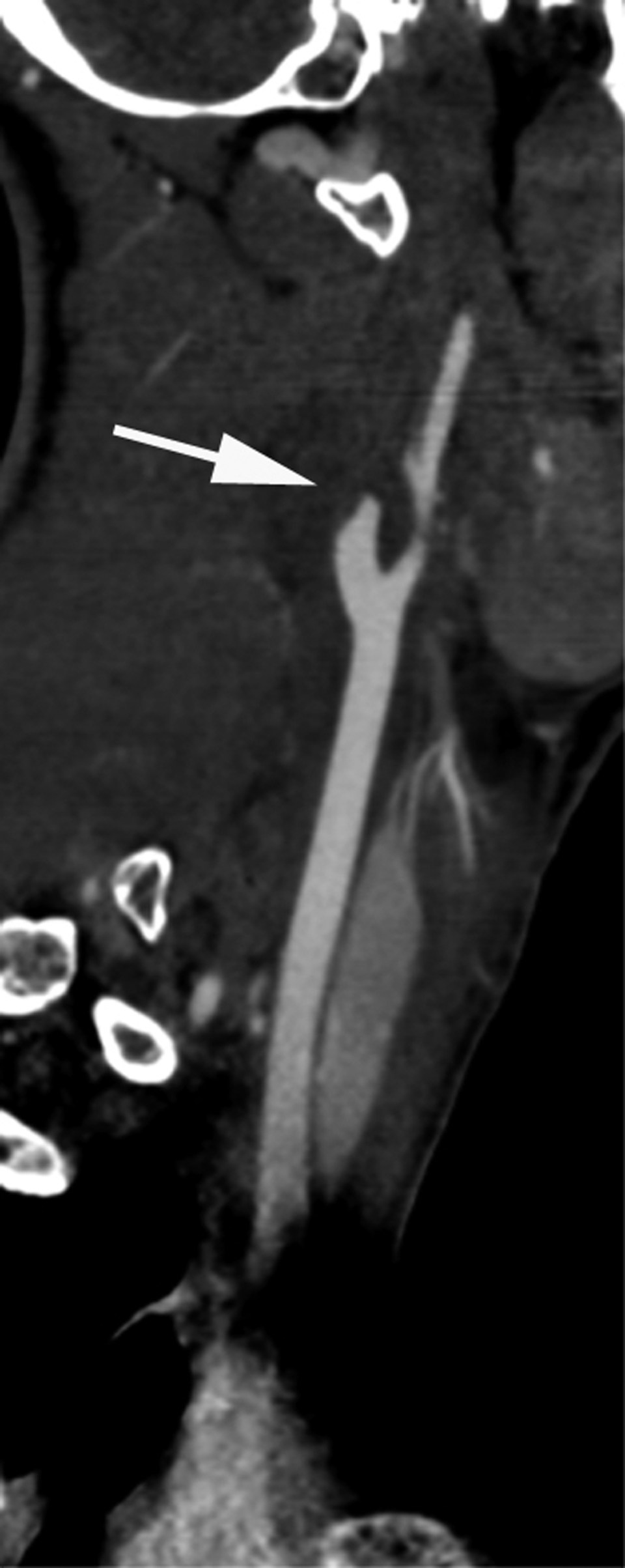
Internal carotid dissection commonly presents as a flame-shaped tapering of the contrast, due to nonenhancing thrombus obstructing the vessel lumen ( Fig. 7 ). Nonobstructive carotid and vertebral artery dissection is more subtle, detected by flattening of the normal round cross-section of the vessel due to eccentric thrombus beneath the intimal flap.

It is important to remember venous thrombosis as a potential source for ischemic and hemorrhagic stroke. Although CTA is optimized for arterial assessment, there often is sufficient enhancement to exclude venous sinus thrombosis. If this is suspected as the etiology, however, it is prudent to notify the radiologist and technologist so they may adapt the bolus-to-scan timing. The empty delta sign is specific to clot within the superior sagittal sinus ( Fig. 8 ); however, the same principle applies to the other sinuses, with the filling defect as a dark area within otherwise enhancing venous sinuses. Normal variants of arachnoid granulations and anatomic asymmetry of cortical and deep venous drainage can mimic thrombus.


