Figure 18.1
Ultrasound image of a thickened tubular structure with gut signature in the right mid-abdomen, distant from the cecum, representing an inflamed Meckel’s diverticulum (Image credit: Kara Gill, MD)
Patients with evidence of lower GI bleeding should undergo a thorough workup for other causes of bleeding, including a detailed history and physical and placement of a nasogastric tube to rule out upper GI source. To evaluate for a Meckel’s diverticulum as a bleeding source, a nuclear medicine Technetium (Tc)-99 m pertechnetate scan is the preferred test (Fig. 18.2). The Tc-99 m is taken up by gastric mucosa, so this scan will only identify a diverticulum containing heterotopic gastric mucosa. Sensitivity for this test ranges from 60 % historically to over 90 % in a recent study [4]. Regardless, some diverticula will be missed with this test, so diagnostic laparoscopy is indicated in a highly suspicious case of painless rectal bleeding, even if the “Meckel’s scan” is negative.
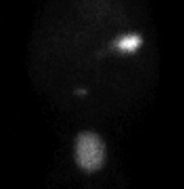
Figure 18.2
Technetium (Tc)-99 m pertechnetate nuclear medicine scan (“Meckel’s scan”). The tracer is taken up by gastric mucosa, which is commonly present in Meckel’s diverticula and causes peptic ulceration and bleeding. Radiotracer is readily seen in the stomach and bladder, with the Meckel’s diverticulum revealed as a small focus of abnormal uptake in the right mid-abdomen after 30 min (Image credit: Kara Gill, MD)
Pre-operative preparation for patients presenting with a symptomatic Meckel’s diverticulum includes intra-venous fluid resuscitation and a 2nd-generation cephalosporin. In cases of diverticulitis, which can be perforated, broad-spectrum gram-negative and anaerobe coverage should be utilized. In the setting of obstruction, nasogastric tube decompression is helpful. If symptomatic from severe anemia, the patient may require packed red blood cell transfusion.
Description of the Procedure
In many cases, the diagnosis is suspected but not known, so it is often advantageous to begin with a diagnostic laparoscopy to confirm the diagnosis [5–8]. Laparoscopic exploration for a suspected or confirmed Meckel’s diverticulum is typically performed with standard umbilical access through either a peri-umbilical or midline umbilical incision. Since the diverticulum may be adherent to the underside of the umbilicus, the fascia and peritoneum should be entered with a careful open cutdown approach. As the umbilical stalk is explored in this manner, a remnant fibrous cord may be identified, allowing an adherent diverticulum to be pulled up and out of the wound.
If the diverticulum is not immediately identified, laparoscopy proceeds using either a single-site or standard three port set up. In the three port approach, the additional 5 mm ports are typically placed in the left lower quadrant and left supra-pubic location, similar to the placement for a laparoscopic appendectomy. The patient is positioned in Trendelenburg and tilted left side down. This set up and position allows easy identification of the cecum and terminal ileum, which is then run proximally to look for the diverticulum. If the diverticulum is densely adherent to the abdominal wall at the umbilicus, the laparoscope can be moved to the left lower quadrant port site to visualize and divide this attachment.
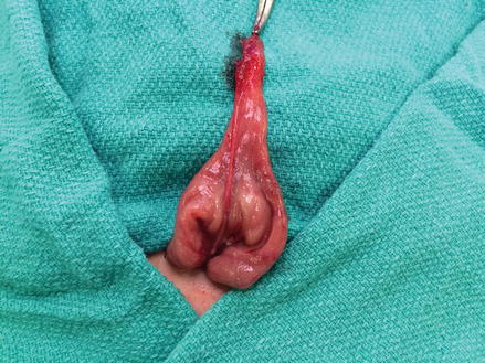
After the diverticulum is identified, options for removal are simple diverticulectomy or segmental small bowel resection. In most cases, a simple diverticulectomy can be performed using a GIA stapling device either intra- or extra-corporeally. The staple line should be oriented transversely to avoid narrowing the ileal lumen. In an infant or small child, the diverticulectomy is more easily accomplished by eviscerating the segment of ileum and amputating the diverticulum with a stapler extra-corporeally (Figs. 18.3, 18.4, and 18.5). In an older child or adult with more abdominal domain, an endo-GIA stapler may be utilized for an intra-corporeal diverticulectomy, similar to a laparoscopic appendectomy. The mesenteric vessel to the diverticulum must be controlled with either suture, staples, or any available energy device. In cases of GI bleeding, it is important to be aware that the bleeding ulcer is often located in the adjacent ileum, so a short segment bowel resection with primary anastomosis is generally recommended to avoid the pitfall of recurrent bleeding. Alternatively, a V-shaped cuff of adjacent ileum can be excised with the diverticulum, and the remaining enterotomy closed transversely with sutures after inspecting the ileum for ulcers. The resected specimen should be opened prior to sending it to pathology to confirm that it contains the ulcer.