Management of Skin Ulceration
Peter C. Schalock
Arthur J. Sober
Skin ulceration can be a troublesome, disabling, and potentially dangerous problem. Cutaneous ulcers commonly encountered in medical practice include leg and pressure ulcerations. Approximately 1% of the population is affected by venous leg ulcers. Diabetics and others with arterial insufficiency are at increased risk for ischemic ulceration and limb-threatening infection. About 20% of bed-bound or immobilized patients have pressure ulcers. The primary physician who recognizes and effectively treats early skin changes can prevent many of the debilitating consequences.
Skin ulceration most commonly results from venous/arterial insufficiency or from prolonged, excessive pressure. Infectious and malignant causes are also encountered, especially in immunocompromised persons.
Venous Insufficiency
The initial manifestation of venous insufficiency is edema, usually absent on waking and severe at the end of the day. Incompetent venous valves can be associated with age, thrombophlebitis, or a hereditary tendency for the development of venous varicosities. In all three conditions, abnormally high venous pressure during ambulation causes fibrinogen to leak from the engorged capillary bed. A precapillary fibrin layer develops that is a sign of abnormal microcirculation. Some believe that this fibrin layer interferes with oxygen and nutrient exchange. Pigmentation, induration, dermatitis (stasis dermatitis), and finally ulceration may develop. The rupture of delicate venules releases hemoglobin, which changes to hemosiderin, producing pigmentation. Scaling and oozing develop when the skin is scratched. Vesicles may indicate a contact dermatitis caused by a topical medication. Secondary bacterial invasion occurs and may lead to cellulitis.
Stasis ulcerations develop within areas of dermatitis or indurated cellulitis. They occur most often above the medial malleolus because of its poor vascular supply and sparse subcutaneous tissue, although the entire medial lower calf has higher venous pressures. Minor trauma can precipitate ulceration. The stasis ulcers vary in size from small erosions to an ulcer that encircles the ankle. They may or may not be painful. The base of the ulcer is usually moist, with exuberant granulation tissue. Purulence indicates secondary infection.
Arterial Insufficiency
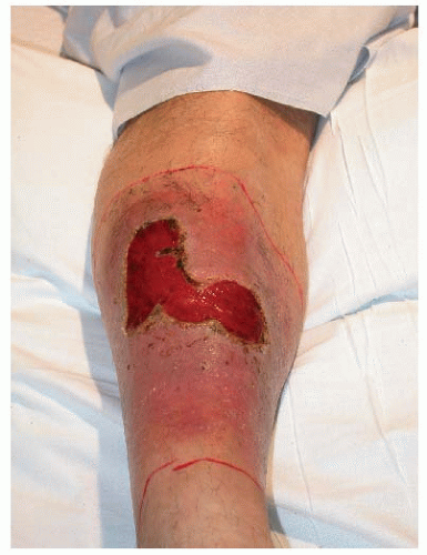
The most common cause of arterial insufficiency is atherosclerotic disease. The leg is cold and appears pale or cyanotic (although dependent rubor may be present), and peripheral pulses are lost or reduced. The ulcers are initially small, punctate, and superficial, but with worsening ischemia, they become larger and deeper (Fig. 197-1). Typically, they occur on the sides of the feet, the heels, the toes, and the nail beds. This type of ulceration makes up approximately 10% of leg ulcerations.
Ischemic ulcers are also associated with hypertensive disease and vasculitis. Those occurring in the context of hypertension characteristically develop over the lateral malleoli and surface of the calf. They begin as painful, blue-red plaques that soon ulcerate. A purpuric halo may surround the ulceration. Vasculitic ulcers occur in the context of connective tissue disease, hematologic and malignant conditions, and hypersensitivity reactions, beginning as palpable purpuric lesions or hemorrhagic vesicles (see Chapter 179).
Decubitus Ulcer
The pressure sore or decubitus ulcer is common in bedridden or semiambulatory patients. Factors contributing to the development of pressure sores include shearing forces, friction, and moisture; age per se does not. Decubitus ulcers usually occur over bony prominences. The pressure gradient occludes lymphatic vessels and overloads the microvascular system, and so waste products accumulate, and ultimately necrosis ensues. The lower part of the body and sacrococcygeal area are the predominant sites, with the hip, malleolus, and heel being other important areas.
The problem may initially present as nonblanchable erythema, soft tissue loss, blisters, or eschar over bony prominences. Pressure ulcer severity is reflected by the stage of the lesion. Stage 1 lesions are manifested by nonblanchable erythema of intact skin. Stage 2 ulcers involve only the epidermis and the dermis. Stage 3 ulcers extend into the subcutaneous tissues and undermine the surrounding skin. Stage 4 lesions extend through deep fascia to involve muscle and may extend to the bone.
 Figure 197-1 Large, later-stage arterial ulceration on the leg. This lesion was extremely painful. Source: All photos are courtesy of Peter C. Schalock. |
Pressure sores can lead to cellulitis, bacteremia, osteomyelitis, and even meningitis. The microbiology of decubitus ulcers is polymicrobial, and the organisms that cause the most problems, including life-threatening bacteremias, are group A streptococci, Staphylococcus aureus, Escherichia coli, and Bacteroides fragilis.
Pyoderma Gangrenosum
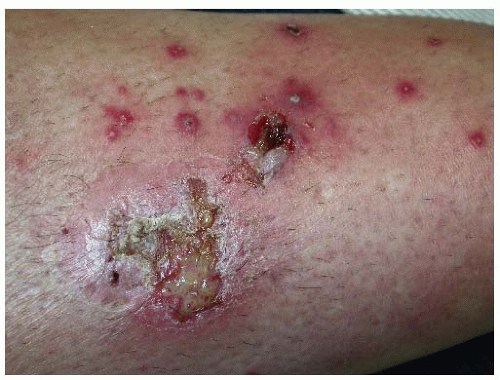
The condition is characterized pathologically by sterile neutrophilic infiltrates and dermal abscesses of the skin leading to tissue breakdown. Its association with minor trauma as well as with inflammatory and neoplastic conditions (e.g., inflammatory bowel and joint diseases, paraproteinemias, myeloproliferative conditions) suggests hyperactive dysfunction of neutrophils in response to an inflammatory or neoplastic stimulus. Clinical hallmarks include rapid development, pain, suppuration, violaceous discoloration, and necrotic borders (Fig. 197-2). Ulcers due to pyoderma gangrenosum are frequently confused with those due to other conditions (see Table 197-1).
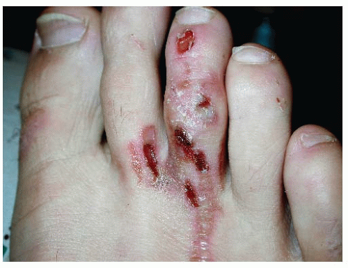
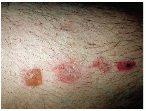
The differential diagnosis of skin ulceration is extensive (Table 197-1), but workup is aided by the appearance and the location of the ulcer and the clinical context in which it occurs. If it involves the lower extremity and the ulcer initially appears small, punctate, and superficial in a patient with absent pulses and a cold, pale leg (or one with dependent rubor), then peripheral atherosclerotic disease is the likely etiology. Vasculitic ulcers are suggested by associated palpable purpura and/or hemorrhagic vesicles in the setting of connective tissue disease, malignancy, or hypersensitivity reaction (see Chapter 179). Ulcers that begin as areas of nonblanchable erythema, soft tissue loss, blisters, or eschar over bony prominences in debilitated persons are likely to be decubital in origin. Ulceration in the lower extremity occurring in the context of chronic edema, hyperpigmentation, induration, and erythema points toward venous insufficiency. Pyoderma gangrenosum is suggested by a painful, violaceous leg ulcer with irregular borders that begins as a pustule in the setting of minor trauma or concurrent inflammatory/neoplastic disease. When diagnosis is uncertain, referral for biopsy can be helpful, especially in cases of suspected vasculitis or pyoderma gangrenosum. About 10% of cases labeled as pyoderma gangrenosum are misdiagnosed. Factitial ulceration is a diagnosis of exclusion. Angulated or square borders and linear patterns may suggest this diagnosis (Figs. 197-3 and 197-4).
TABLE 197-1 Some Important Causes of Skin Ulcers | |||||||||||
|---|---|---|---|---|---|---|---|---|---|---|---|
|
 Figure 197-3 Ulcerations on the dorsal foot. These were caused by heating a piece of metal with a lighter and burning the foot. |
 Figure 197-4 Quadratic bullae on the leg of the same patient as in Figure 197-3. These were also factitial. |


