Background/ Epidemiology
The lower leg is commonly injured in active people.
Impact-related activities create repetitive low levels of trauma that build over time resulting in a variety of injuries.
A review of 100 US high schools reported that over the course of one year there were over 800,000 lower extremity injuries reported.1
The most common sport involved was soccer, but injuries also occur commonly in basketball, football, and track (running/ jogging).
Emergency physicians (EP) must be aware of some basic principles of injury to the lower leg to effectively examine and assess for pathology.
Anatomic Considerations/ Pathophysiology
The anatomy of the lower leg is comprised of the larger anteromedial tibia that bears the majority of the body’s weight and the laterally positioned fibula, which contributes mostly to leg stability and rotational strength.
Proximally, the tibia interfaces with the femur to create the knee joint, while the fibula connects laterally to the proximal tibia.
The proximal tibiofibular joint has very little motion, but is important in the distribution of rotational forces from the ankle to the leg.2
Rarely, this joint may be symptomatic from increased motion leading to instability or cystic structure formation.
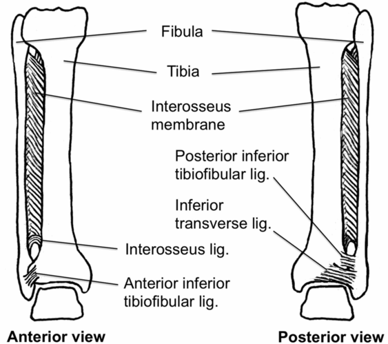
The interosseus membrane (Figure 7.1) is a thick fibrous connection between the tibia and fibula that effectively creates a ring structure of the lower leg.
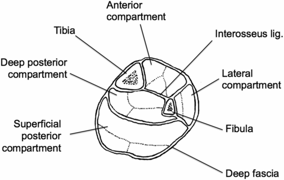
Fascial layers divide the musculature of the leg into four distinct compartments.
Anterior, lateral, superficial posterior, deep posterior (Figure 7.2)
The causes of pain in the lower leg may be from a wide variety of sources. Most commonly the following processes are involved:
Bone stress: May be due to acute traumatic injuries resulting in acute fractures or chronic injuries from repeated low level impacts causing stress reactions or fractures.
Inflammation: Often due to muscle units pulling on the periosteum of the lower leg; this leads to focal pain, especially with repeated activities
Elevated compartment pressures: Muscle action during athletic or work activities requires increased blood flow, which increases the pressures within facial compartments of the lower leg; these pressures may increase to the point of causing injurious compression of vascular, neurologic, and muscular structures.
Nerve entrapment: Scar tissue or other structures may impinge nerves traversing the lower leg leading to distal leg/ foot pain or weakness.

Interosseus membrane and syndesmosis of the lower leg.

Fascial compartments of the lower leg.
Focused History and Physical Exam
A thorough history will include focused questions on the onset, timing, character of pain, injury mechanism, exacerbating/ relieving factors and history of prior injuries.
A recent traumatic event with sufficient forces involved increases the possibility of acute fracture or muscle tears, while a more insidious onset and development of pain may involve stress injuries, nerve or vessel entrapment, or inflammatory conditions.
Other contributing factors may include training regimens, dietary restrictions, and comorbid conditions.
A broad review of systems may be helpful in identifying potentially associated symptoms.
At times a musculoskeletal complaint or a limitation of athletic performance may be due to a systemic metabolic condition leading to early limitation of activities or other painful conditions (i.e., pulmonary disease may limit aerobic performance).
Examining the lower leg in a systematic fashion will ensure a thorough exam to provide evidence of injury or disease
Inspection
Look for any asymmetric swelling, ecchymosis, deformity, soft tissue swelling, or discoloration.
Since the leg is involved in locomotion, it is imperative to evaluate the leg at rest and during standing and walking.
Evidence of varus or valgus alignment from the knee down may indicate knee pathology or the propensity for such pathology.
Observe any evidence of tibial torsion (in-toeing or out-toeing) or pes planus (flat feet). These conditions contribute to alterations in gait that may cause injury.
Observe the patient’s gait; watching for asymmetry between legs or potential weaknesses of certain muscle groups.
Palpation
Because the entire lower leg is connected in a ring-like structure, the entire tibia and fibula should be palpated, in addition to the area of identified pain.
The anterior tibial shaft is very shallow beneath the skin and stress injuries, and fractures or inflammation may be focally tender to palpation.
The full length of the fibula should be palpated for any focal or diffuse tenderness as well as any areas of step-off or mass.
The gastrocnemius, soleus, and plantaris muscle bellies, musculotendinous junctions, and Achilles tendon should be palpated for tenderness, swelling, gaping, or nodularity. These muscle groups should be assessed for overall bulk and tone.
Finally, the four major compartments of the leg should be palpated for tightness, firmness, or pain. Rarely, a mass may be felt consistent with muscle hernia through a fascial defect.
Range of motion (ROM)
While the leg does not have any testable motions outside of the knee or ankle joints, the muscles that cause the joint motion are attached to particular points on the leg.
While observing active motion of the knee and particularly the ankle, the examiner may watch for muscle action, bulk, and symmetry during particular motions.
This may provide clues to muscular involvement leading to weakness or other pathology.
Special tests
Due to the stability of the lower leg, there are not many specialized tests; however, it is important to differentiate between a bony injury and soft tissue inflammation.
When there is concern for a stress injury, placing a vibrating tuning fork or ultrasound on the site of pain may elicit pain over a stress fracture.
A positive test is indicated by an increase in pain.
A recent systematic review found that while some studies reported relatively acceptable test performance characteristics of tuning fork and ultrasound tests, overall these tests didn’t perform well enough to be used alone without confirmatory imaging.3
The single leg hop test is another useful test for stress fractures.
The patient is asked to hop on the affected leg; a positive test occurs when the patient has extreme pain and cannot hop more than once or twice.
Generally, patients with medial tibial stress syndrome (MTSS) can hop up to ten times.
Neurovascular exam
Sensory dermatomes of the L4-S1 nerve roots are located in the lower leg.
The L4 dermatome is on the medial aspect of the leg.
The L5 dermatome is on the proximal lateral leg.
The S1 dermatome is located on the distal posterior calf, heel, and lateral malleolus.
Vascular structures traversing the lower leg are important to assess, but are most easily assessed at the knee or ankle.
The easiest way to assess circulation is to palpate the dorsalis pedis pulse.
Overall leg perfusion should be generally assessed, as well as evaluating for swelling or edema that may indicate other pathology, like thrombotic disease.
Differential Diagnosis-Emergent and Common Diagnoses
General
A list of differential diagnoses for leg pain includes the common issues of shin splints (medial tibial stress syndrome), acute fractures, arthritis, cysts, stress fractures, proximal and midshaft fibular fractures, and the less common but important diagnoses of CECS nerve entrapments, and gastrocnemius musculotendinous tears.
Shin Splints
Background
The term shin splints is commonly used in active runners and may, at times, be a vague “catch-all” term for any lower leg pain.
MTSS is a more specific term for this condition.
It is most common in runners, but may occur in other athletes.
| Emergent Diagnoses | Common Diagnoses | |
|---|---|---|
| Leg | Open fractures | Shin splints |
| Fractures with neurovascular compromise | Arthritis | |
| Acute compartment syndrome | Stress fractures | |
| Displaced fractures | Chronic exertional compartment syndrome | |
| Nerve entrapments | ||
| Gastrocnemius strains/tears | ||
| Ankle | Ankle dislocation | Ankle instability |
| Displaced fractures | Ankle sprain | |
| Open fractures | Tendinopathy | |
| Osteochondral lesions | ||
| Arthritis |
Mechanism
MTSS is caused by excessive pull of lower leg muscles on the periosteal lining of the tibia.
These muscles generally include the tibialis posterior, soleus, and flexor digitorum longus.
The excess traction causes a periosteal reaction along the tibial shaft that may be diffusely tender to palpation.
Excessive foot pronation (flat feet), training errors, and inadequate shoe support may contribute to the development of this condition.
Presentation
Patients will present with a gradual onset of pain that will be worse in the morning and after exercise.
Symptoms will often improve with rest, adequate warmup before exercise, and stretching.
Physical Exam
The patient will complain of diffuse pain along the medial tibial border.
The area will be diffusely tender to palpation; occasionally mild swelling may be visible in the area.
Any focal tenderness along the tibial shaft should cause the examiner to consider a diagnosis of stress fracture.
Essential Diagnostics
Radiographs of the tibia and fibula are not mandatory, but may be useful to rule out fracture or stress reaction.
ED Treatment
ED treatment consists of pain control, thorough evaluation for other conditions (i.e., fractures), and reassurance.
Often times, symptoms may be improved with altering training regimens (introducing cross training, changing running surface), improving shoe type, or using shoe arch supports.
Symptomatic treatments that may be helpful are focal ice massage and nonsteroidal antiinflammatory drug (NSAID) creams.
These are particularly useful just after activity.
In recalcitrant cases there may be more severe gait abnormalities and referral for a formal gait analysis may guide treatment, especially with custom orthotics.
Severe cases may benefit from foot/ankle bracing to limit motion and allow any inflammatory processes to heal.
Disposition
Patients may be discharged home with primary care or sports medicine follow-up.
Patients may need referral for physical therapy to work on stretching, soft tissue treatments, and foot/ankle strengthening.
Athletes may continue activities as tolerated by pain, but need to modify their activities to prevent progression to stress fracture.
Complications
Continued pain may force the athlete to modify or stop their activities.
Persistent symptoms raise the concern for the development of a stress fracture.
Pediatric Considerations
This condition may be common in children, especially those just starting to run or those running intermittently (i.e., physical education classes).
Usually, an evaluation to rule out other pathology as well as reassurance and conservative treatments are adequate to return patient to activity.
Pearls and Pitfalls
Whenever the diagnosis of MTSS is considered, other diagnoses like stress fractures, muscle imbalance, neuropathies, or CECS should be considered.
Often times these diagnoses have other historical or physical exam findings that point toward the alternate diagnosis, but they should be adequately evaluated for and ruled out, especially in MTSS cases that are not improving.
Stress Fracture
Background
Stress fractures in the tibia (and slightly less in the fibula) are a feared complication of running and impact activities.
Stress fractures occur as a result of multiple repetitive smaller forces directed at the bone resulting in a stress injury or fracture.
Stress injury refers to the early signs and symptoms prior to the development of a discrete fracture line.
A fracture line in the anterior tibial cortex is referred to as “the dreaded black line” because it is often difficult to heal and requires significant change in activities for the patient.
Stress fractures may occur anywhere in the body, but most concerning in a running population are those in the hip, tibia, navicular, and metatarsals.
Mechanism
Bone metabolic processes include the continual reshaping of bony structures to adapt to ongoing forces applied to the bone.
When a bone is placed under repetitive stresses that exceed its capacity to withstand such stress, it begins a process of remodeling.
This initiates with resorption of bony material along the site of stress.
This weakens the area and results in eventual fracture of the cortex.
Finally bone is laid down around the fracture area, starting directly below the periosteum.
This causes the periosteum to elevate directly over the fracture area. This may be seen on x-rays as a hazy heaped-up line running parallel to the cortex.
This is referred to as a periosteal reaction and is indicative of a stress fracture.
Presentation
Stress fractures occur in patients who have experienced a relative increase in impact activities over a period of time sufficient to induce bony changes.
Often the patient may remain asymptomatic for a period of time, and only after one to two months of sustained activity does the patient begin to manifest symptoms.
Patients will complain of gradual development of pain over focal area of bone.
As time progresses, the pain will worsen with activity and eventually become symptomatic during routine walking.
The stress injury may also produce a deep achy pain at night.
Other important considerations are overall patient health, gender, diet, calcium intake, and age.
With female patients in particular, identification of an eating disorder and a lack of regular menses may indicate conditions of increased risk for potential stress fractures.
Physical Exam
Patients will have a focal area of pain directly over the bone, usually the tibia.
This is in contradistinction to MTSS, which will generally have a diffuse area of tenderness.
As time goes on and a bony callous grows, an area of swelling and bony growth may occasionally be palpated.
Other tests which may be performed include:
Placing a vibrating tuning fork, or active ultrasound probe, directly on the painful area
Single leg hop test
This is performed by having the patient hop on the affected leg while evaluating for reproduction of their pain.
Essential Diagnostics
A standard two view x-ray of the tibia and fibula should be obtained any time the diagnosis of stress fracture is considered.
These should be evaluated for evidence of a periosteal reaction, callous development, or frank fracture.
In some cases, a bone scan or MRI may be needed to determine the presence of a stress injury or fracture.
These studies are generally not necessary while the patient is in the ED.
ED Treatment
When a stress fracture is suspected, the patient should be placed into a fracture walker boot, even if initial x-rays are negative.
This will cushion the leg and prevent ongoing trauma.
The patient may be weight-bearing as tolerated.
On occasion, patients may be symptomatic enough, or the fracture may be tenuous enough (i.e., anterior tibia), that the patient should be made non-weight bearing.
Patients should be advised to avoid further impact activities.
Patients should also be advised regarding adequate calcium, vitamin D, and protein intake.
In addition, any other ongoing comorbid conditions (i.e., female athlete triad) should be identified and adequately managed.
Disposition
Patients should be advised to follow-up with orthopedics or sports medicine within one week from ED discharge.
Patients will need ongoing care and repeat imaging to ensure healing over time.
After a period of immobilization, progressive weight-bearing and return to activities may be incorporated, provided the patient is pain free with walking and other activities.
Complications
Once a stress injury progresses to a stress fracture the major concern becomes adequately stabilizing the area and removing the stress to ensure appropriate healing.
Stress fractures of the anterior tibial cortex are particularly concerning due to their risk of progressing to complete fractures.
They are also at risk for delayed healing and nonunion.
In these cases, the patient may need surgery to stabilize the area.
Pediatric Considerations
Due to active bone growth and turnover, children could be considered to be at an increased risk of stress injury.
However, stress fractures tend to be fairly uncommon in children.
Pearls and Pitfalls
Given the high morbidity of displaced fractures and the often vague presentation, the risk of missing this diagnosis is substantial
X-rays findings may be very subtle and easily missed
Serial imaging and a low threshold for removing patients from activities or even making them non-weight bearing should be considered if there is a high index of suspicion
It may take ten to fourteen days for a fracture line to manifest itself on x-rays.
Proximal and Midshaft Fibular Fractures
Background
Isolated fibular proximal and midshaft fibular fractures are rare.
By definition, there are no other associated bony or ligamentous injuries.
The fibula typically bears only about 5–15 percent of the body’s total weight and may sometimes be overlooked as a site of injury.
However, its integrity is important to the stability of the lower leg and serves as a critical source of attachment of muscles and ligamentous structures.
Mechanism
Fibular fractures may occur by direct or indirect measures.
A direct blow to the lateral leg may fracture the proximal or midshaft of the fibula.
An indirect twisting force to the foot or leg may cause a spiral fracture, which may be unstable.
This force may also cause disruption of the ankle ligaments, the tibiofibular syndesmosis, or a fracture of the medial malleolus.
This type of fracture is called a Maisonneuve fracture.
Although much less common, stress injuries may occur in the fibula in runners or individuals who sustain a significant amount of repetitive impact force to leg.
Presentation
Patients may present with localized pain in the lateral leg with a history of a direct blow.
They may be able to weight bear with minimal or no pain.
Physical Exam
It is always important to palpate the entire length of the fibula, including the proximal fibular head, in any injury to the leg.
Patients may have tenderness over the site of fracture, swelling, and ecchymosis and have variable degrees of difficulty walking after acute injuries.
More chronic injuries may be subtle and only have symptoms during exercise, so any exacerbating symptoms should be asked about.
It is important to also evaluate the ankle for any signs of injury and exclude an unstable Maisonneuve injury.
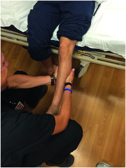
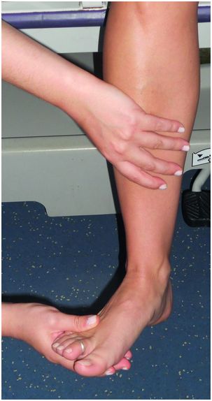
The integrity of the medial and lateral ankle, as well as the tibiofibular syndesmosis, should be evaluated with the compression and dorsiflexion eversion tests (Figures 7.3 and 7.4). See ankle section for descriptions of tests.
It is also important to pay particular attention to the integrity of the peroneal nerve.

Compression test.

Dorsiflexion eversion test.
Essential Diagnostics
Two-view x-rays of the tibia and fibula should be obtained to fully evaluate for fracture.
Ankle and/or knee x-rays may also be needed, depending on the clinical suspicion of concomitant injury.
ED Treatment
Patients should have the affected leg elevated, iced, and splinted in a position of comfort until definitive x-rays are obtained.
Once a fibular fracture is determined, the stability of the ankle should be evaluated.
Stable, isolated proximal or midshaft fibula shaft fractures may be treated based upon patient’s symptoms:
If there is no or minimal pain with ambulation, a simple compression dressing may be used.
If there is difficulty with ambulation, then a splint, cast, or walking boot may be used.
Rest, ice, elevation, and weight-bearing as tolerated may be recommended.
Unstable Maisonneuve fractures should be splinted with a posterior splint with the ankle at 90° and the patient made non-weight bearing.
Disposition
Most isolated proximal and midshaft fibula fractures may be referred for outpatient management.
Stable, isolated fractures may be followed up by sports medicine or primary care physicians.
Patients may begin to return to play with a rehabilitation program once they are symptom free, usually after four to six weeks. Return to contact activities usually takes longer due to increased risk of re-fracture.
Unstable Maisonneuve injuries need close follow-up within two to three days with orthopedic surgery.
Open fractures or grossly displaced fractures that are unable to be reduced in the ED need emergent orthopedic consultation.
Complications
Painful nonunion or malunion are possible, but given the small amount of weight-bearing by the fibula, this is usually not a major problem.
Instability of the ankle from a Maisonneuve-type injury may lead to foot and ankle dysfunction and posttraumatic arthritis.
Neurovascular injury may occur.
Pediatric Considerations
Children are not at any special risk for these types of injury.
Similar patterns of injury as adults should be considered.
Pearls and Pitfalls
The major issue to consider with proximal or midshaft fibular fractures are associated ligamentous ankle injuries resulting in Maisonneuve injuries.
These are unstable and easy to miss if the provider only evaluates the ankle and/or leg in isolation.
Consideration should be given to obtaining leg or knee films in these situations.
Pay particular attention to the integrity of the peroneal nerve.
Chronic Exertional Compartment Syndrome
Background
Chronic exertional compartment syndrome (CECS) is an entity not much different from acute compartment syndrome during its acute phase, but quickly resolves once activities stop.
CECS is due to acute swelling of muscles during exercise within defined fascial compartments that leads to tissue compression causing pain, numbness, or weakness.
Usually, the pain causes the patient to stop the activity and the symptoms resolve spontaneously (typically within 30 minutes).
The compartments most commonly affected in the lower leg are the anterior and lateral compartments, although any of the four compartments may be involved (Figure 7.2).
Mechanism
Exercise induces increased blood flow to active muscles, which effectively swell within the enclosed fascial compartments.
As the pressure increases, muscular pain increases.
Eventually, pressures may become high enough to compress vascular and neurologic structures, which further exacerbates pain and leads to other symptoms like pallor, coolness, loss of pulse, or neuropathies.
The fascial compartments are likely abnormally stiff or noncompliant in these patients, thus not allowing for adequate expansion during exercise.
Presentation
Symptoms will usually develop after a particular period of continuous exertion.
This period may be variable, depending on the patient and the severity of disease.
Most commonly described is an achy constant pain in the affected compartment that slowly develops as the patient exercises.
The pain usually gets intense enough that the patient will have to stop exercising.
With rest, the pain gradually improves but may take over 30 minutes to resolve.
The patient’s symptoms may be variable, depending on the structure(s) compressed.
Patients may simply have pain from muscle compression, but also temporary paresthesias and weakness during the acute phase of the disease.
Physical Exam
While the patient is at rest, he/she will have a normal physical exam.
However, during exertion the muscles of the affected compartment may become visibly enlarged, and the compartment will become tense and firm to palpation.
Occasionally, muscle herniations and swelling may be seen.
One way to provoke this is to have the patient lay supine and actively dorsiflex and plantarflex the ankles against resistance. Reproduction of pain, swelling, or paresthesias is considered a positive test.
Absences of pulses should prompt investigation for arterial insufficiency.
Essential Diagnostics
Most commonly this is initially a clinical diagnosis that is based upon a typical history and exam features.
Other etiologies such as MTSS and stress fracture should also be evaluated.
Imaging diagnostics like x-ray or MRI should be considered to evaluate for other causes of pain (fractures or soft tissue masses causing external compression).
No workup is indicated in the ED if CECS is suspected, as long as there is not concern for acute compartment syndrome or fracture.
In the clinic setting, compartment pressures of the anterior, lateral, and sometimes posterior superficial compartments should be obtained.
This is usually done in a clinical setting where the patient may run or exercise enough to bring on symptoms.
The patient usually runs at least 5 minutes into the “pain zone,” and then the compartment pressures are tested in the standard fashion.
Typically compartment pressures are obtained at rest prior to exercise, at 1 minute post exercise, and again at 5 minutes post exercise.
ED Treatment
These patients will not routinely present to the ED, however, they may present while in the acute phase of the disease.
In these cases they may be treated conservatively with ice, compression, and anti-inflammatories.
If there is concern for acute compartment syndrome, they should be treated with imaging, pain control, compartment pressure testing, and orthopedic consultation for possible fasciotomy.
Disposition
Patients with CECS may be sent home with follow-up with orthopedics or sports medicine.
A period of rest and a reduced exercise program should be advised.
Patients should avoid exercises that provoke symptoms as there is a small risk of causing acute compartment syndrome.
Physical therapy may be beneficial to work on soft tissue mobilization and stretching/ strengthening of the affected compartments.
Any biomechanical problems (e.g., pes planus) should be corrected in an attempt to offload the overstressed muscles and improve symptoms.
Once conservative therapies fail, fasciotomy or fasciectomy may be considered to alleviate symptoms.
Complications
Severe, untreated CECS could potentially progress to full-blown acute compartment syndrome with the ensuing complications of muscle necrosis, nerve damage, contractures, or even loss of limb.
Pediatric Considerations
This condition is uncommon in children, but not impossible.
Other conditions like fractures, tumors, or metabolic disorders should be considered.
Pearls and Pitfalls
As with acute compartment syndrome, CECS is a difficult diagnosis to make and takes a thorough history and an astute physical exam.
It is typically a diagnosis that takes several encounters to make and confirm.
Unfortunately, it is a difficult entity to treat and may often cause patients to alter their exercise or work patterns due to pain and other debilitating symptoms.
Once acute compartment syndrome and fracture has been ruled out, patients may be referred to sports medicine or orthopedics for further evaluation and workup.
Nerve Entrapment
General Description
As nerves traverse the lower leg, they may become entrapped at a number of different sites from a variety of conditions.
Typically this is a condition that occurs with the slow growth of masses or scar tissue.
However, casts and other compressive braces may cause focal impingement that may cause the patient to be symptomatic.
The location and extent of entrapment will determine the extent of symptoms.
Mechanism
Prior trauma, surgery, tight-fitting casts, braces, contusions, or arthritis may cause the development of scar tissue, cysts, masses, or osteophytes that effectively entrap nerves.
Symptoms usually develop over time, as the structure is progressively impinged.
Presentation
Patients generally complain of insidious onset of burning pain or paresthesias in a particular dermatome that maps to the affected nerve.
They may also present with weakness of the innervated muscles or subtle muscle atrophy.
Physical Exam
A key to diagnosis is to determine the specific area involved to further assist in the identification of the particular nerve that is entrapped.
The common peroneal nerve may become entrapped at the fibular head causing neuropathic pain on the anterolateral aspect of the leg that extends into the foot.
They may also have a foot drop and recurrent ankle injuries.
The distal branches of the common peroneal nerve may also be entrapped and symptomatic.
The sural and saphenous nerves may also become entrapped and cause pain.
Any overlying restricting devices should be removed and the area of complaint fully examined for scars, masses, or deformities that may cause pressure or stretching of nerves.
A Tinel’s sign may be performed at the presumed site of impingement.
A recurrent tapping on the nerve will cause a distinct shooting pain into the affected area that will reproduce the patient’s symptoms.
Essential Diagnostics
Imaging is important to rule out bony or soft tissue structures that are compressing the nerve.
This may begin with x-rays but an MRI scan may be needed to fully assess for soft tissue masses.
Nerve conduction studies may be useful in determining the location and severity of nerve entrapments.
ED Treatment
The EP should evaluate for acute causes of compression, such as fractures, masses, or cysts, utilizing the physical exam and standard x-rays.
In most cases, more advanced imaging may be done as an outpatient.
Generally, conservative treatment is all that is needed in the ED.
Patients should be advised to rest, remove any compressive devices, use NSAIDs for pain and avoid activities that exacerbate symptoms.
Disposition
Patients will need further imaging and possible nerve conduction studies and should be referred to sports medicine or orthopedics for further workup.
Complications
Progressive weakness, paresthesias, and loss of function are the most common outcomes in untreated disease.
Pediatric Considerations
There are no specific pediatric considerations.
Pearls and Pitfalls
Don’t forget other causes of neuropathy, like diabetes or multiple sclerosis.
In addition, it is important to rule out a more proximal cause of symptoms such as impingement from the low back or spine.
A broad differential diagnosis should always be considered and evaluated with a thorough history and physical exam.
The diagnosis of nerve entrapment is often a difficult one to make and it may be missed on the first encounter.
Gastrocnemius Musculotendinous Tears
General Description
Acute strains and tears of the gastrocnemius musculotendinous complex usually occur on the medial aspect.
Patient are often over 30 years of age.
Mechanism
A strong force is applied across the calf musculature, as often occurs in running up a hill, playing tennis, or jumping.
Presentation
Patients feel an acute tearing or stabbing sensation in the posterior mid calf.
They may complain of weakness after the injury and will have moderate swelling and ecchymosis of the area.
Occasionally, they may hear a distinct popping sound at the time of injury.
Stay updated, free articles. Join our Telegram channel

Full access? Get Clinical Tree


