10 Lower Extremity Joint Injections
Lower extremity intraarticular injections with corticosteroids and anesthetics are useful treatment options for patients with hip, knee, ankle, or foot pain.4 Injections are given to treat acutely painful joints refractory to rest and oral medications. The indications, patient selection, complications,1,2,6,7,9,14,16,19 and general technique of joint injections10,25 were covered in the previous chapter. This chapter will focus on the technique of specific lower extremity intraarticular joint injections. Knowledge of multiple techniques is helpful.26
Hip Joint
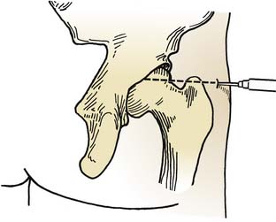
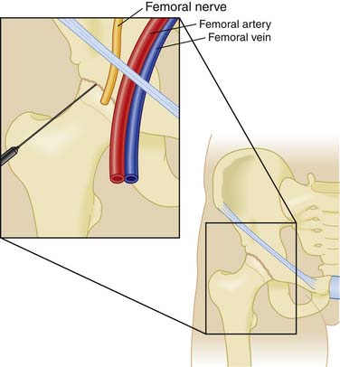
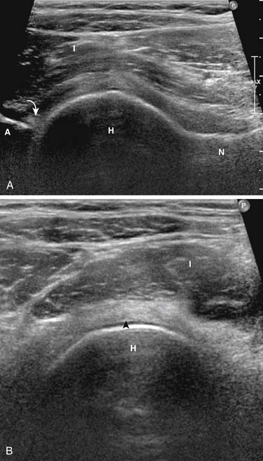
The hip joint is often difficult to infiltrate or aspirate because of its depth and the surrounding tissue. Fluoroscopic guidance with injection of contrast material or ultrasound guidance is often necessary to confirm proper needle placement. This joint may be infiltrated by an anterior or lateral approach (Figs. 10-1, 10-2, 10-3). The anterior approach is preferred.18,23,24 With the anterior approach, the patient is in the supine position with the lower extremity externally rotated. The length of the needle will depend on the patient’s size. The anatomic landmarks for the anterior approach are 2 cm distal to the anterior superior iliac spine and 3 cm lateral to the palpated femoral artery at a level corresponding to the superior margins of the greater trochanter. After superficial anesthesia is administered, the needle is advanced at an angle 60 degrees posteromedially through the tough capsular ligaments, advanced to bone, and slightly withdrawn. This technique places the tip of the needle directly into the joint, and aspiration or injection may be performed. This approach is much simpler using image guidance to direct the needle posteromedially into the joint. When the capsular ligaments have been penetrated, 2 to 4 mL of anesthetic and corticosteroid suspension may be introduced.
Knee
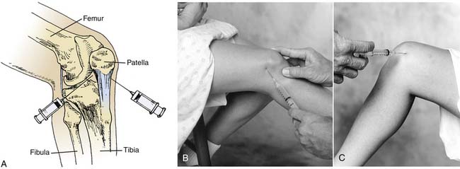
The knee is the most commonly aspirated and injected joint in the body. It contains the largest synovial space and demonstrates the most visible and palpable effusion (when present). A patient is usually most comfortable lying supine with sufficient pillows. The knee is prepared using an aseptic technique. If a large effusion is present, whether medially or laterally, the site of entry should be over the maximal expansion of the effusion in order to cause the least discomfort during the procedure. For injections in which a large effusion is not present, the lateral, medial, suprapatellar, or anterior approach may be used (Fig. 10-4). Before injecting or aspirating the knee, the patella should be grasped between the examiner’s thumb and forefinger and rocked gently from side to side to ensure that the patient’s muscles are relaxed.
Stay updated, free articles. Join our Telegram channel

Full access? Get Clinical Tree


