ANATOMY
The tibia provides primary support for weight bearing. The tibia has a thick cortex, and significant force is required to fracture it. Proximally, the tibia splays out to form the medial and lateral plateaus that articulate with the femoral condyles. The lateral plateau is higher and smaller than the medial and is more susceptible to fracture. The distal tibia articulates with the fibula laterally and the talus inferiorly. A dense interosseous membrane connects the tibia and fibula. The distal tibial articulation is supported by the ankle syndesmosis, a series of ligaments inferior to the interosseous membrane. The fibula has a small diameter and lies lateral and posterior to the tibia. It bears little weight but is more easily fractured than the tibia.
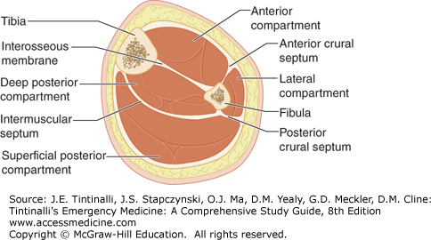
The lower leg is divided into four compartments, each coursing parallel to the tibia (Figure 275-1). The compartments are enclosed by nonexpandable bones and connective tissue that limit the compartment size and prevent compartment expansion if its volume increases. Each compartment contains muscles and nerves that may sustain permanent damage with elevated tissue compartment pressure (Table 275-1). (See also chapter 278, “Compartment Syndrome.”)
| Compartments | ||||
|---|---|---|---|---|
| Anterior | Lateral | Superficial Posterior | Deep Posterior | |
| Muscles | Dorsiflex foot and ankle | Plantarflex and evert foot | Flex knee and ankle | Plantarflex toes, inversion of foot |
| Nerve | Deep peroneal | Superficial peroneal | Sural | Posterior tibial |
| Sensation | First dorsal web space | Dorsum of foot | Lateral aspect of foot and distal calf | Sole of foot |
| Artery | Anterior tibial | — | — | Posterior tibial |
A cross-section at the midcalf level shows the anterior compartment enclosed by the tibia, interosseous membrane, and anterior crural septum (Table 275-1 and Figure 275-1). Muscles in the anterior compartment group dorsiflex the foot and ankle. The deep peroneal nerve courses within the anterior compartment and exits to provide sensation to the dorsal web space between the first and second toes.
The lateral compartment is bordered by the anterior crural septum, the fibula, and the posterior crural septum. Its muscles plantarflex and evert the foot. The superficial peroneal nerve in this compartment provides sensation to the dorsum of the foot. The superficial posterior compartment contains muscles that flex the knee and the tibiotalar joints. Its sural nerve provides sensation for the lateral aspect of the foot and the distal calf. The muscles of the deep posterior compartment plantarflex the foot and toes and invert the foot. The posterior tibial nerve that exits this compartment provides sensation to the sole of the foot.
CLINICAL FEATURES
The history may give clues about the mechanism of injury and nontraumatic soft tissue injuries. Evaluate the nerves by checking sensation in the web space, lateral heel, and sole of the foot. Plantarflex and dorsiflex the foot, and evert the foot to test motor function. Evaluate the extent of soft tissue injury visually and by palpating the compartmental muscle groups. It is often the extent of soft tissue injury, rather than the fracture itself, that determines the outcome. Palpate the knee, and the tibia and fibula along their entire lengths. Palpate the popliteal, dorsal pedal, and posterior tibial pulses. An absent or decreased pulse may indicate the need for urgent fracture reduction and further vascular evaluation.
DIAGNOSIS
Anteroposterior and lateral radiographs of the leg that include the knee and ankle are sufficient to evaluate bony injuries. If ankle or knee injuries are suspected, then further imaging is needed. If a tibial shaft fracture is suspected, splint the leg with a radiolucent device to control pain and prevent further soft tissue damage before obtaining films. Check pulses, movement, and sensation before and after splinting the leg.
TREATMENT
Cleanse wounds and debride loose tissue and foreign material. Administer tetanus immunization as indicated. Splint fractures before obtaining radiographs; this will prevent further damage to soft tissue caused by movement of bone fragments. Irrigate open wounds and administer parenteral antibiotics (such as cefazolin, 1 gram IV) for open fractures. If compartment syndrome is suspected, measure compartment pressure (see chapter 278, “Compartment Syndrome”). Treatment of compartment syndrome is fasciotomy of the involved compartment. Details of suturing pretibial lacerations are provided in the chapter 44, “Leg and Foot Lacerations.”
COMPLICATIONS
Wounds that are not adequately cleansed and debrided are prone to infection. Patients with compartment syndromes may develop permanent disability if elevated tissue pressures are not suspected or diagnosed in a timely fashion. Fractures that are not adequately aligned or immobilized heal poorly or not at all.
SPECIFIC INJURIES
The tibia is the most commonly fractured long bone. Fractures often result in open injuries because of the minimal amount of subcutaneous tissue between the tibia and the skin. The fracture pattern seen on radiographs will give a clue to the force that caused the injury. Transverse shaft fractures typically result from a direct blow to the bone. Spiral fractures are the result of rotational forces. A comminuted fracture suggests the mechanism had a very-high-energy impact. A force powerful enough to shatter the dense cortex of the tibial shaft will often be transmitted through the interosseous membrane to the fibula, fracturing that bone as well.
Open tibial shaft fractures have been classified (Table 275-2).1
Stay updated, free articles. Join our Telegram channel

Full access? Get Clinical Tree


