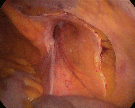
Figure 31.1
Right indirect inguinal hernia

Figure 31.2
Peritoneal incision
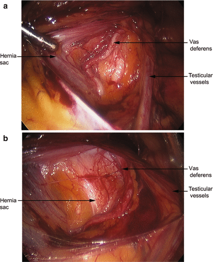
We begin our dissection in the indirect space where the testicular vessels and vas deferens are identified and bluntly dissected off the underlying peritoneum (Fig. 31.3a, b). In the female patient, the round ligament is generally quite adherent to the peritoneum and in order to avoid peritoneal tears, the round ligament is clipped and divided. The preperitoneal space is then bluntly dissected along the medial portion of our incision. The bladder is mobilized in the space of Retzius and Cooper’s ligament is identified on both the right and left sides (Fig. 31.4). Direct defects will be found in this location and can be reduced by pulling the perperitoneal fat off of the overlying transversalis fascia. Indirect defects should then be reduced by pulling the peritoneal hernia sac medially and bluntly pushing the vas deferens and testicular vessels laterally (Fig. 31.5).


Figure 31.3


(a) Testicular vessels are seen dissected off the peritoneum while vas deferens are still attached. (b) Both testicular vessels and vas deferens are mobilized off the peritoneum
Stay updated, free articles. Join our Telegram channel

Full access? Get Clinical Tree


