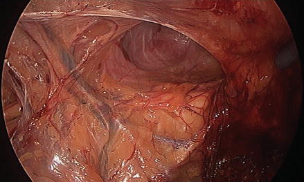
Figure 32.1
View through dissecting balloon. Note pubis and Cooper’s ligament bilaterally
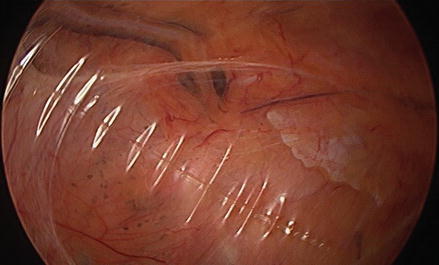
Figure 32.2
View through dissecting balloon showing left inferior epigastric vessels and direct hernia
Direct inguinal hernias are typically seen and oftentimes reduced as the balloon dissector inflates. The same is true of femoral and obturator hernias. Unreduced direct hernias should be carefully reduced, clearing Cooper’s ligament and allowing for visualization of lateral structures (Figs. 32.3 and 32.4). Prior to lateral dissection, the position of the inferior epigastric vessels should be identified as occasionally, these vessels will be pushed posteriorly by the dissecting balloon. It is important that lateral peritoneal dissection occur posterior to the plane of the epigastric vessels. In rare circumstances, the epigastric vessels may be clipped and divided should they dissect free from the rectus bed.
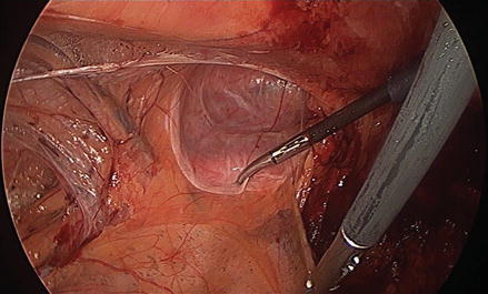
Figure 32.3
Attenuated transversalis fascia of left direct hernia, medial to inferior epigastric vessels