Fig. 4.1
A dilating balloon is passed over the guide wire and through the cystic ductotomy. The position of the balloon is checked fluoroscopically to ensure that it is traversing the entire length of the cystic duct. The balloon is then held inflated for at least 3 min to maximally dilate the duct and obliterate any values and tortuosity
Choledochoscope Setup and Insertion
Connect the choledochoscope to a pressurized saline bag. Check to ensure adequate flow through the working channel, and then clamp the irrigation during insertion. Use a picture-in-picture setup on the main monitor, so that the laparoscopic and choledochoscopic images can be viewed simultaneously. Feed the guide wire through the scope’s working channel and then pass the scope along the wire and through the cystic ductotomy. Use one hand to advance the scope and control torque, while the other operates the scope flexion dial. Once the scope is within the cystic duct, turn on the irrigation and scope light source. Usually an illumination intensity of only 30 % is required. Slowly advance the choledochoscope, while using torque and flexion to keep the ductal lumen centered. Usually the scope can be advanced by manipulating it external to the trocar; however, if this is not possible, a padded laparoscopic grasper can be used to gently pass it internally (Fig. 4.2). Once the scope is within the common duct, remove the guide wire from the working channel. Advance the scope until the first common duct stone is encountered.
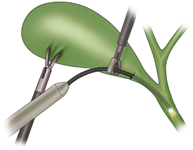
Fig. 4.2
A padded grasper is used to gently advance the choledochoscope distally under direct visualization. It is important to guide the scope in parallel with the cystic and common bile ducts to avoid ductal injury. The scope should not be advanced unless the lumen of the duct is clearly visualized endoscopically
Stone Capture and Extraction
Pass a three- or four-wire stone extraction basket through the working channel of the choledochoscope. Advance the closed basket beyond the stone and then have your assistant open it. Slowly trawl the open basket back toward the scope until the stone falls within it (Fig. 4.3). Often a back-and-forth jiggling motion is required to make the stone drop within the wires of the basket. Once the stone is centered within the wires, slowly close the basket around it. Withdraw the scope from ducts, while keeping the stone pinned against the face of the scope. Often there will be some resistance at the cystic-common duct junction, which can be overcome with steady, gentle pressure. Once the stone is delivered out of the cystic ductotomy, it can be deposited in the peritoneal cavity and removed using a stone-grasper or with the gallbladder in an endoscopic bag after completion of the cholecystectomy. If multiple stones were present on cholangiogram, reintroduce the scope through the cystic ductotomy and repeat the process of capture and extraction. Usually once the cystic duct has been dilated, the scope can be reintroduced manually without the need to regain guide wire access.
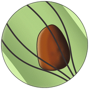
Fig. 4.3
The wire basket is advanced distal to the stone and opened. It is then slowly pulled back, and in this figure we can see the stone has fallen within the wires of the basket. The basket is then closed to capture the stone against the face of the choledochoscope
Completion Cholangiogram and Cystic Duct Ligation
Even if all common duct stones were thought to have been removed, it is important to perform a repeat cholangiogram after LCBDE to confirm and document ductal clearance and antegrade flow of contrast into the duodenum. As the cystic duct was dilated to at least 8 mm, use two suture loops to ligate it (rather than clips). Then proceed with the cholecystectomy as usual. It is not necessary to leave a drain after transcystic LCBDE.
Transcholedochal LCBDE
If a transyctic approach is not feasible based on stone size or patient anatomy, a transcholedochal approach can be utilized. However, this should not be attempted unless the surgeon has adequate experience with advanced intracorporeal suturing, as well as the ability to perform an open repair of the common bile duct. Alternatively, the patient can always be referred first for ERCP without “burning any bridges,” with transcholedochal LCBDE reserved in case the duct cannot be cleared endoscopically.
Related posts:


Stay updated, free articles. Join our Telegram channel

Full access? Get Clinical Tree


