A line should be drawn along the posterior border of the SCM. The cricoid cartilage should be identified and a line drawn along a skin crease at this level to approximate C6 level. Standing next to the head of the bed, palpate the quadrant closest to the clavicle posterior to the SCM while the patient takes a forceful inspiration through the nose. This should delineate the interscalene groove and allow marking of the anterior and middle scalene muscles.
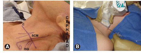
A. Surface anatomy relevant to the supraclavicular block. The “X” marks the suggested site for needle insertion when performing the plumbbob technique with nerve stimulation. SCM, sternocleidomastoid muscle; C, clavicle. B. A HF linear transducer is placed perpendicular to skin medial to the clavicle. The block needle is inserted lateral to the transducer and directed anteromedially for inplane needle guidance.
Approach and Technique
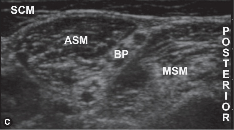
 With a high-frequency (HF) linear transducer positioned perpendicular to skin overthe posterior border of the SCM,
  the brachial plexus is visualized in short-axisbetween the anterior and middle scalene.
After sterile skin preparation, a local anesthetic skin wheal is raised lateral to the ultrasound transducer.
US-GUIDED INTERSCALENE BLOCK
Related posts:


Stay updated, free articles. Join our Telegram channel

Full access? Get Clinical Tree


