31 Intercostal Nerve Block
Anatomy of Intercostal Nerves
The twelve pairs of thoracic spinal nerves (T1-12) are divided into ventral and dorsal rami after they pass through the intervertebral foramina. The ventral rami of T1-T11 form the intercostal nerves that enter the intercostal spaces. The ventral ramus of T12 forms the subcostal nerve that is located inferior to the 12th rib. The dorsal rami of T1-T12 pass posteriorly to supply sensation to skin, muscles, and bones of the back.1
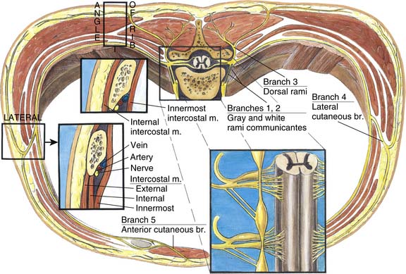
Intercostal nerves are composed of dorsal horn sensory afferent fibers, ventral horn motor efferent fibers, and postganglionic sympathetic nerves. The major branches of intercostal nerves are anterior and lateral cutaneous branches (Fig. 31-1). These branches divide and innervate the skin and intercostal muscles of an individual segment along with variable collateral innervation of the adjacent segments. Because of such collateral innervation, it is necessary to block a level above and below the desired level when an intercostal nerve block is performed.
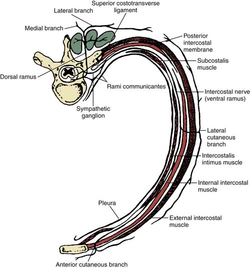
Figure 31-1 Anatomy of an intercostal nerve.
(Adapted from Ferrante FM, VadeBoncouer TR: Postoperative Pain Management. New York, Churchill Livingstone, 1993. Elsevier.)
Throughout its course, each intercostal nerve is associated with an artery and a vein. The intercostal arteries are derived directly from the aorta. The intercostal veins are derived from the confluence of venules along the chest and end in the azygous and hemiazygous veins. The intercostal nerve travels inferior to the vein and artery of the same segment (Fig. 31-2).