Infectious Diseases


Infectious Diseases


20-1 Rocky Mountain Spotted Fever

Rakesh D. Mistry


Clinical Presentation

The clinical presentation of Rocky Mountain spotted fever (RMSF) is characterized by fever, headache, and a petechial rash. In the early phases, patients may develop high fever followed by a severe headache (85%). Between 50% and 60% of patients develop gastrointestinal symptoms.1 Other early symptoms may include myalgias, sore throat, cough, abdominal pain, malaise, and anorexia.1,2 Two to 3 days after onset of the illness, patients develop the characteristic rash on the ankles and wrists, initially resembling pinpoint macules. The macules quickly become petechial in nature, spread to the palms and soles, and then move centrally to involve the trunk. In severe cases, this diffuse petechial rash resembles meningococcemia. The rash may coalesce, and necrosis may follow in regions supplied by terminal arterioles. The rash is not always petechial in nature, and delayed presentation of the rash is possible. Absence of an exanthem is described in 10% of cases.3 Other signs and symptoms that may develop during the course of RMSF include diarrhea, hepatosplenomegaly, edema, lymphadenopathy, and conjunctivitis.


Pathophysiology

The causative organism is Rickettsia rickettsi, an obligate, intracellular, gram-negative bacillus. The ixodid species of ticks serves as both reservoir and vector for R. rickettsi. The most common of these is the dog tick, Dermacentor virabilis. RMSF is endemic in much of North and Central America, yet, contrary to its name, the majority of cases occur in the south and Atlantic coastal regions of the United States. The bacterium is introduced into humans via a tick bite. After an incubation period of 2 to 14 days, rickettsial organisms spread via blood and lymphatics. Rickettsia then invade and proliferate in
endothelial and mesothelial cells of blood vessels. Cell death ensues, after which the rickettsia contiguously spread and infect adjacent cells. The infection stimulates a severe inflammatory response. This widespread, systemic vasculitis is directly responsible for almost all of the symptoms of RMSF.


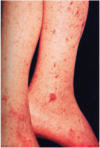
FIGURE 20-1 Rocky Mountain spotted fever. There is a generalized petechial eruption that involves the entire cutaneous surface, including the palms and soles. (From Habif, with permission.)


Diagnosis

The diagnosis is primarily clinical, because microbiologic identification of the R. rickettsi organism is difficult. The “classic” triad of symptoms (fever, headache, and rash) is present in fewer than 50% of patients.1 Only 60% of patients recall being bitten by a tick.1,2

The indirect fluorescent antibody (IFA) test, the most widely used test for R. rickettsi, can detect serum immunoglobulin M or G antibody with a sensitivity of greater than 90%.1 However, IFA serves only as a confirmatory test for RMSF, because antibody titers do not rise until late into the second week of illness, by which time resolution or death has already occurred. Alternatively, skin biopsies may be obtained for direct immunofluorescence or immunoperoxidase staining for R. rickettsi.

Laboratory findings may include hyponatremia, thrombocytopenia, elevated transaminases, and leukopenia or leukocytosis. Renal insufficiency, cerebrospinal fluid pleocytosis, and electrocardiographic changes may be seen. Although none of these findings are specific, their presence may be suggestive of RMSF.


Clinical Complications

Complications include encephalitis, pulmonary edema, acute respiratory distress syndrome, arrhythmias, gastrointestinal bleeding, coagulopathy, and death. Even with treatment, case-fatality rates are approximately 5%; they may be as high as 25% in untreated patients. The majority of deaths occur during the second week of illness, although fulminant cases resulting in rapid death may occur. Long-term sequelae in survivors include paraparesis, hearing loss, peripheral neuropathy, and cerebellar, vestibular, and motor dysfunction.


Management

Initial treatment of RMSF is often empiric, based on the history and physical findings. Studies have demonstrated that antibiotic therapy is most effective if instituted before the fifth day of illness. Consequently, treatment should not be withheld if RMSF is suspected. Tetracycline antibiotics remain the mainstay of treatment for RMSF. Doxycycline is the preferred agent. Doxycycline therapy usually is continued for 7 days, or 2 days after the patient becomes afebrile. Alternative treatment consists of chloramphenicol, but this drug is reserved for pregnant women, in whom doxycycline is contraindicated.

Patients with mild cases of RMSF in the early phases of illness may be treated on an outpatient basis with oral antibiotics and close follow-up. If the disease is diagnosed in the later stages, inpatient therapy and intravenous antibiotics are reasonable. Patients who appear ill or toxic, have severe vomiting, or are at risk for poor compliance should be admitted. Intensive care admission should be considered for patients with neurologic symptoms or renal insufficiency.4


REFERENCES

1. Sexton DJ, Kaye KS. Rocky Mountain spotted fever. Med Clin North Am 2002;86:351-360.

2. Thorner AR, Walker DH, Petri WA Jr. Rocky Mountain spotted fever. Clin Infect Dis 1998;27:1353-1360.

3. Sexton DJ, Corey GR. Rocky Mountain “spotless” and “almost spotless” fever: a wolf in sheep’s clothing. Clin Infect Dis 1992;15:439-448.

4. Conlon PJ, Procop GW, Fowler V, et al. Predictors of prognosis and risk of acute renal failure in patients with Rocky Mountain spotted fever. Am J Med 1996;101:621-626.


20-2 Lyme Disease

Christopher J. Russo


Clinical Presentation

In the United States, most patients with Lyme disease (LD) present with a slowly expanding skin lesion (erythema migrans), which occurs at the site of the tick bite. Erythema migrans typically manifests after an incubation of 7 to 14 days (range, 3 to 32 days). Its classic presentation is a red, circular, macular lesion with central clearing resembling a ring or bull’s-eye. Constitutional symptoms often occur in early stages of the disease and include fatigue, muscle aches, fever, headaches, and arthralgias. With dissemination, spirochetemia may result in multiple erythema migrans lesions.1

Neurologic manifestations in U.S. patients include symptoms of acute neuroborreliosis in approximately 15% of untreated patients; these symptoms include lymphocytic meningitis, subtle encephalitis, cranial neuropathy (unilateral or bilateral peripheral facial nerve palsy), motor or sensory radiculoneuritis, cerebellar ataxia, or myelitis. Untreated, these neurologic abnormalities typically improve or resolve within months; however, up to 5% of untreated patients develop chronic neuroborreliosis.1 Untreated, 5% of patients demonstrate acute cardiac manifestations, including atrioventricular block and acute myopericarditis.1 Joint involvement is common among untreated patients, with 60% reporting intermittent attacks of joint swelling and pain that often involves large joints (most commonly the knee). Swelling is generally out of proportion to pain, which is typically mild to moderate.2


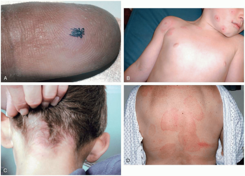
FIGURE 20-2 A: Ixodes tick. An adult tick is the size of the head of a match. (From Goodheart, with permission.) B: Multiple target erythema migrans lesions. (Courtesy of Alfredo Sabbaj, MD.) C: Target lesion of Lyme disease. (Courtesy of Christy Salvaggio, MD.) D: Erythema migrans lesion of Lyme disease. (From Goodheart, with permission.)


Pathophysiology

LD, the most common tick-borne disease in the United States, is caused by the spirochete Borrelia burgdorferi. Currently, about 15,000 cases are reported each year.1 Usually, prolonged tick attachment (36 hours or longer) is required for B. burgdorferi transmission. After infection,
spirochetes disseminate to various tissues; virulence factors influence which tissues are affected. B. burgdorferi remains in a constant state of antigenic flux during its life in the mammalian host, continuously triggering an inflammatory response, which may vary in severity. Chronic infection in susceptible individuals may result from this inflammatory response.3


Diagnosis

Positive cultures of the organism B. burgdorferi have been obtained only early in the course of illness, primarily from skin biopsy specimens of erythema migrans lesions. Less invasive is the “two-step” approach, which combines an enzyme-linked immunosorbent assay (ELISA) with confirmatory immunoblotting (Western blot). These tests are insensitive during the first several weeks of infection. A Lyme urine antigen test is available, but it remains unreliable and should not be used.


Clinical Complications

LD may result in chronic neurologic or rheumatologic sequelae. Patients may have recurrent episodes of arthritis, even after appropriate treatment. A small percentage of appropriately treated patients continue to have subjective symptoms (musculoskeletal pain, neurocognitive difficulties, or fatigue)—so-called “chronic Lyme disease.” In a large study, however, the frequencies of these symptoms were not different from those in age-matched control subjects without LD.1


Management

For early localized or disseminated infection, doxycycline therapy for 14 to 21 days is recommended for all patients older than 8 years of age except pregnant women. Amoxicillin and cefuroxime axetil are accepted alternatives. For patients with neurologic abnormalities (except isolated facial nerve palsy, which is treated as described above), intravenous ceftriaxone is recommended for 2 to 4 weeks. A vaccine is available for high-risk individuals aged 15 to 70 years. Prevention of tick bites should be emphasized. After a tick bite, even in Lyme-endemic areas, neither tick analysis nor prophylactic antibiotic treatment is routinely recommended.1,2,3


REFERENCES

1. Steere AC. Lyme disease. N Engl J Med 2001;345:115-125.

2. Sood SK. Lyme disease. Pediatr Infect Dis J 1999;18:913-925.

3. Weis JJ. Host-pathogen interactions and the pathogenesis of murine Lyme disease. Curr Opin Rheumatol 2002;14:399-403.


20-3 Malaria

Michele Lambert


Clinical Presentation

Malaria should be considered in any patient with fever who has recently returned from, or resides in, an endemic area.1 Although signs and symptoms may be nonspecific, the hallmark of malaria is a high, spiking, recurrent fever. Classically, the length of time between fevers may help differentiate the offending species (a 72-hour cycle for Plasmodium malariae and a 48-hour cycle for Plasmodium falciparum, Plasmodium vivax, and Plasmodium ovale).2 Patients infected with P. falciparum may present with encephalitis. More than 90% of travelers with P. falciparum infection become ill within 6 weeks after travel in endemic regions.2 Patients with P. vivax infection may present as long as 6 months to years after their travel.


Pathophysiology

Malaria causes between 300 and 500 million new infections annually and kills 1.5 to 2.7 million people worldwide yearly.2,3 Four Plasmodium species are known to infect humans, causing two types of disease: “relapsing” (P. vivax and P. ovale) and “nonrelapsing” (P. falciparum and P. malariae). P. falciparum is the most common cause of malaria worldwide and is the species most likely to cause serious illness (see Table 20-3).


Diagnosis

Diagnosis is made by the examination of thick blood smears (which allows for evaluation of many cells); identification of the Plasmodium species and the degree of parasitemia (percentage of neutrophils containing malarial pigment) is made on thin blood smears. Blood should be obtained while the patient is febrile. If the diagnosis is highly suspected (e.g., the patient is a recent traveler to a highly endemic or hyperendemic region), negative smears should be repeated every 8 to 12 hours for a total of three smears.2 Physical examination may reveal hepatosplenomegaly, scleral icterus, or jaundice (20% of patients). Laboratory evaluation may reveal anemia, leukopenia, or thrombocytopenia. Markers for serious infection include respiratory distress, hypoglycemia, hypotension, renal failure, and greater than 5% parasitemia.1,2


Clinical Complications

Cerebral malaria (altered mental status, seizures, or coma) is a complication of P. falciparum with a high mortality rate. Other complications include hypoglycemia, hypotension, renal failure, and severe anemia. Complications are more likely in patients with greater than 5% parasitemia, pregnant women, children, and the elderly.1,2,3


Management

Treatment depends on the species and the severity of disease. Patients with greater than 5% parasitemia or other signs of severe disease (encephalitis, end-organ involvement, shock, acidosis, or hypoglycemia) should be admitted to the intensive care unit and treated with parenteral quinidine. Other patients should be closely monitored and treated with oral medications, depending on local resistance patterns of the area in which they traveled. For all species of Plasmodium except chloroquineresistant P. falciparum and P. vivax, the oral treatment of choice is chloroquine. If drug resistance is suspected, multiple medications may be needed.1,2,3


REFERENCES

1. White NJ. The treatment of malaria. N Engl J Med 1996;335:800-806.

2. Suh KN, Kozarsky PE, Keystone JS. Travel medicine: evaluation of fever in the returned traveler. Med Clin North Am 1999;83:997-1017.

3. Baird JK, Hoffman SL. Prevention of malaria in travelers. Med Clin North Am 1999;83:923-944.


20-3 Malaria

Michele Lambert


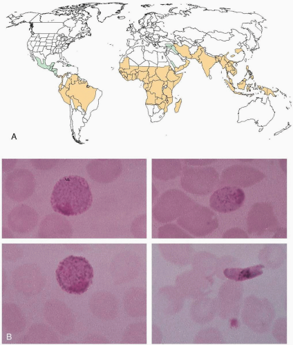
FIGURE 20-3 A: World distribution of malaria. Black areas indicate distribution of chloroquine-susceptible Plasmodium falciparum malaria, and gray areas indicate distribution of chloroquine-resistant Plasmodium falciparum malaria. (Modified with permission from Barat LM, Bloland PB. Drug resistance among malaria and other parasites. Infect Dis Clin North Am 1997;11:969-987.) B: Macrogametocytes of Plasmodium vivax (top left), Plasmodium malaria (top right), Plasmodium ovale (bottom left), and Plasmodium falciparum (bottom right). (From Smith JW. Atlas of diagnostic medical parasitology: blood and tissue parasites. Chicago: American Society of Clinical Pathologists, 1976.)


TABLE 20-3 Selected Clinical Characteristics of Four Types of Malaria


Characteristic


Plasmodium falciparum


Plasmodium vivax


Plasmodium ovale


Plasmodium malariae


Usual incubation period (d)


8-11


10-17 or longer


10-17 or longer


18-40 or longer


Severity of primary attack


Severe in nonimmune


Mild to severe


Mild


Mild


Periodicity (h)


None


48


48


72


Duration of untreated primary attack (wk)


2-3


3-8


2-3


3-24


Duration of untreated infection


6-17 mo


5-7 yr


12 mo


20+ yr


Average parasitemia (per mm2)


≥20,000


10,000


9,000


6,000


Anemia


Frequent and severe


Mild


Mild


Mild


CNS involvement


Yes, severe


Rare


Rare


Rare


Nephritic syndrome


Rare


Rare


No


Frequent


From McClatchy, with permission.


d, days; h, hours; wk, week; mo, months; CNS, central nervous system.


20-4 Toxic Shock Syndrome

Michael Greenberg


Clinical Presentation

Patients with toxic shock syndrome (TSS) may present with malaise, chills, high fever (greater than 102° F), hypotension, mental status changes, vomiting, diarrhea, myalgias, conjunctival injection, “strawberry” tongue, and desquamation of the skin, usually involving the hands and feet.1,2,3


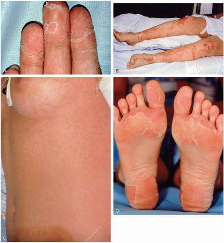
FIGURE 20-4 A: Desquamation of the palmar surface of the fingers. B: Petechiae and ecchymoses of the lower extremities. C: Petechiae of the trunk. D: Desquamation of the soles. (AD, Courtesy of B. Zane Horowitz, MD.)


Pathophysiology

TSS is caused by various strains of staphylococcal and streptococcal bacteria capable of producing TSS toxins. Seventy-five percent of TSS cases involve TSS toxin-1, whereas 20% to 25% involve enterotoxin B. A small percentage of cases involve enterotoxin C.1,2,3


Despite the fact that the first reported cases of TSS appeared to exclusively afflict menstruating women who used tampons, recent work has shown as many as one third of cases are probably not related to menstruation.1,2,3 Most cases are associated with local skin or wound infections, infected surgical wounds, or infections occurring in the postpartum period. Some cases have been associated with burns, retained nasal packing, or the use of barrier contraceptive devices.1,2,3 Necrotizing fasciitis may be present in half of all TSS cases. A strong correlation exists between TSS and Staphylococcus aureus grown from vaginal cultures of TSS patients.1,2,3 TSS occurs most often in women of childbearing age, especially teenage girls who use “hyperabsorbent” tampons.1,2,3


Diagnosis

The diagnosis of TSS is clinical and is based on the history and physical examination findings. Table 20-4 lists the current case-defining parameters for TSS.


TABLE 20-4 Case-Defining Parameters for Toxic Shock Syndrome (TSS)


Patients diagnosed as having TSS presented with shock and at least two of the following:


Acute renal insufficiency


Hematologic alterations:


Thrombocytopenia (≤100,000 platelets/mm3)


Disseminated intravascular coagulation


Hepatic injury:


Elevated aminotransferases or total bilirubin (greater than two times normal values)


Respiratory distress


Erythematous macular rash (desquamative or not desquamative)


Skin/soft-tissue necrosis


Adapted from Bernaldo De Quiros JC, Moreno S, Cercenado E, et al. Group A streptococcal bacteremia: a 10-year prospective study. Medicine (Baltimore) 1997;76:238-248.


Clinical Complications

Complications include septic shock, multiorgan system failure, extremity amputations, scarring, and death.1,2,3


Management

Aggressive treatment promptly initiated in the emergency department, including invasive hemodynamic monitoring and intravenous antibiotics, may be life-saving. Usual drug regimens include clindamycin, penicillins, cephalosporins, and aminoglycoside antibiotics in combination.1,2,3 In addition, emergency and extensive surgical débridement of infected areas may be life-saving. Some cases have been successfully treated with the use of intravenous immune globulin. Patients with suspected TSS require immediate hospitalization in an intensive care setting and emergency consultation with infectious disease specialists.1,2,3


REFERENCES

1. Bisno AL, Stevens DL. Streptococcal infections of skin and soft tissues. N Engl J Med 1996;334:240-245.

2. Bernaldo De Quiros JC, Moreno S, et al. Group A streptococcal bacteremia: a 10-year prospective study. Medicine (Baltimore) 1997;76:238-248.

3. Chadwell JS, Gustafson LM, Tami TA. Toxic shock syndrome associated with frontal sinus stents. Otolaryngol Head Neck Surg 2001; 124:573-574.


20-5 Staphylococcal Scalded Skin Syndrome

Michael Greenberg


Clinical Presentation

Patients with staphylococcal scalded skin syndrome (SSSS) present with fever and a diffuse, erythematous, tender rash of the face, trunk, and extremities. This rash rapidly develops into fluid-filled blisters that enlarge and then slough. Most cases of SSSS occur in children, but adult cases have also been reported.1,2,3


Pathophysiology

SSSS is caused by staphylococcal exfoliative toxins A (ETA) and B (ETB) that are secreted from phage II staphylococci. These toxins spread via the bloodstream.1,2,3 ETA exhibits “superantigen” characteristics, including epidermolysis and lymphocyte mitogenicity. This leads to separation of the stratum granulosum and stratum spinosum, giving rise to a positive Nikolsky’s sign. Immature renal function and inefficient clearing of exotoxins may predispose neonates to SSSS. Outbreaks of SSSS in neonatal intensive care units (NICUs) are often related to the presence of medical and nursing staff who are infected or colonized with coagulase-positive Staphylococcus aureus. This organism may be cultured from up to 30% of NICU staff members in hospitals. Culture of blister fluid usually is not helpful.2,3


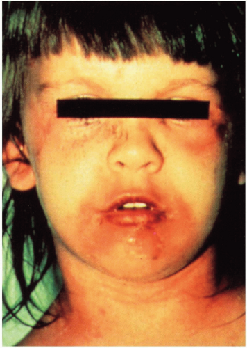
FIGURE 20-5 Staphylococcal scalded skin syndrome. Erythema is prominent on the neck and around the eyes and mouth. Crusting is also apparent in the periorificial areas. Over the chin, a bulla has ruptured, leaving a moist erosion. (From Mandell, Essential Atlas of Infectious Diseases, with permission.)


Diagnosis

SSSS must be considered in any infant with exfoliate dermatitis. The diagnosis is based on clinical findings. Laboratory and imaging studies are of limited help in confirming the diagnosis.


Clinical Complications

Complications include temperature instability, fluid losses, secondary skin infections, sepsis, and death.2,3


Management

The nares, conjunctiva, and skin around blisters of SSSS patients should be cultured. Prompt treatment with β-lactamase-resistant penicillin is usually effective. Supportive care aimed at limiting fluid and electrolyte losses is critically important. Topical antibiotics usually are not effective and therefore are not indicated.1,2,3 Meticulous local wound care is essential to prevent secondary infection. The use of an incubator for infants with SSSS may help to decrease insensible fluid loss through denuded skin. All staff must adhere to strict infection control techniques, and hand washing for staff and parents is essential.1,2,3


REFERENCES

1. Prabhash K, Babu KG, Ravi S, et al. Staphylococcal scalded skin syndrome. Lancet Infect Dis 2003;3:442.

2. Makhoul IR, Kassis I, Hashman N, Sujov P. Staphylococcal scalded skin syndrome in a very low birth weight premature infant. Pediatrics 2001;108:E16.

3. Prevost G, Couppie P, Monteil H. Staphylococcal epidermolysins. Curr Opin Infect Dis 2003;16:71-76.


20-6 Acute Rheumatic Fever

Kyung Rhee


Clinical Presentation

Acute rheumatic fever (ARF) manifests as fever, abdominal pain, polyarthritis, carditis, erythema marginatum, and subcutaneous nodules in children between 5 and 15 years of age. Symptoms usually develop 2 to 4 weeks after an episode of acute group A β-hemolytic streptococci (GABHS) pharyngitis, although one third of patients cannot recall a recent pharyngitis. Polyarthritis (60% to 75% of cases) typically affects multiple joints (mean, six joints). The carditis is a pancarditis that may manifest with fever or only a murmur. Erythema marginatum (5%) is typically transient (present for only hours) and is found on the trunk and proximal extremities, sparing the face. Subcutaneous nodules (5%) are firm, painless nodules on extensor surfaces and bony prominences. Children may also have anemia, prolongation of the PR interval (35%), and elevation of acute phase reactants (erythrocyte sedimentation rate (ESR), C-reactive protein (CRP), and white blood cell count). Sydenham’s chorea (10% to 30%) occurs 1 to 6 months after the ARF and is self-limited (usually 1 to 2 weeks, occasionally 3 to 6 months).1,2


Pathophysiology

ARF is a systemic inflammatory disease associated with previous pharyngeal GABHS infection.1 ARF is the leading cause of acquired heart disease in children on a worldwide basis. ARF is an autoimmune response to proteins in the GABHS organism that are immunologically cross-reactive with human tissues (e.g., heart, joints).1


Diagnosis

Diagnosis is based on the Jones criteria, which include five major criteria and four minor criteria. To make the diagnosis, there must be evidence of a recent GABHS infection and the fulfillment of two major, or one major and two minor, criteria.1,2 Evidence of recent infection usually involves rising antistreptolysin O (ASO) titers or anti-DNase B tests. A throat culture may be done at the time of ARF illness; however, it is positive in only 10% to 30% of cases. Additional laboratory findings include an elevated ESR, CRP, and leukocytosis.


Clinical Complications

Complications include valvular heart disease and heart failure.


Management

Patients with ARF should be treated with oral penicillin V for 10 days or with one dose of intramuscular benzathine penicillin G (or erythromycin, if allergic to penicillin).1 Family members should also be cultured for GABHS. Antiinflammatory agents (e.g., aspirin) may be used to treat carditis, arthritis, and chorea.1 Glucocorticoids may be used in severe cases. Chorea is usually self-limited and does not necessarily warrant therapy. Severe cases may be treated with bed rest and anticonvulsants.


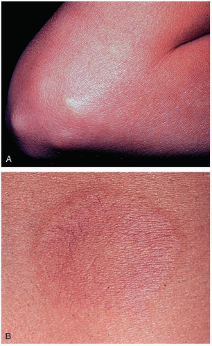
FIGURE 20-6 A: Subcutaneous nodules are generally associated with severe carditis. They are painless, firm, movable, measure approximately 0.5 to 2 cm, and usually are located over extensor surfaces of the joints, particularly knees, wrists, and elbows. B: Closer view of erythema marginatum in the same patient. (A and B, From Binotto MA, Guilherme L, Tanaka AC. Rheumatic fever. Images Paediatr Cardiol 2002;11:12-25.)


REFERENCES

1. Stollerman GH. Rheumatic fever. Lancet 1997;349:935-942.

2. Alsaeid K, Majeed HA. Acute rheumatic fever: diagnosis and treatment. Pediatr Ann 1998;27:295-300.


20-7 Tetanus

Michael Greenberg


Clinical Presentation

Tetanus is an uncommon disease in the United States but a substantial cause of death worldwide.1,2 Patients may present after obvious injuries, including thermal burns, snakebites, otitis media, skin ulcers, gangrene, septic abortions, childbirth, intramuscular injections, surgery, and puncture wounds or other wounds contaminated with soil, manure, or rusty metal.1,2 Up to 50% of cases occur after apparently insignificant and often clinically inapparent injuries.1,2 Between 15% and 25% of cases are not associated with a recent wound.1,2 The earliest symptoms of tetanus involve neck stiffness, sore throat, and difficulty opening the mouth.1,2


Pathophysiology

Tetanus is caused by Clostridium tetani, a gram-positive bacillus known to secrete two toxins, tetanospasmin and tetanolysin, under anaerobic conditions.1,2 Tetanospasmin is responsible for causing the clinical syndrome of tetanus. Tetanospasmin is a two-chain polypeptide, the light chain of which acts presynaptically to prevent neurotransmitter release after the heavy chain facilitates entry of these molecules into cells. Tetanospasmin cleaves synaptobrevin, which is a protein required for neurotransmitter release. The incubation period ranges from 1 to 60 days (average, 7 to 10 days). Recovery from tetanus occurs as a result of toxin destruction and regrowth of axon terminals.1,2


Diagnosis

The diagnosis is made on the basis of index of suspicion and clinical findings alone. No specific laboratory tests rule out tetanus. Tetanus may involve a clinical triad that includes muscular rigidity, muscle spasms, and autonomic dysfunction. It affects the head and neck muscles first and then spread caudally.1,2 Masseter spasm results in so-called “lockjaw” (trismus) and may progress to the facial muscles, causing “risus sardonicus” and difficulty swallowing. Neck muscle rigidity may cause head retractions and opisthotonus.1,2 The differential diagnosis includes strychnine poisoning, dystonia, hypocalcemia, and hysterical conversion.2


Clinical Complications

Potential complications are legion and may include aspiration, airway obstruction, acute respiratory distress syndrome, cardiac arrhythmias, congestive heart failure, renal failure, gastric stasis, ileus, diarrhea, gastrointestinal bleeding, thromboembolus, sepsis, multiorgan failure, vertebral fractures, and tendon avulsions during spasms.1,2


Management

Three concomitant modalities are employed to (1) neutralize unbound toxin by the administration of human tetanus immune globulin; (2) remove the source of infection by the use of metronidazole, erythromycin, tetracycline, chloramphenicol, or clindamycin; and (3) control rigidity, spasms, and autonomic dysfunction by the use of benzodiazepines, propofol, neuromuscular blocking agents, and/or baclofen.1,2


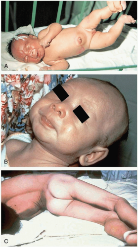
FIGURE 20-7 A: Neonatal tetanus at 6 days. The umbilical stump was treated with ashes. Note risus sardonicus and opisthotonus. (From Ostler, with permission.) B: “Lockjaw.” (Courtesy of the World Health Organization.) C: Opisthotonus. (Courtesy of the Centers for Disease Control and Prevention.)


REFERENCES

1. Hsu SS, Groleau G. Tetanus in the emergency department: a current review. J Emerg Med 2001;20:357-365.

2. Cook TM, Protheroe RT, Handel JM. Tetanus: a review of the literature. Br J Anaesth 2001;87:477-487.


20-8 Botulism

Anthony Morocco


Clinical Presentation

Symptoms of botulism begin 2 hours to 8 days after exposure. Patients who develop botulism by inhalation or ingestion of toxin exhibit the same neurologic symptoms but not the gastrointestinal symptoms that may be present in the naturally occurring disease. Invariably, a descending paralysis develops, beginning with bulbar findings, including ptosis, gaze paralysis, and dilated or fixed pupils. Patients complain of blurred vision, dysarthria, dysphonia, and dysphagia. Most patients exhibit an alert mental status. Gradual decrease or loss of reflexes and respiratory muscle paralysis may occur.1


Pathophysiology

Botulinum toxin is a potential weapon of bioterrorism via aerosol release or food supply contamination. Botulinum toxin exists in seven serotypes, designated by the letters A through G. Types A, B, E, and, rarely, F are known to cause the natural disease. Toxin is absorbed from the gastrointestinal tract and lungs, but it does not penetrate intact skin. The lethal human oral dose is estimated to be 70 ng. The toxin prevents the release of acetylcholine by blocking the fusion of neurotransmittercontaining vesicles with the presynaptic membrane, primarily at the neuromuscular junction.1


Diagnosis

The diagnosis of botulism should be strongly suspected in any patient with descending paralysis and a clear sensorium. Bioterrorism should be suspected in the following circumstances: an outbreak with a common geographic area but no common food source; identification of toxin types C, D, E (without an aquatic food source) F, or G; or occurrence of a large number of cases. The mouse bioassay to confirm the diagnosis and toxin type is available through the Centers for Disease Control and Prevention (CDC). Electromyography may help to confirm the diagnosis and differentiate botulism from potential misdiagnoses, such as Guillain-Barré and Miller-Fisher syndrome.


TABLE 20-8 Differential Diagnosis of Botulism


Disease


Fever


Eye signs


Ascend descend


Symmetric
asymmetric


Motor sensory
autonomic


Comment


Botulism


Yes


Descend bulbar


Symmetric


Motor > autonomic


DTR absent late, ptosis late


Guillain-Barré


+


No


Ascend bulbar


Symmetric


Motor > sensory


Abnormal CSF, DTR absent


Fisher type


Yes


Autonomic


Early, previous URI


Poliomyelitis


+


No


Ascend bulbar


Asymmetric focal


Motor


Abnormal CSF, DTR absent early


Paralytic shellfish


No


Ascend


Symmetric


Motor = sensory


History, onset within 30-60 min


Tick paralysis


No


Ascend


Symmetric


Motor > sensory


Presence of tick


Diphtheria


+


No


Ascend


Symmetric


Motor


Membrane in pharynx


Myasthenia gravis


Yes


Descend bulbar


Symmetric


Motor autonomic


Ptosis early, fatigue


Lead


No


Ascend


Symmetric


Motor


History


Arsenic


No


Ascend


Symmetric distal


Sensory > motor


History


Periodic familial paralysis


No


Ascend


Symmetric


Motor


Family history


Does not affect muscles of respirations


+, present; -, absent; CSF, cerebrospinal fluid; DTR, deep tendon reflexes; URI, upper respiratory infection.


Adapted from Mofenson HC, et al., eds. PP/T News. MMWC Poison Control Center 1989;8:139.


Clinical Complications

Patients may require prolonged mechanical ventilation, resulting in complications such as nosocomial infections.1


Management

Treatment consists primarily of supportive care. Recovery occurs in weeks to months as new axons sprout to create new neuromuscular junctions. An equine antitoxin is available through the CDC. Antitoxin may limit progression of the disease, but it does not improve existing paralysis, so it should be administered as early as possible. This trivalent antitoxin is effective for types A, B, and E only. Secondary infections should not be treated with clindamycin or aminoglycosides, because they may worsen neuromuscular blockade.


REFERENCES

1. Arnon SS, Schecter R, Inglesby TV, et al. Botulinum toxin as a biological weapon: medical and public health management. JAMA 2001;285:1059-1070.


20-9 Hepatitis and Jaundice

Jonathan Glauser


Clinical Presentation

Patients with hepatitis or jaundice may present with a prodrome of anorexia, malaise, and low-grade fever. Dark urine and light stools may be present. Clinical jaundice requires a bilirubin concentration of 3 to 4 mg/dL. Immune complex formation may cause morbilliform rash, arthralgias, or arthritis.1,2,3


Pathophysiology

Jaundice is generally categorized as unconjugated or conjugated. Unconjugated hyperbilirubinemia may result from overproduction, as in hemolysis, or from decreased conjugation by the liver. Conjugated hyperbilirubinemia typically entails acquired liver disease, as in hepatitis, or biliary obstruction, as from a tumor or a common bile duct stone.2

Hepatitis A is spread by oral contact, is generally nonlethal, and does not produce a carrier state. Chronic hepatitis B virus (HBV) infection affects an estimated 1.25 million people in the United States. Hepatitis C virus (HCV) affects approximately 1.8% of the American population, causing 8,000 to 10,000 deaths annually, and is the leading cause for liver failure that necessitates transplantation. Because of their shared route of transmission, HCV infection is common among persons infected with the human immunodeficiency virus (HIV).1 The risk of infection after a needlestick injury if the source is positive for the hepatitis B e antigen, Hb(e)Ag, is greater than 30%. In contrast, the average infection rate if the source is HCV positive is 1.8%.2

A variety of other viruses may cause hepatitis, including cytomegalovirus, herpes simplex, and Epstein-Barr virus.1,2 Drugs and toxicants associated with hepatitis are listed in Table 20-9.


TABLE 20-9 Drugs and Toxicants Associated with Hepatitis


Acetaminophen


Amanita phalloides mushrooms


Halothane


Isoniazid


Lamotrigine


Lovastatin


α-Methyldopa


Nonsteroidal antiinflammatory drugs


Phenytoin


Sertraline


Terbinafine


Troglitazone


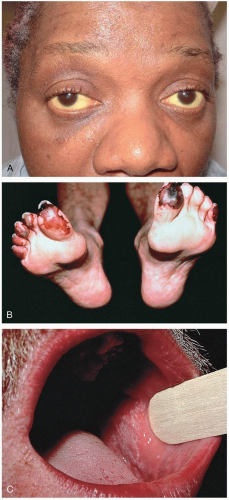
FIGURE 20-9 A: Icteric sclera in a patient with hepatitis C. (Courtesy of Mark Silverberg, MD.) B: Cryoglobulinemia produces acral vasculitic infarcts. Hepatitis C is a leading cause. (From Yamada, with permission.) C: Lichen planus produces lacy mucosal plaques and pruritic papules on the skin. Patients should be evaluated for hepatitis C infection. (From Yamada, with permission.)


Diagnosis

Transaminase levels are elevated (more than 10 times normal). Serologic testing for hepatitis A, B, or C may yield the cause. Conjugated and unconjugated bilirubin levels may suggest whether biliary obstruction is present. Serum albumin and prothrombin time may indicate synthetic function of the liver. Computed tomography and ultrasonography are helpful in diagnosing biliary obstruction.1,2,3


Clinical Complications

Hypoglycemia, coagulopathy, portal hypertension, gastrointestinal bleeding, or encephalopathy may result from hepatic insufficiency. HBV or HCV infection may cause fulminate liver failure, chronic liver disease and cirrhosis, hepatocellular carcinoma, or death.1,2,3


Prophylaxis

Hepatitis B vaccination for high-risk adults could prevent up to 800 cases of hepatitis and 10 deaths from hepatitis per 10,000 vaccinations.3 Hepatitis A vaccine is recommended for some travelers outside the United States and in some high-risk locations in the United States.3


Management

Admission and antibiotic therapy is mandatory if bacterial cholangitis is suspected. Otherwise, the decision for admission of a patient with hepatitis or jaundice depends on management of pain, hydration, vomiting, or complications of liver disease (e.g., gastrointestinal bleeding, altered mental status). Therapy for hepatitis B may include adefovir, lamivudine, or interferon alfa-2b. For HCV infection, pegylated interferon alfa-2a or -2b plus ribavirin has been used.2,3


REFERENCES

1. Sulkowski MS, Thomas DL. Hepatitis C in the HIV-infected person. Ann Intern Med 2003;138:197-207.

2. Goldmann DA. Blood-borne pathogens and nosocomial infections. J Allergy Clin Immunol 2002;110(2 Suppl):S21-S26.

3. Rich JD, Ching CG, Lally MA, et al. A review of the case for hepatitis B vaccination of high-risk adults. Am J Med 2003;114:316-318.


20-10 Necrotizing Infections

Gail Rudnitsky


Clinical Presentation

The affected area in a patient with a necrotizing infection may initially resemble cellulitis, with redness and edema at the site of infection. There is no clearcut line of demarcation or lymphangitis. As the infection progresses to necrosis, the skin turns purple or black. Hemorrhagic bullae may develop.1,2,3 The subcutaneous tissues and the fascia underneath become gangrenous, and myonecrosis may develop. Bacteremia and signs of toxic shock syndrome may be present. These patients generally appear ill, with high fevers, chills, hypotension, and other constitutional symptoms. Hypotension and frank shock may develop.1,2,3


Pathophysiology

Necrotizing infections are soft-tissue infections that cause extensive local tissue damage and may lead to toxemia or death. Two common terms for these types of infections are Fournier’s gangrene (localized to the perineum) and necrotizing fasciitis (deep subcutaneous infection).1,2,3 Necrotizing fasciitis is frequently caused by group A β-hemolytic streptococci (GABHS), although Staphylococcus aureus and Clostridium perfringens have also been implicated.1,2,3 Patients with diabetes, trauma, or immunocompromise are particularly susceptible. The organisms spread through the deep tissue above the fascia, causing thrombosis of vessels, which leads to gangrene of the subcutaneous tissues. Fishermen in warm-water areas may develop necrotizing fasciitis from Vibrio species. Fournier’s gangrene is most commonly a polymicrobial infection, with gram-negative organisms predominating.1,2,3


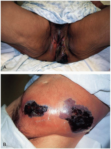
FIGURE 20-10 A: Fournier’s gangrene. (Courtesy of Mark Silverberg, MD.) B: Necrotizing fasciitis of the anterior abdominal wall. (From Isaacs L. Necrotizing fasciitis: diagnosis and treatment. Emerg Med News 2002;24[8]:4, with permission.)


Diagnosis

The clinical diagnosis may be supported by diagnostic studies. Patients generally have a leukocytosis with a left shift. A Gram stain from the site of infection or from a blood culture may help identify the organism. Computed tomography and magnetic resonance imaging may be used to determine the extent of the infection.


Clinical Complications

Complications include metastatic abscesses in distant organs, septic shock, and multisystem organ failure. If left untreated, this disease has a high mortality rate.


Management

Patients with necrotizing infections should be admitted to the intensive care unit, and all necessary supportive measures should be instituted. The mainstays of treatment are high-dose antibiotics and surgical débridement. Broad-spectrum antibiotics are used until cultures and sensitivity results are obtained. Surgical débridement should include areas of thrombotic tissue to prevent further necrosis. Fasciotomy may be necessary. Hyperbaric oxygen therapy may improve the mortality rate. Intravenous immunoglobulin may be beneficial in necrotizing infections caused by GABHS.


REFERENCES

1. Trent JT, Kirsner RS. Diagnosing necrotizing fasciitis. Adv Skin Wound Care 2002;15:135-138.

2. Seal DV. Necrotizing fasciitis. Curr Opin Infect Dis 2001;14:127-132.

3. Headley AJ. Necrotizing soft tissue infections: a primary care review. Am Fam Physician 2003;68:323-328.


20-11 Meningococcemia

Douglas Thompson


Clinical Presentation

Patients with meningococcemia present with fever, vomiting, headache, abdominal pain, lethargy, and myalgia. The characteristic petechial rash may be subtle or absent on presentation but may progress to purpura and necrosis.1,2,3 Specific signs of meningitis may not be evident.1,2,3


Pathophysiology

The spectrum of disease ranges from a self-limited illness to rapid progression to septic shock, coma, and death in as little as 12 hours. Cardiovascular collapse, change in mental status, acute respiratory distress syndrome, renal failure, hepatitis, disseminated intravascular coagulopathy (DIC), and ischemic extremities are evidence of multisystem organ dysfunction.1,2,3 Massive adrenal hemorrhage in association with cardiovascular collapse is referred to as Waterhouse-Friderichsen syndrome. Arthritis occurs in a small percentage of patients.

The peak incidence occurs in late winter and early spring. Children younger than 2 years of age account for almost half of the infections, with the peak attack rate in those younger than 4 months. Individuals in crowded military barracks and college dormitories represent a much smaller peak. Those with terminal complement and properdin deficiencies are also at risk.1,2,3

Between 5% and 15% of individuals in nonendemic areas are nasopharyngeal carriers of meningococci. Serogroups B, C, and Y account for most cases of disease in the United States. Transmission occurs via small droplets in close contacts, with the development of disease usually occurring within 2 weeks after acquisition. Passive cigarette smoke and concurrent viral respiratory tract infection are risk factors for bacterial translocation across the nasopharyngeal mucosa, leading to bacteremia and release of a lipopolysaccharide endotoxin into the bloodstream. Subsequent complement activation and release of cytokines and other inflammatory mediators results in the systemic inflammatory response syndrome that is responsible for the clinical manifestations. Capillary leak, vasodilation, and myocardial dysfunction all contribute to cardiovascular collapse. The procoagulant state resulting from the inflammatory cascade leads to systemic microthrombi and petechiae, purpura, DIC, extremity ischemia, and multisystem organ dysfunction.1,2,3

Only gold members can continue reading. Log In or Register to continue

Stay updated, free articles. Join our Telegram channel

Jun 25, 2016 | Posted by in EMERGENCY MEDICINE | Comments Off on Infectious Diseases

Full access? Get Clinical Tree

Get Clinical Tree app for offline access