Fig. 24.1
Canadian C-spine rule. Abbreviations: Glasgow Coma Scale (GCS), Motor Vehicle Collision (MVC), Emergency Department (ED). (Adapted from Vaillancourt et al. Evaluation of the safety of c-spine clearance by paramedics: design and methodology. BMC Emerg Med. 2011 Feb 1;11:1. doi: 10.1186/1471-227X-11-1. With permission from BioMed Central)
The chest x-ray is a critical imaging study for all trauma patients. Certain key findings mandate treatment prior to leaving the trauma bay. Patients with a significant hemo- or pneumothorax should have a chest tube inserted prior to leaving the trauma bay as these injuries can worsen rapidly (Fig. 24.2). A widened mediastinum would also raise the possibility of blunt aortic injury. With intubated patients, the chest film is also a helpful adjunct to confirm adequate endotracheal tube position prior to transport. As mentioned in Chap. 13, while useful to locate the tip of the endotracheal tube and rule out a right main stem intubation, it is important to remember that a chest x-ray cannot differentiate between an esophageal or tracheal intubation and other clinical signs and confirmatory tests must be employed. Ultrasound can also play a key role in diagnosing and ruling out hemo- or pneumothorax. Additionally, ultrasound can reliably detect esophageal intubation and main stem intubation at the bedside, in real time.
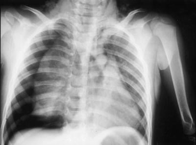
Fig. 24.2
Chest x-ray—large right pneumothorax. (Reprinted with kind permission from Springer Science + Business Media: Pediatric Surgery International, Complete bronchial rupture in a child; report of a case. 21(8); 2005, Okumus M, Fig. 1)
The pelvic film is also often being replaced by CT. In the clinically stable pelvis, the pelvic film is sometimes omitted if the patient is hemodynamically stable and going for CT which will include the pelvis. However, if the patient is not going for CT imaging or if there is any suggestion of hemodynamic instability, the pelvic film should be performed. In the clinically unstable pelvis, a binder should be applied and a pelvic film should be obtained prior to leaving the trauma bay.
Extremity films may be considered as adjuncts to the ATLS secondary survey as such films can often guide the urgency of orthopedic intervention [1]. With limb-threatening orthopedic injuries, pre- and postreduction films may be necessary prior to leaving the trauma bay. In the hemodynamically stable patient, extremity films may also be helpful in guiding the astute trauma practitioner to add extremity CT scans for injuries in which the orthopedics team may require them (such as injuries involving major joints). This can help eliminate the need for additional trips to the radiology suite after the patient reaches definitive care. However, timely transit to CT should not be delayed in favor of getting extremity films.
Focused Assessment with Sonography for Trauma (FAST)
As outlined in the preceding chapters, the FAST exam has become a critical adjunct to the ATLS primary survey [1, 5–7]. While the primary function of the FAST exam is to exclude hemoperitoneum and hemopericardium in the unstable patient, it remains a mandatory adjunct in the stable patient as well. Stable patients with a positive abdominal FAST should still proceed to CT scan. However, if a patient with a positive FAST becomes unstable after leaving the trauma bay, they can be quickly rerouted to the operating room for definitive management (Fig. 24.3). On the other hand, stable patients with a positive pericardial FAST should most likely proceed directly to the operating room.
Fig. 24.3
Ultrasound probe positioning for FAST. (Reprinted with kind permission from Sprin ger Science + Business Media: Notfall and Rettungsmedizin, Aktueller Stellenwert der konventionellen Radiographie und Sonographie in der frühen Versorgung traumatisierter Patienten, 13(6); 2010, Geyer, L.L, Fig. 3)
Many trauma providers are becoming facile with the extended FAST (E-FAST) exam. In addition to the four abdominal views, the E-FAST includes bilateral assessment for pneumothorax and hemothorax. Like the standard FAST, the primary utility of the E-FAST views is to guide chest intervention in the unstable trauma patient. The E-FAST views can still be a helpful tool in the stable trauma patient where the clinical exam is equivocal and the chest x-ray is either delayed or equivocal. An important consideration to the use of E-FAST in the stable patient is its high specificity; that is, the E-FAST may detect small volumes of air or fluid which are not apparent on chest x-ray [8]. Such occult injuries are often managed conservatively, and hence chest tube placement may be delayed until CT imaging is obtained or deferred entirely.
CT Scan
CT is the mainstay imaging modality for the stable trauma patient and careful consideration should be given to each body area which may require CT imaging. Once the primary and secondary surveys are complete, the patient may be prepared to travel to the CT suite. Mechanism of injury and physical exam findings generally dictate which areas should be further imaged with CT. However, certain injuries warrant special mention.
Much like with C-spine, the Canadian CT Head Rule helps determine who requires a CT head [9] (Table 24.1). CT angiogram of the chest has become the gold standard to diagnose blunt thoracic aortic injury. Such a diagnosis should be suspected in patients that suffer sudden acceleration-deceleration-type injuries (e.g., head-on motor vehicle collision, fall from height). Most stable trauma patients with abdominal pain and/or a positive FAST should undergo CT imaging of abdomen and pelvis (Fig. 24.4). CT scans of the thorax, abdomen, and pelvis require good arterial phase intravenous contrast. Delayed films for venous phase or to evaluate urinary tract injuries may also be helpful depending on injuries found.
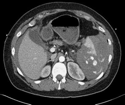
Table 24.1
Canadian CT Head Rule
CT Head is only required for patients with minor head injuriesa with any one of the following: |
High risk (for neurological intervention) |
• GCS score <15 at 2 h after injury |
• Suspected open or depressed skull fracture |
• Any sign of basal skull fracture (hemotympanum, “raccoon” eyes, cerebrospinal fluid otorrhea/rhinorrhea, Battle sign) |
• Vomiting ≥two episodes |
• Age ≥65 years |
Medium risk (for brain injury on CT) |
• Amnesia before impact >30 min |
• Dangerous mechanism (pedestrian struck by motor vehicle, occupant ejected from motor vehicle, fall from height >3 feet or five stairs) |
Fig. 24.4
CT scan of the abdomen—blunt splenic injury with blood tracking around the spleen and liver. (Reprinted with kind permission from Springer Science + Business Media: European Journal of Trauma and Emergency Surgery, Correlation of operative and pathological injury grade with computed tomographic grade in the failed nonoperative management of blunt splenic trauma, 38, 2012, Carr JA, Fig. 5)
Many trauma patients require a “pan-scan” which includes the head, C-spine, chest, abdomen, and pelvis. CT pan-scan detects up to an additional 40 % of injuries, 20 % of which are clinically significant, when compared to selective imaging [10, 11]. Furthermore, there is a distinct survival benefit to patients who undergo pan-scan [12]. This method also allows one to radiographically examine the entire spine. CT pan-scan should be strongly considered for any stable blunt trauma patient with significant mechanism of injury and/or decreased level of consciousness.
Stable patients sustaining penetrating trauma may be candidates for more selective imaging. Consideration must be given to the possible path of the missile and all body cavities at risk should be included if CT imaging is sought [13]. Penetrating wounds to the neck are triaged based on clinical exam into bedside exploration, contrast CT, or operative exploration [14]. There are several approaches to penetrating thoracoabdominal trauma. Gunshot or stab wounds to the chest are often imaged with CT. Any injury to the thoracoabdominal junction needs to be evaluated for diaphragmatic injury. While CT technology is improving, the radiographic detection rate is still not ideal [15] and laparoscopic or thoracoscopic evaluation of the diaphragm is usually warranted. Low velocity (eg. stab wounds) penetrating injuries to the anterior abdomen can often be managed by serial physical exams as bowel injuries can be missed on early CT scanning. Penetrating injuries to the flank should undergo triple contrast CT scan (IV, oral, and rectal contrast) as serial exams may miss retroperitoneal injury [13]. CT imaging may be warranted in blunt or penetrating extremity trauma based on clinical exam and contrast studies are often helpful to exclude major peripheral vascular injury.
Related posts:


Stay updated, free articles. Join our Telegram channel

Full access? Get Clinical Tree


