43 Imaging for Chronic Spinal Pain
Back pain remains the most common and most expensive cause of work disability in the United States.1 Recent data suggest that approximately 26% of U.S. citizens have experienced low back pain, and 14% have experienced neck pain, within the past 3 months.2 The course of acute or persistent low back pain is not as optimistic as has been generally conveyed.3 Von Korff and colleagues studied patients with a recent (within 6 months) history of low back pain and found that at 6 months, 76% of patients either had no pain (21%) or mild pain and low disability, but that 14% had high disability with moderate-to-severe limitation of function.4 A British study of acute low back pain noted full recovery in only 21% and 25% at 3 and 12 months, respectively.5 A Dutch study found that 70% of acute low back pain patients still had pain at 4 weeks, with a median time to recovery of 7 weeks; however, back pain tended to persist with residual prevalence of 35% at 12 weeks and 10% at 1 year, and 76% sustained a median number of two relapses of pain within that 1 year.6 An Australian study was more encouraging; at 3 months 49% to 67% (two study groups) had recovered, at 12 months 56% to 71% had recovered, with a relapse rate within that 1 year of 7% to 27%.7 Back pain accounted for more than 890 million physician office visits in the United States in 2002; however, the back pain proportion of total physician visits has remained stable since the early 1990s.2 Despite this, utilization of imaging has increased dramatically; U.S. Medicare use of lumbar magnetic resonance imaging (MRI) rose 307% in the 12-year interval 1994 to 2005.8 Imaging utilization is often unreasoned. There are large variations in rates of imaging across the United States; from one-third to two-thirds of spine computed tomography (CT) and MRI studies are inappropriate when measured against established guidelines.8 The relatively unregulated U.S. medical marketplace has served as a laboratory for the mass consumption of imaging technology, with no detectable benefit in patient outcomes. Despite incurring the greatest per capita expenditures for the diagnosis and treatment of back pain, including dramatically increased use of imaging, the United States continues to have the highest rate of back pain-related work disability of all industrialized societies.
It is well established that there is no role for imaging early in the course of a low back or limb pain syndrome. In a recent meta-analysis in Lancet, Chou and coworkers identified six randomized controlled trials evaluating the early use of imaging versus clinically directed care.9 In the pooled data, there were no significant outcome differences in pain or function in imaged versus nonimage patients in the short- (3 months) or long-term (6 to 12 months). These data apply primarily to acute or subacute back pain in primary care settings, and encompass radiographs and advanced imaging. Multiple studies have included economic analyses, which make it clear that radiographs obtained early in the course of a back pain syndrome are not cost effective; a study by Jarvik and associates suggested that radiographs performed on an initial visit for acute back pain in the absence of signs of systemic disease will ultimately cost $2000 (1982 U.S. dollars) to alleviate a single day of pain.1
A study by Carragee10 reinforced the futility of early advanced imaging. This 5-year prospective observational study obtained baseline MRI examinations on a large cadre of asymptomatic patients at risk for back pain due to strenuous vocations. These patients were then contacted periodically; when a subset ultimately presented to their physician with back or leg pain, a second lumbar MRI was performed. The study noted that fewer than 5% of the MRI scans obtained at the time of acute presentation showed clinically relevant new findings; the vast majority of the “abnormalities” were present on the imaging performed when the patient was asymptomatic. Only direct evidence of neural compression in patients with corresponding radicular pain syndromes was useful. Also of note, psychosocial factors were the primary predictors of the degree of disability, not the morphology identified on imaging.
Early Imaging Recommendations
Based on such data, there is broad international consensus on the lack of efficacy of imaging in acute pain syndromes of spinal origin. The American College of Radiology11,12 considers that imaging is inappropriate in acute low back pain unless there are complicating (“red flag”) features including the following:
Similarly, the Agency for Healthcare Research and Policy (U.S.) guidelines advises no imaging in patients younger than 50 years of age in the absence of systemic disease, or until pain persists for at least 4 weeks.13 The Australian National Health and Medical Research Council recommends against imaging in nonspecific low back pain of less than 12 weeks’ duration in the absence of red flag features suggesting underlying systemic disease.14 The American College of Physicians and the American Pain Society released a joint guideline in 2007 stating that imaging should not be obtained in patients with nonspecific low back pain unless there is a severe or progressive neurologic deficit, or when serious underlying systemic disease is suspected.15 Furthermore, patients with signs or symptoms of radiculopathy or spinal stenosis should be imaged with MRI (preferred) or CT only if they are candidates for surgery or epidural steroid injection.15
The primary role of imaging in the back pain patient is to aid in the detection of systemic disease that is causal of back or back-related leg pain. In essence, imaging is undertaken to detect neoplasm, infection, or manifestations of unsuspected traumatic injury. In a patient presenting with back pain to a primary care physician, these conditions are highly uncommon. In this population, only 0.7% of patients will have metastatic neoplasm, 0.01% spine infection, 0.3% ankylosing spondylitis, and 4% will have osteoporotic compression fractures.1 The differential diagnosis of back pain, and the relative frequency of causal conditions, are displayed in Table 43-1.1 The imperfect sensitivity and specificity of all imaging modalities makes diagnostic accuracy problematic, given the low pretest probability of systemic disease. According to Jarvik and Deyo, there are three essential questions that must be answered for the patient with back pain: (1) is there evidence of underlying systemic disease? (2) Is there neurologic impairment requiring surgical intervention? (3) Are social/psychological factors exacerbating the pain? Imaging may help to identify systemic disease and anatomic neural impingement, but its role should not be overstated.1 As Jarvik and colleagues note, depression is a more significant predictor of new low back pain than MRI imaging findings.16
Table 43-1 Differential Diagnosis of Low Back Pain∗
| Mechanical Low Back or Leg Pain (97%)† | Nonmechanical Spinal Conditions (−1%) | Visceral Disease (2%) |
|---|---|---|
| Lumbar strain or sprain (70%)‡ | Neoplasia (0.7%) | Pelvic organ involvement |
| Degenerative processes of disc and facets (usually related to age) (10%) | Multiple myeloma | Prostatitis |
| Herniated disc (4%) | Metastatic carcinoma | Endometriosis |
| Spinal stenosis (3%) | Lymphoma and leukemia | Chronic pelvic inflammatory disease |
| Osteoporotic compression fracture (4%) | Spinal cord tumors | Renal involvement |
| Spondylolisthesis (2%) | Retroperitoneal tumors | Nephrolithiasis |
| Traumatic fractures (<1%) | Primary vertebral tumors | Pyelonephritis |
| Congenital disease (<1%) | Infection (0.01%) | Perinephric abscess |
| Severe kyphosis | Osteomyelitis | Aortic aneurysm |
| Severe scoliosis | Septic discitis | Gastrointestinal involvement |
| Transitional vertebrae | Paraspinous abscess | Pancreatitis |
| Spondylolysis§ | Epidural abscess | Cholecystitis |
| Internal disc disruption or discogenic back pain¶ | Shingles | Penetrating ulcer |
| Presumed instability∗∗ | Inflammatory arthritis (often HLA-B27 associated) (0.3%) | |
| Ankylosing spondylitis | ||
| Psoriatic spondylitis | ||
| Reiter syndrome | ||
| Inflammatory bowel disease | ||
| Scheuermann disease (osteochondrosis) | ||
| Paget disease |
∗ Diagnoses in italics are often associated with neurogenic leg pain. Figures in parentheses indicate estimated percentage of patients with these conditions among all adult patients with signs and symptoms of low back pain. Percentages may vary substantially according to demographic characteristics or referral patterns in a practice. For example, spinal stenosis and osteoporosis will be more common in geriatric practices and spinal infection will be more common in injection drug users.
† The term mechanical is used here to designate an anatomic or functional abnormality without an underlying malignant, neoplastic, or inflammatory disease.
‡ Strain and sprain are nonspecific terms with no pathoanatomic confirmation. Idiopathic low back pain may be a preferable term.
§ Because spondylolysis is equally common in asymptomatic persons and those with low back pain, its etiologic role remains ambiguous.
¶ Internal disc disruption is diagnosed by provocative discography (injection of contrast material into a degenerative disc, with assessment of pain at the time of injection). However, discography often generates pain in asymptomatic adults, and many patients with positive discogram results improve spontaneously. Thus, the significance and appropriate management of this disorder remain unclear. Discogenic back pain is often used synonymously with internal disc disruption.
∗∗ Presumed instability is loosely defined as >10 degrees of angulation or 4 mm of vertebral displacement on lateral flexion and extension radiographs. However, diagnostic criteria, natural history, and surgical indications remain controversial.
Data obtained from Deyo,4 Hart et al.,6 Deyo et al.,7 and Deyo et al.8 Reproduced with permission Deyo RA, Weinstein JN: Low back pain. N Engl J Med 2001;344:363-370. Copyright© 2001. Massachusetts Medical Society. All rights reserved.
Risk/Benefit Analysis
There are, however, risks involved in imaging. These include the labeling effect, radiation exposure, monetary cost, and the provocation of intervention. The labeling effect refers to the inevitable identification of degenerative phenomenon on any spine imaging test. Degenerative findings on spine imaging are almost always inconsequential. If this is not made very clear to patients, they may identify themselves as suffering from a degenerative process from which there is no ultimate recovery. This can lead to fear avoidance behaviors, deconditioning, and depression. The pain practitioner is obligated to educate the patient that degenerative findings seen on imaging have little if any prognostic value, and actively contradict common misconceptions regarding back pain. A recent Cochrane review concluded that intensive patient education is effective in patients with acute and subacute low back pain; its value in chronic pain patients is less clear.17 The Australian media studies of Buchbinder also document the effectiveness of carefully planned educational efforts in changing patient and physician attitudes and reducing disability related to back pain.18
Radiation exposure from radiographs or CT must also be carefully considered. The effective radiation dose (biologic effect) is measured by the Sievert (Sv). The average natural background exposure is approximately 3.0 mSv per year.19 A three- view lumbar spine radiographic series exposes the patient to approximately 1.5 mSv; lumbar spine CT carries an exposure of approximate 6 mSv. An abdominal and pelvic CT delivers an average of 14 mSv; a technetium bone scan results in an average of 6.3 mSv exposure. Frontal and lateral chest radiographs, by contrast, expose the patient to 0.1 mSv.19 Radiation exposure is cumulative; repeated radiographic or CT examinations without clear expectation of patient benefit through improved decision-making cannot be condoned.
Imaging is costly. In the United Kingdom, imaging is known to comprise approximately 5% of the direct medical costs related to back pain.20 This is likely significantly higher in the less regulated U.S. marketplace. The 2009 Medicare reimbursements for lumbar spine imaging in the United States are: lumbar radiographs, $36; lumbar spine CT (noncontrast), $241; CT myelography, $525; MRI (noncontrast), $402, and bone scan with SPECT, $239.21 Nominal fees may be three to five times Medicare reimbursements. These costs directly affect our patients and our society and should be incurred only through reasoned decision-making.
Finally, imaging is likely to provoke intervention. Jarvik noted that early MRI imaging of the spine leads to increased surgical interventions despite equivalent pain and disability profiles when compared with nonimaged patients.22 Similarly, a study by Lurie and associates found that the intensity of CT and MRI use can account for virtually all of the extensive (12-fold) regional variation in surgical rates for spinal stenosis in the United States.23 When we image, we intervene. This occurs with standard of care interventions (many of which have little evidence basis) and totally unproven interventions with significant risks that have found their way into our highly imperfect medical marketplace. The single most important imaging precept in the back pain patient is this: treat the patient, do not treat the images.
Specificity and Sensitivity Considerations
Specificity shortcomings of spine imaging apply to all modalities from simple radiographs to the most elaborate MRI study, and have been well documented for decades. Hitselberger and associates noted in 1968 that myelographic studies were abnormal in 24% of asymptomatic volunteers.24 Wiesel and associates subsequently showed that CT demonstrated “significant” abnormalities in 50% of asymptomatic volunteers over the age of 40.25 Boden and associates showed that MRI of asymptomatic volunteers over the age of 60 demonstrated “significant” findings in 57% of asymptomatic volunteers.26 Numerous studies of asymptomatic volunteers can be summarized as follows: (1) imaging demonstrates abnormalities in the lumbar spine of 1/3 to 2/3 of asymptomatic persons; (2) annular fissures, disc bulges or protrusions, facet degeneration, and low-grade antero- or retrolisthesis are common, usually asymptomatic, and their prevalence increases with increasing age; (3) disc extrusions, severe central canal stenosis, and direct evidence of a neural compression are more likely to be symptomatic.16,25,27–38 Only clear concordance of an individual patient’s pain syndrome and imaging findings can suggest causation. Imaging cannot prove causation.
Imaging also suffers from sensitivity shortcomings. It is well documented that neuroclaudicatory pain is exacerbated by extension and axial load. The cross-sectional area of the lumbar and cervical central canal, lateral recesses, and neural foramina are known to decrease with extension and axial load.39 Studies by Danielson and Willem have shown that axial loading and extension will reduce the cross-sectional dimension of the lumbar dural sac in 56% of asymptomatic persons, but in up to 80% of patients with neuroclaudicatory pain syndromes.40,41 Unfortunately, most advanced imaging occurs with the patient in a supine, psoas relaxed (nonextended) position. This position diminishes imaging sensitivity to dynamic processes in which neural compression and pain are experienced only in specific postures. Upright and seated MRI imaging of the spine is commercially available, but suffers from modest image quality at best, due to the low field strength magnets available in this configuration. This may ultimately prove to be a valuable tool, but current devices excessively compromise image quality, and their cost precludes their use being appropriately limited to a problem-solving application.
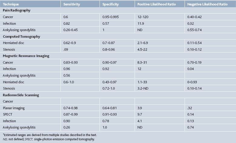
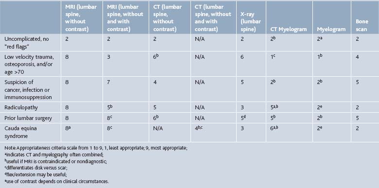
Multiple imaging modalities can be used to investigate the spine pain patient, from simple radiographs to elegant MRI scans. The efficacy of the several imaging modalities used in evaluating common spine disorders has been summarized by Jarvik and Deyo in Table 43-2.1 The American College of Radiology Appropriateness Criteria grade the utility of these imaging modalities in common clinical scenarios, presented in Table 43-3.42
Radiographs
In the setting of clinical red flags suggestive of systemic disease, pain unresponsive to conservative measures, or progressive neurologic deficit, imaging should commence with plain radiographs.11 Radiographs are a modest sensitivity screening tool for sinister processes such as neoplasm, infection, or fracture. They also provide vertebral enumeration, and allow assessment of sagittal and coronal balance. To provide the physiologic information of balance, radiographs should be obtained while the patient is weight-bearing. Single frontal and lateral views of the lumbar spine are adequate. Spot images of the lumbosacral junction, oblique views, and flexion-extension views should not be obtained on a routine basis. They provide little additional information, and significantly increase radiation exposure.43 The significance of vertebral enumeration should not be underestimated. There are anomalies of segmentation at the lumbosacral junction in 7% to 12% of the back pain population.44 Radiographs should establish the convention of vertebral numbering for all subsequent advanced imaging in the individual patient, preventing wrong level minimally invasive or surgical interventions.
Radiographic identification of morphologic changes of disc degeneration or osteoarthritis in lumbar zygapophyseal or sacroiliac joints has no value in identifying the cause of an individual patient’s pain.45 Radiographic manifestations of disc degeneration include loss of disc space height, nitrogen gas within the disc, and end plate hypertrophs; these findings have no predictive value for discogenic pain. Similarly, radiographic findings of osteoarthritis, including hypertrophic change, sclerosis, joint space narrowing, gas within the joint space and subchondral cyst formation do not correlate with response to intraarticular anesthetic injections. Morphologically degenerated joints are most commonly asymptomatic, and joints that appear normal radiographically may be painful.
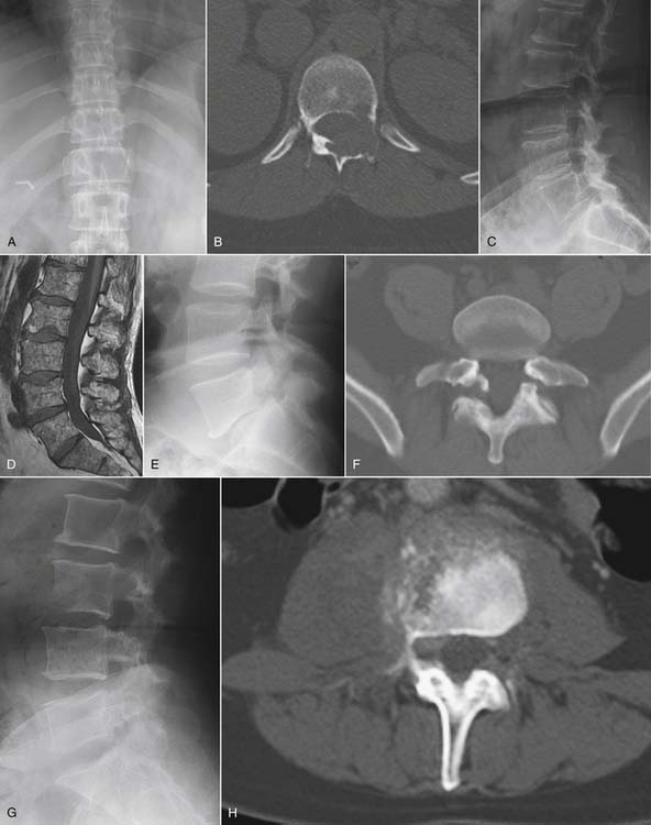
Radiographs provide a modest sensitivity screen for systemic disease causal of back pain, which may require advanced imaging to characterize and establish extent of disease (Fig. 43-1). The most common neoplastic condition is metastatic disease. Skeletal metastases are primarily blastic (bone forming), lytic (bone destroying), or of mixed type. Radiographs primarily image cortical bone. Metastases most commonly involve trabecular bone; at least 50% of trabecular bone mass must be destroyed to be visible on a radiograph. Radiographs therefore have only modest sensitivity (60%) in the detection of metastatic disease.1 Myeloma may also be initially suggested by radiographic evaluation; myeloma typically manifests as small discrete lytic lesions without sclerotic margination. When diffuse, myeloma may be indistinguishable from metabolic osteoporosis. Primary bone tumors are rare causes of spine-related pain. Osteoid osteoma deserves mention because it commonly presents with nocturnal pain.46 This neoplasm of adolescents and young adults has a posterior element predilection, and it generates a lesion consisting of a central lytic nidus with considerable surrounding sclerotic reaction. The diagnosis is seldom made with plain films; CT imaging, with image directed biopsy and radio frequency ablation constitute standard of care. Osteoid osteomas may also be identified on MRI examinations or bone scans as focal intensely enhancing (or metabolically active) posterior element lesions.47
Radiographs may also suggest infectious or inflammatory disease. Radiographic findings of infectious spondylodiscitis (disc space infection) only become apparent several weeks after the onset of disease.48 Because disc space infection in the adult begins as a hematologically seeded vertebral osteomyelitis, radiographs initially demonstrate rarefaction in the anterior subchondral bone adjacent to the end plates within a single disc space. Over time, there will be frank end-plate destruction with loss of disc space height.48 These findings should provoke an urgent MRI examination with gadolinium enhancement. Granulomatous disease (TB, brucellosis) may be indistinguishable from pyogenic disc space infection. Granulomatous discitis more commonly spares the disc space with vertebral collapse and kyphotic deformity at the site of infection.48
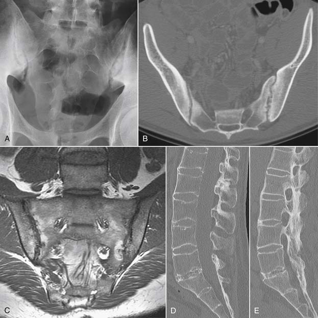
Inflammatory spondyloarthropathies may be suggested or diagnosed with plain radiographs. Rheumatoid arthritis primarily involves the cervical spine, with inflammatory destruction of the transverse ligament binding the dens to C1 with consequent atlantoaxial subluxation.48 There is frequently multilevel cervical disc space narrowing with low grade anterior subluxations resulting in a stair-step appearance to the cervical spine. The seronegative spondyloarthropathies are more commonly apparent in the sacroiliac joints, lumbar and thoracic regions (Fig. 43-2). Narrowing of the sacroiliac joints with associated indistinct cortical surfaces and adjacent sclerosis may herald ankylosing spondylitis, psoriatic arthritis, or Reiter syndrome. The ankylosing spondylitis patient may also have fusion of portions of the thoracolumbar or cervical spine with vertically oriented, gracile syndesmophytes bridging adjacent vertebral bodies. The long-segment spine rigidity in these patients makes them susceptible to catastrophic fracture dislocation, often with neurologic compromise, even with modest trauma. Such fractures may be difficult to visualize with plain radiographs, and the threshold for use of advanced imaging should be very low in ankylosing spondylitis patients suffering trauma.49
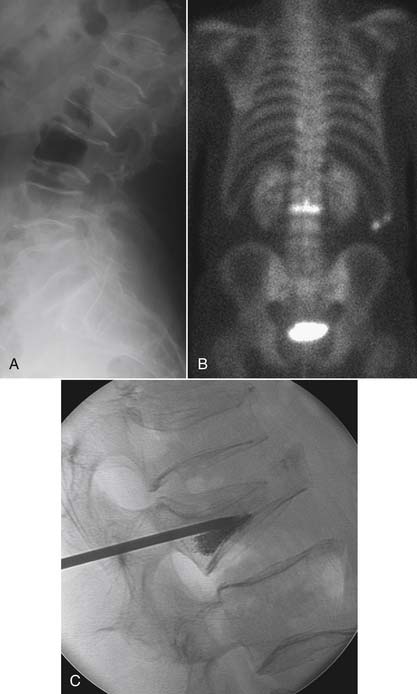
Compression fractures can be identified on plain radiographs, but the acuity of a fracture may not be apparent in the absence of serial images. MRI or technetium bone scan can characterize fractures as acute or subacute, metabolically active, and therefore more likely to respond to bone augmentation procedures such as vertebroplasty (Fig. 43-3). However, recent randomized controlled trials have raised questions regarding the efficacy of vertebroplasty.50,51 Similarly, pelvic insufficiency fractures may be apparent on radiographs within the sacral ala, pubic rami, pubic symphysis, and periacetabular regions of the pelvis. The plain film finding of a vertical sclerotic band paralleling the sacroiliac joint in the sacral ala strongly suggests insufficiency fracture. CT, technetium bone scan, or MRI are all more sensitive than radiographs in detection of insufficiency fractures.52,53
Another traumatic entity in which plain films play a role is spondylolysis. Defects in the pars interarticularis may be detected on lateral plain films, with a prevalence of approximately 3% to −10% of the population.54 Most cases are asymptomatic. If pars defects are suspected as the cause of an axial pain syndrome, a localized CT will confirm or refute the presence of such a defect and aid in characterizing the defect as acute or chronic. There is no role for oblique radiographs, which double the gonadal radiation dose over the standard images, and do not advance the diagnosis.43
Stay updated, free articles. Join our Telegram channel

Full access? Get Clinical Tree


