Fig. 35.1
A number 15 blade is used to make a transverse incision just above the midway point between the xiphoid and umbilicus. The incision should be below the liver edge, identified by palpation
The filmy omentum is carefully swept inferiorly. The stomach is reasonably sturdy but the epiploic vessels may be avulsed from the greater curve by vigorous retraction. Once a Babcock clamp or Singley forcep pulls the stomach into the field it should be removed from the organ completely. Any further manipulation should be manual, aided by a gauze sponge to allow a firm but gentle grasp to coax the pylorus into view. An experienced surgeon recognizes that tearing of the serosa is a sign that the incision has to be enlarged.
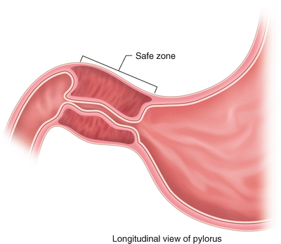
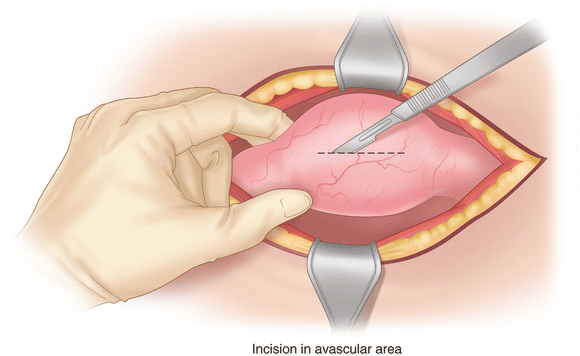
A shallow longitudinal incision is made just through the serosa. The duodenum is closest to the serosa immediately adjacent to the pylorus, so it is prudent to begin the incision 2 mm proximal to its junction with the stomach (Fig. 35.2). The incision extends well onto the antrum, a distance of about 2 cm. There is a path for the incision free of visible vessels over the anterior aspect of the pylorus and antrum (Fig. 35.3). Bleeding is scant and generally does not require cautery.


Fig. 35.2
The margin of the duodenal lumen at left has a superficial position. An incision placed too far distally may unintentionally enter the lumen at that point. The “safe zone” therefore begins 2 mm proximal to the duodenal–gastric junction

Fig. 35.3
The incision in the serosa of the pylorus starts distally about 2 mm proximal to the duodenal junction and extends in an avascular path between visible vessels from the superior and inferior margins. It extends well onto the antrum, a total distance of about 2 cm
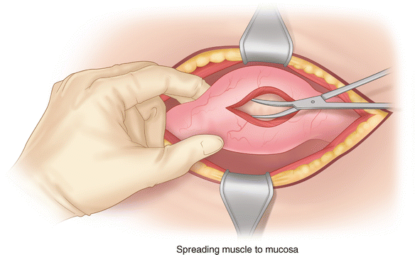
An instrument is placed into the incision and used to break the fibers of the hypertrophied muscle. A “gritty” sensation may be felt through the instrument. A blunt scalpel handle or the serrated edge of one jaw of a clamp is appropriate to begin splitting the muscle. Once the muscle begins to divide the muscularis is split to the level of the submucosa (Fig. 35.4). Appropriate instruments for this task include a pyloric muscle spreader—a simple blunt-tipped instrument with serrations on the outside aspect of the jaws to hold the exposed muscle edges; a Mosquito clamp, tips directed upward so that the mucosa is not perforated; or continued blunt splitting using the scalpel handle. The muscle is split until the pale pyloric submucosal layer is seen over the length of the incision, indicating complete division of the muscle fibers. An adequate pyloromyotomy is confirmed by assuring that the superior and inferior edges of the myotomy move independently.


