Fig. 13.1
Antegrade nephrostogram of upper ureteral injury
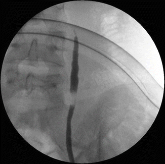
Fig. 13.2
Retrograde ureterogram of the same injury
13.1.1 Teaching Points
The patient underwent open exploration of the upper ureter through a lumbotomy approach. Significant scarring of the retroperitoneum was found. Following dissection of the scar tissue, the upper and lower ends of the ureter were isolated, leaving an 8 cm gap between them (Fig. 13.3).
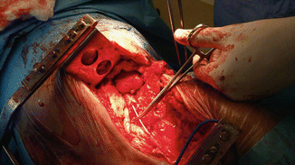

Fig. 13.3
Retroperitoneal dissection of the injured ureter. The two ends were mobilized, and an internal double-J stent was placed. Note the significant gap
In order to re-approximate the ureteral ends, the distal ureter was mobilized all the way to the bladder, and the left kidney was completely mobilized and descended. These maneuvers allowed the formation of a tension-free uretero-ureteral anastomosis (Fig. 13.4). The anastomoric site was further wrapped with retroperitoneal fat in order to minimize the risk of retroperitoneal fibrosis. The internal stent and nephrostomy remained for an additional 8 weeks and then removed.
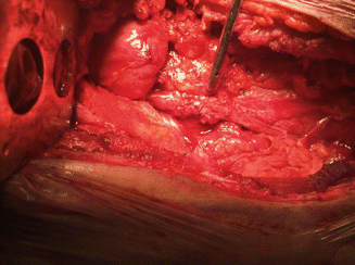

Fig. 13.4
The uretero-ureteral anastomosis
13.1.2 Clinical Implications
The long-term follow-up of ureteral reconstruction is usually good. Yet the majority of ureteral injuries are associated with iatrogenic damage related to electric spark injuries, transection, or ligation of the ureter during elective or urgent surgery (e.g., caesarian section). Differing by the level of injury, Syrian war injuries were characterized by severe local damage to soft tissues, associated damage to adjunct viscera, significant blood loss, and following preliminary treatment and damage control procedures—an extensive locoregional fibrosis.
13.1.3 Damage Control and Final Reconstruction
During an emergency surgery, no attempt should be made to re-anastomose the ureter; the tissue is burned, unprepared for reconstructive surgery due to local spillage; and associated life-threatening injuries are often present. The fastest way to secure the ureter is to perform a simple tube ureterostomy. In this fast procedure, a simple 5–8 Fr. feeding tube is inserted through a simple skin incision. The tube is inserted through the ureteral stump up to the renal pelvis and secured with a vicryl 3:0 suture on the stump and a 0 silk suture on the skin level. The procedure is rapid. It can be performed by one surgeon, while others continue their life-saving procedures. Alternatively, the ureter can be ligated and marked with a metal clip, and percutaneous nephrostomy can be placed later in the angiography suite or bedside.
Following recovery, a reconstructive surgery can be performed safely between 3 and 6 months from the injury.
The factors which predict the success of ureteral anastomosis are the tension over the anastomosis, the presence of active infection, the blood supply to the ureteral stumps, and the development of re-fibrosis around the anastomotic site.
During surgery, careful dissection of the ureteral stumps, combined with renal mobilization, can produce a tension-free anastomosis. Appropriate drainage and antibiotic coverage will decrease the risk of reinfection. Wrapping the ureter with local fat tissue (preferably the omentum) will decrease the risk of recurrent periureteral fibrosis and external compression. Under such conditions, the long-term rate of patent anastomosis is excellent.
13.2 Lower Ureteral Injury: Case Presentation
A 32-year-old male suffered multiple gunshot wounds to the left lower abdomen and pelvis. He was operated urgently in a frontline facility where he underwent ligation of bleeding vessels in the pelvis, colectomy, Hartman’s procedure, and temporary ileostomy. The left ureter was lacerated and sutured over a feeding tube. The patient was transferred to our center with significant ischemia of the left leg and pelvis and underwent left hemipelvectomy. During the procedure, the feeding tube was found floating in the pelvis. Due to hemodynamic instability, further dissection of the ureter was not attempted. On postoperative day no. 6, urine was noted to appear in the surgical drain.
13.2.1 Teaching Points
The patient underwent left retrograde pyelography which demonstrated complete disruption of the ureter with contrast leak from the ureteral stump to the drain (Fig. 13.5). An antegrade pyelogram showed complete transection of the distal ureter (Fig. 13.6). A percutaneous nephrostomy was placed and the urinary leak subsided. The patient is currently in rehabilitation and will be further referred to ureteral reconstruction.
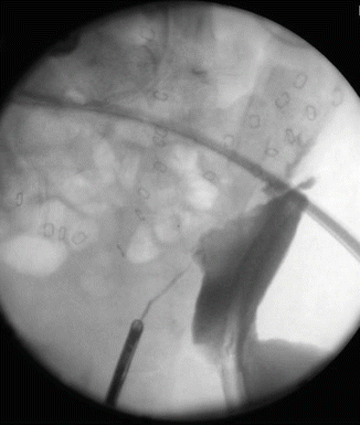
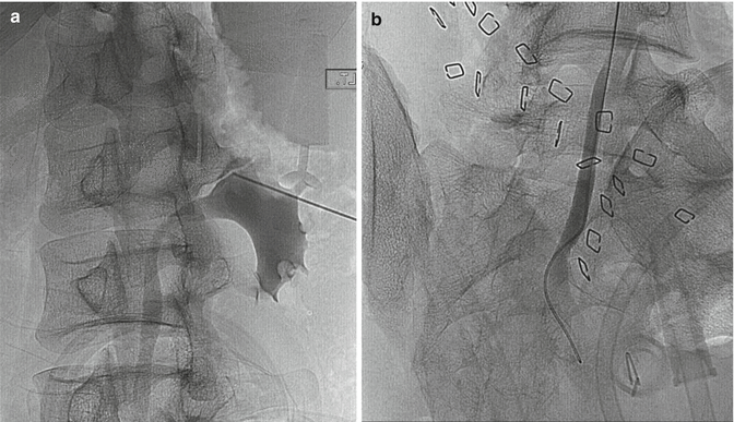

Fig. 13.5
Retrograde pyelography of the disrupted ureter

Fig. 13.6
Antegrade pyelography of the same patient showing mild hydronephrosis (a) and complete transection of the lower ureter (b)
13.2.2 Clinical Implications
As mentioned in the previous case, urgent reconstruction of the ureter is nearly always fruitless. The tissues are contaminated and edematous, and the blood supply is often not adequate, resulting in tissue necrosis, sloughing, and breakdown of the anastomosis. The appropriate and easy to perform percutaneous tube ureterostomy offers a rapid long-standing drainage of the ureter, allowing later reconstruction. Once a ureteral damage has been diagnosed after the laparotomy, the best management is placing a percutaneous nephrostomy until the patient heals.
Stay updated, free articles. Join our Telegram channel

Full access? Get Clinical Tree


