Inclusion criteria
Amputation of lower or upper limb
Chronic pain from amputation for ≥6 months
Pain refractory to conventional medical management
Severe pain in an amputated limb (worst pain intensity ≥7 on 0–10 Numerical Rating Scale [NRS])
Frequent occurrence of severe pain (≥2 episodes/week on average by recall, confirmed by screening diary of 2–4 weeks)
Significant pain reduction (≥50 % on NRS) after local anesthetic injection for temporary nerve block; no significant pain reduction after saline injection as a placebo injected prior to the anesthetic
Exclusion criteria
Pacemaker or other implanted devices
Debilitating pain other than pain in amputated limb
Pregnancy
Inability to accurately and consistently report pain intensity and related information
High risk of infection due to comorbidity or compromised immune state (e.g., chemotherapy)
High risk of mortality for general anesthesia (e.g., severe cardiopulmonary disease)
Infectious etiology for amputation (e.g., osteomyelitis, cellulitis)
Uncontrolled diabetic vascular disease or neuropathy
Skin graft or severe scarring over targeted implant site

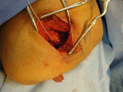
Fig. 30.1
The stimulating nerve cuff placed around the sciatic nerve in an above-the-knee amputee
30.4.2 Preimplant Testing
In our first-in-human and pilot studies, the screening local anesthetic peripheral nerve blocks used as a trial for HFAC block were done under ultrasound guidance. This method allowed the practitioner to visualize the neuroma at the end of the severed nerve and allowed identification of the location, depth, and diameter of the targeted nerve, to help facilitate surgical placement of a peripheral nerve cuff (Fig. 30.2). Other traditional peripheral nerve block modalities would also be acceptable ways to complete the “trial” local anesthetic block, however. These could include direct infiltration of local anesthetic proximal to the neuroma via palpation technique, or the use of a Stimuplex® needle (B. Braun Medical; Bethlehem, PA) to complete the peripheral nerve block.


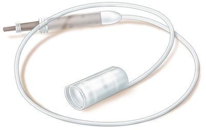
Fig. 30.2
The peripheral nerve cuff electrode
The location and placement of the cuff electrode depends on which of the severed peripheral nerves are transmitting the pain signals. Local anesthetic peripheral nerve blocks help to identify which nerves are involved in pain transmission. Once the practitioner achieves significant pain reduction after local anesthetic blocks to a nerve or group of nerves, he or she has a roadmap showing the nerves to which the nerve cuff electrode should be attached to achieve effective pain relief. The metric of success is pain reduction of more than 50 % after two local anesthetic peripheral nerve blocks.] Two local anesthetic blocks are recommended to help rule out a placebo effect from a single, one-time injection. It is also recommended that each block be performed on a different day, with the patient filling out a pain diary charting his or her pain reduction and duration of pain relief.
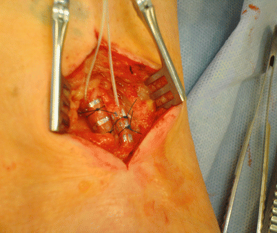
After the peripheral nerve is identified and the patient has had two successful screening injection blocks with more than 50 % pain relief, peripheral nerve cuffs are placed around the appropriate nerves. In below-the-knee amputation patients, painful neuromas typically occur near the common peroneal and tibial nerves (Fig. 30.3). In patients with above-the-knee amputations, the sciatic nerve is usually the pain generator.


Fig. 30.3
Peripheral nerve cuffs placed around the common peroneal and tibial nerves in a below-the-knee amputee
For upper extremity amputations, painful neuromas typically occur around the radial, ulnar, median, or musculocutaneous nerves.
30.4.3 Implantation Technique
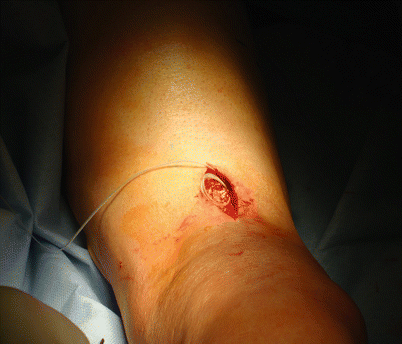
The implantation of the high-frequency PNS device requires specialized training in the field of neuromodulation so that implantation can be achieved in a timely and minimally invasive fashion. The electrode implantation is achieved via surgical dissection to adequately place the nerve cuff electrode around the peripheral nerve(s). Meticulous attention is required in this surgical dissection to ensure that vascular or adjacent nerve structures are not damaged when placing the peripheral nerve cuff. These risks can potentially be decreased by perioperative ultrasound mapping of the patient’s vascular and nerve structures. In clinical studies, a preoperative ultrasound was completed in the preoperative holding area to mark the location and depth of the targeted nerves. This technique allowed faster and more efficient cuff placement, providing the implanting physician with accurate knowledge of the nerve depth and location prior to incision. Additionally, during the preoperative ultrasound mapping, the surgeon can choose to inject 0.1 mL of methylene blue near the targeted nerve so that, after the incision is made and dissection is carried out to the desired depth, the surgeon can see the methylene blue surrounding the targeted nerve. These techniques allow for not only more rapid nerve exposure (average time for exposure of the targeted nerve was less than 10 min) but also a smaller incision (Fig. 30.4).


