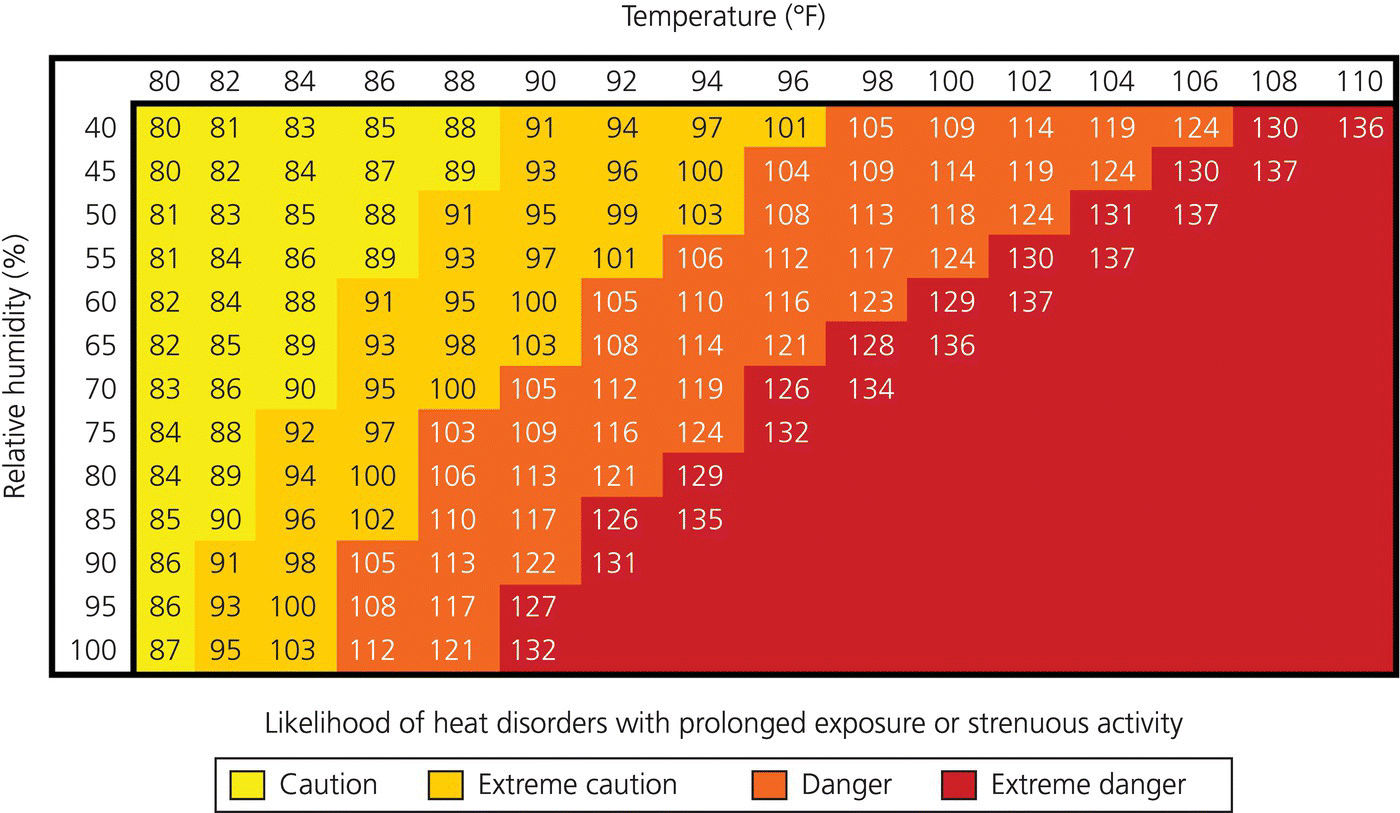
Chapter 49 Gerald (Wook) Beltran Heat-related illnesses are a spectrum of disorders more commonly seen when the patient is in a warm environment, has underlying comorbidities, is physically active, and/or has attire which does not readily permit easy removal of body heat (such as firefighter “turnout gear”). If not diagnosed and managed appropriately, significant morbidity and mortality may result. This chapter will discuss the physiology of thermoregulation and the various heat-related illnesses, including management strategies. According to the Centers for Disease Control and Prevention (CDC), an average of 688 people die annually from heat-related illness [1]. Between 1999 and 2003, an estimated 3,442 deaths were attributed to heat injury. Males accounted for 66% of the deaths. Fifty-three percent of those who died were between the ages of 15 and 64 years, while 40% were greater than 64 years of age. Emergency medical services protocols should reflect the most up-to-date science on the management of patients with heat-related illness. The best management strategies incorporate expected transport times given the acuity of the patient’s illness, local geography, weather, hospital proximity, and local traffic patterns. Normal oral temperature has been demonstrated to be 33.2–38.2 °C [2]. Rectal temperature averages 34.4–37.8 °C, while the normal tympanic temperature is 35.4–37.8 °C. The human body generates heat through metabolism. Basal metabolic rate is a measure of the number of calories expended at rest while sedentary. The maintenance of body temperature to a narrow window, or thermoregulation, is a complex task controlled by the hypothalamus. The body’s extremities have a greater variation in temperature, dependent on environmental factors, including clothing. Generally, the extremities tend to be cooler than the rest of the body, while the core temperature fluctuates very little. While metabolism of the various body tissues generates heat, the majority of body heat comes from skeletal muscle activity. Endocrine function can also affect metabolic rate and increase temperature. For example, epinephrine and norepinephrine can increase basal metabolic rate and subsequent heat generation. Similarly, thyroid hormones can produce an elevation of metabolic rate as well as an increase in body temperature. Body temperature homeostasis occurs with heat generation in balance with heat dissipation. The narrow homeostatic range prevents enzymatic and cellular dysfunction or injury. Several reflexes or semi-reflexes help to maintain temperature. The reflexes or semi-reflexes activated by cold include shivering, hunger, increased voluntary activity, curling up, decreased heat loss, cutaneous vasoconstriction, epinephrine and norepinephrine release, and erection of the short body airs (i.e. “goose bumps”). The reflexes or semi-reflexes activated by heat include cutaneous vasodilation, sweating, decreased voluntary movement, anorexia, decreased heat production, and increased respiration. The posterior hypothalamus controls the reflexes activated by cold, while those for warmth are located in the anterior hypothalamus. Sweating and cutaneous vasodilation occur with activation of the anterior hypothalamus [3]. Sensors in the spinal cord, skin, deep tissues, hypothalamus, and extrahypothalamic regions of the brain provide feedback to the hypothalamus on body temperature [3]. The hypothalamus is activated by core temperature increases of less than 1 ºC [4]. Lesions of the anterior area of the hypothalamus cause hyperthermia. Heat is dispersed from the body by several different mechanisms including conduction, convection, evaporation, and radiation. Heat loss by conduction occurs through direct contact with an object or environment that is cooler. Skin temperature greatly affects the amount of body heat lost or gained through conduction. The amount of heat dispersed from the body’s core to the skin is reliant on cutaneous blood flow. When the cutaneous vessels dilate, more blood flows to the skin, allowing for greater heat transfer from the deeper tissues (tissue conductance). Horripilation is the erection of the cutaneous hairs which helps to trap air near the skin and inhibit heat transfer from the skin. Clothing also inhibits tissue conductance by limiting transfer of heat from the skin to the environment. Convection refers to the removal of heat as cooler air passes over exposed skin. The more air passes over the skin (e.g. from a fan), the more heat can be dispersed. Evaporation is the heat lost via converting a liquid to a gas. In the human body, about 600 kcal/hour in ideal conditions can be removed through evaporation [5]. Radiation is the transfer of heat by infrared electromagnetic waves. Approximately 250–300 kcal/hour can be transferred to the human body by solar radiation, with clothing acting as a barrier and providing some protection, estimated at 100 kcal/hour [6]. Additionally, the body disperses heat by radiating to cool objects in the vicinity [3]. At an ambient temperature of 21 °C and while at rest, heat loss from the body occurs through radiation and conduction (70%), evaporation (27%), respiration (2%), and urination and defecation (1%) [3]. As the ambient temperature increases, radiation losses decline and heat loss through evaporation increases [3]. At higher ambient temperatures, evaporation plays a more critical role in body heat removal. The removal of heat is reliant upon the gradients of moisture and temperature. As the environmental temperature and humidity increase, the exchange of heat becomes impaired. Therefore, hot humid environments confer the greatest risk to patients for heat-related illness (Figure 49.1). Figure 49.1 NOAA’s National Weather Service Heat Index. Source: National Weather Service National Oceanic and Atmospheric Administration: www.nws.noaa.gov/os/heat/images/heatindex.png The body adapts over time to more efficiently manage heat stress, primarily through salt retention and increased fluid secretion from sweat glands to increase the rate of evaporation [6]. Other adaptations include increased circulating plasma volume, improved renal filtration, and increased resistance by the kidney to exertional rhabdomyolysis [7]. Adaptation through production of acute-phase reactants also protects tissues from heat stress [8]. Individual cells produce intracellular heat shock proteins which protect them from sudden heating [9,10]. The mechanism is believed to occur through the binding of heat shock proteins to cellular proteins which inhibit the cellular proteins from denaturing (or unfolding) in hot environments. Regardless of the etiology, if hyperthermia is not addressed, tissue and cellular swelling and disruption will occur with widespread hemorrhage. Heat injury causes denaturation of proteins, a severe inflammatory response, and disruption of the coagulation cascade. The denaturation of proteins causes direct injury to the cells and cellular function. Pyrexia above 41.6 °C for a few hours can cause cellular damage. Temperatures above 49 °C can cause nearly immediate cell death. Organs most susceptible to apoptosis secondary to hyperthermia include the mucosa of the small intestine, thymus, lymph nodes, and spleen. Injury caused by inflammatory response is due to the release of several inflammatory cytokines including tumor necrosis factor-alpha, interleukin (IL)-1 (beta), and interferon gamma. Several anti-inflammatory cytokines are also released during heat stress, including IL-6, IL-10, and tumor necrosis factor receptors p55 and p75. Animal models with injection of IL-1 and tumor necrosis factor-alpha have demonstrated changes similar to heat stroke [11]. Heat injury causes activation of the coagulation cascade, injury to the vascular endothelium, and increased permeability of the vasculature. This has been demonstrated through surrogate markers of endothelial damage or activation as seen in heat stroke patients. Some of these markers include von Willebrand factor antigen, endothelin, and intracellular adhesion molecule 1 [11–14].
Heat-related illness
Introduction
Physiology of thermoregulation

Pathophysiology
Stay updated, free articles. Join our Telegram channel

Full access? Get Clinical Tree


