Head and Spinal Cord Trauma


Head and Spinal Cord Trauma


Robert C. Tasker

P. David Adelson


The practicalities of critical care in children with traumatic brain injury (TBI) or traumatic spinal cord injury (TSCI) are discussed in this chapter. The developmental neuroscience of acute neurotoxicity, vascular control, and cerebral hemodynamics and physiology is discussed in other chapters within this section of the book. The principal aim of this chapter is to review the recent clinical literature (in the main, between 2005 and 2014) toward informing the practice of neurocritical care.

Central to our understanding and treatment of TBI is the fact that mechanical forces at the time of accident—direct or contact force, acceleration and deceleration forces, and rotational or torsional forces—are responsible for primary injury. As a consequence of these forces, a variety of primary and secondary brain injuries occur (Table 61.1). The pathophysiology of these forms of injury is dealt with in other chapters.


EPIDEMIOLOGY


Traumatic Brain Injury

TBI is a major public health problem. Worldwide, TBI is the leading cause of death and disability for children (>1 year old) and young adults. In 2009, the US Centers for Disease Control and Prevention estimated that at least 2.4 million emergency department visits, hospitalizations, or deaths were related to a TBI, either alone or in combination with other injuries (1). Approximately 75% of TBIs are mild concussions. Children, adolescents, and older adults are most likely to sustain a TBI. Nearly one third of all injury deaths included a diagnosis of TBI. In addition, an estimated 5.3 million people in the US are living with TBI-related disabilities, including long-term cognitive and psychologic impairments. A severe TBI not only affects a person’s life and family, but also has a large societal and economic toll. The economic costs of TBIs in 2010 were estimated at $76.5 billion, including $11.5 billion in direct medical costs and $64.8 billion in indirect costs (e.g., lost wages, lost productivity, and nonmedical expenditures).


TABLE 61.1 PRIMARY AND SECONDARY BRAIN INJURY


PRIMARY BRAIN INJURY


SECONDARY BRAIN INJURY


Diffuse and focal axonal injury


Diffuse and focal hypoxicischemic injury


Diffuse and focal vascular injury


Diffuse and focal brain swelling


Focal brain contusion


Intracranial hypertension


Focal brain laceration


Hydrocephalus


Infection and fever


Seizures


Metabolic disturbance, e.g., hyponatremia


Spinal Cord Injury

The burden of acute TSCI among US children and adolescents (≤17 years) using emergency department data from the Nationwide Emergency Department Sample (2007-2010) is, on average, 17.5 per million population (2). The median age at presentation in this sample of over 6000 cases was 15 years, with the majority males (72.5%). Children ≤5 years are more likely to have been injured from a road traffic accident (RTA) (50.9%), present with cervical spine C1-C4 injuries (47.4%), and have concurrent TBI (24%) compared to older children and adolescents.

In this chapter we will focus on the intensive care management of infants, children, and adolescents with severe TBI and those with acute TSCI. The prevalence rate for severe TBI in children (0-14 years of age) who are subsequently admitted to intensive care (the majority of whom are intubated and ventilated) is 5.4 per 100,000 population annually (3).


HEAD INJURY PATTERNS

image In general, head injuries conform to one of three types: blunt head injury, sharp head injury, or compression injury.


Blunt Head Injury

A blunt injury occurs when the head comes into forcible contact with a flat, smooth surface. This injury can be caused by a fall when the head hits the ground or a blow to the head by a blunt object. In both instances, the curvature of the skull at the point of impact tends to flatten. The area of impact is therefore spread over an area proportional to the deformation of the skull. If the deformity produces a fracture, its direction and extent will be related to the thickness of the scalp, the elasticity of the bone, and local weaknesses in the skull. In addition, the head and its contents will be subjected to either significant deceleration in the case of a fall or significant acceleration in the case of a blow.

When the deformity of the skull exceeds the limit of tolerance, fracture lines begin. In children, the unfused cranial sutures may be involved and produce the “bursting fracture” of childhood. When the distorting force is spent, the elasticity of the skull causes it to move back toward its original shape. The acceleration and deceleration produced by the changes in speed and direction of motion of the head are important factors in the production of brain damage. An immediate and steep rise in pressure occurs at the point of impact, while a fall in pressure that can equal a negative pressure of one atmosphere occurs at the opposite pole. The increased positive pressure may have little effect on the brain, but the negative pressure, if it exceeds one atmosphere, may produce small areas of cavitation and focal hemorrhages in the superficial cortex. The injury combinations that are likely to follow blunt head injury to the vault are summarized in Table 61.2.


Sharp Head Injury

The area of impact and extent of skull distortion are small in sharp head injury. Laceration of the scalp, local depression or fragmentation of the skull, tearing of the dura, and bruising and laceration of the underlying brain may be seen. An example of this injury is a blow by a thrown hard ball. When the area of impact is small, the effect upon the underlying bone is almost explosive; fragments may be sprayed out into the brain beneath. Intracerebral hemorrhage in these injuries usually arises from torn superficial vessels of the cortex.


Compression Head Injury

A compression or crush injury is unusual. Severe injuries may occur without initial loss of consciousness. Fractures tend to involve the foramina at the base of the skull, producing cranial nerve (CN) palsies (Table 61.3). Occasionally, the internal carotid artery is torn as it passes through the base of the skull, and fatal hemorrhage may occur. Less-severe cases may be associated with vessel dissection and cerebrovascular stroke (see Chapter 64). Side-to-side compression causes fractures through the middle fossa across the sella turcica to the opposite side. In these cases, the pituitary is at risk from direct trauma.


Penetrating/Blast Injuries

Given the recent military conflicts and the proliferation of guns and other weapons, there has been a rise in children injured from blast injuries and/or penetrating cranial and/or spinal trauma. Although not as common as closed head injuries, pediatric penetrating craniocerebral injuries account for significant morbidity and mortality within the pediatric population. As would be expected, the extent of brain tissue damage as well as compromise of vascular supply may lead to worsened secondary mechanisms and injury and increased morbidity and mortality. Many neurosurgical techniques and principles apply to the management of these injuries including evacuation of mass lesions, debridement of devitalized tissue, and decompression. For blast injuries, the pressure wave and forces applied to the tissues can be disruptive of connections and lead to significant distortion creating several unique factors that require further consideration and understanding.


Development and Head Injury Patterns

The stage of development of the skull, brain, and intracranial vasculature directly accounts for the types of injuries seen in different pediatric age groups. The infant’s disproportionately large head and weaker neck muscles place them at more risk of rotational and acceleration-deceleration injuries. The relatively softer cranial vault, anatomy of the dura, and rich vascular supply of the subarachnoid space all place young children at risk for intracranial injury and bleeding—even when a skull fracture is not present. Last, the high water content and viscosity of the young brain means that it may be more at risk for axonal injury. With skull and brain maturation, adult patterns of intracranial injury are seen.

The developmental pattern of injury is reflected in the findings on brain imaging at presentation. The CT scan characteristics in moderate or severe pediatric TBI do differ from those identified in adult TBI (4). At one level, we might expect for a given severity of injury—as measured by the Glasgow Coma Scale (GCS) score (5,6)—similarity in CT scan findings in children and adults. However, from the perspective of biomechanics of TBI, there is evidence of unique age-dependent responses (7,8), which may be related to anatomical differences in skull thickness, overall mass of the skull and brain, the ratio of brain-volume-to-cerebrospinal-fluid-volume, and the physics of dissipating rotational forces to bridging veins (9,10). The net effect is that pediatric patients with thinner skull anatomy are more prone to skull fracture and epidural hematoma (EDH), whereas adult patients are more likely to demonstrate complex patterns such as subdural and intraparenchymal hemorrhages with >5 mm midline shift (4,11).


TYPES OF INTRACRANIAL INJURY

Blunt head injury is the most common reason for admission to the pediatric intensive care unit (PICU). The three main mechanisms by which such injury can cause intracranial damage are (a) focal hemorrhagic and nonhemorrhagic lesions that mainly involve the cortical gray matter, (b) diffuse traumatic axonal injury (TAI), and (c) secondary injury caused by edema and space-occupying hemorrhages (Table 61.1).


Hemorrhage and Other Focal Brain Tissue Effects

Focal injury is thought to occur when the brain impacts against the rigid inner table of the skull, with resulting areas of direct cortical contusion. Focal brain injury may also produce mass effects from hemorrhage, contusion, or hematoma that can induce herniation and brainstem compression.

EDHs complicate 2%-3% of all head injury admissions in children and are more frequent with advancing age; the peak is in the second decade. In infants, EDH of venous or bony

origin is found in the posterior fossa adjacent to the venous sinuses. These venous EDHs often have a delayed presentation because the infant has significant intracranial reserve from unfused sutures and open fontanelles. In older children, EDHs arise from arterial bleeding. Patients may have a short, lucid interval after injury, but they will deteriorate rapidly with an increasing intracranial mass.


TABLE 61.2 FEATURES ASSOCIATED WITH VAULT FRACTURES IN BLUNT HEAD INJURY


SITE OF IMPACT


FEATURES ASSOCIATED WITH FRACTURE LINES


Mid-frontal


Clinical


CSF rhinorrhea


Meningitis, pneumocephalus


Anosmia


Brain


General concussion


Direct bruising of underlying cortex


Laceration of subfrontal cortex


Hemorrhage


SAH, SDH


Lateral-frontal or temporofrontal


Clinical


CSF rhinorrhea and meningitis in anterior fractures


Blindness in medial fractures


EDH in posterior fractures


Brain


General concussion


Motor aphasia from a blow to the left side


Hemorrhage


SAH, SDH, EDH


Lateral or temporoparietal


Clinical


Movement of the brain is restricted by dural folds, but their sharp edges may cut into the brainstem


If fracture lines involve the base of the skull, cranial nerves V, VI, VII, and VIII may be involved, as well as the sella turcica


Involvement of middle meningeal vessels with EDH


Middle-ear involvement with CSF otorrhea and meningitis


Brain


Concussion is not that severe in general


Local contusions beneath impact may cause aphasia or contralateral weakness if the Rolandic fissure is involved


Hemorrhage


SAH is uncommon


SDH follows small lacerations related to point of impact


Posterolateral or occipitoparietal


Clinical


CSF otorrhea, meningitis, and hearing loss when the petrous temporal bone is involved


Brain


Concussion is severe


Distant injury with laceration of the frontal and temporal poles


Hemorrhage


EDH may occur in fractures in the middle fossa or posterior fossa


High risk of tearing of vessels: SAH and SDH formation


Midline posterior or occipital


Clinical


Often associated fracture of cervical spine


Lower cranial nerve palsies


Brain


Concussion is severe


Distant subfrontal or temporal contusions and laceration


Hemorrhage


Subfrontal or temporal SAH and SDH


CSF, cerebrospinal fluid; SAH, subarachnoid hemorrhage; SDH, subdural hemorrhage; EDH, extradural hemorrhage.


TABLE 61.3 COMPRESSION FRACTURES


TYPE


CLINICAL PROBLEMS


Side-to-side


Injuries include:


Fracture passes through the middle fossa across the sella turcica to the opposite side


Anterior group of cranial nerves may be involved, in particular, sixth nerve


Internal carotid artery may be torn


Front-to-back


Disruption of:


Wide fissures through the frontal sinus extending back through the cribriform and ethmoid regions


Frontal sinus


Cribriform plate


Roof of the orbit


Subdural hemorrhage (SDH) is a common problem in children, especially in those who suffer abusive TBI (see Chapter 62). The clinical presentation depends on the size and location of the hemorrhage and the presence of associated brain injuries. It is the associated brain injuries that account for immediate unconsciousness at the time of accident and any focal neurologic deficits (e.g., hemiparesis, pupillary abnormalities, and seizures).

Traumatic intraparenchymal hematomas, or contusions, are not common in children, but their frequency increases with age. These lesions most commonly involve white matter of the frontal and temporal lobes, the body and splenium of the corpus callosum, and the corona radiata. In the cortex, contusions frequently involve the inferior, lateral, and anterior aspects of the frontal and temporal lobes (12). These patterns and distribution of primary lesions are similar to those expected from mechanical modeling (13,14) and are found as well in children with severe TBI (15). It is likely that occult, diffuse whitematter changes may also be present—even in regions of the brain that appear normal on conventional imaging—and such findings are consistent with early generalized cellular injury (16) and later loss of white matter (17,18,19).

Involvement of the frontal lobes (as part of a focal frontallobe compartment syndrome) is suggested by reports of frontal hypoperfusion using single photon emission tomography (20) and subsequent white-matter loss in this brain region (17,18,19). This problem may also occur in children.


Diffuse Injury Involving Axons

Diffuse injury that involves axons results from shearing forces that act at interfaces of the brain with differing structural integrity, such as the gray-white-matter boundaries. The neuronal axons that cross multiple brain regions are particularly vulnerable. TAI may vary from small foci of axonal injury to a more severe form of diffuse TAI, in which injury is widespread throughout the brain, including the brainstem. In fatal injuries, the extent and distribution of TAI throughout the brain appears to be similar in children and adults (21,22). Lesser degrees of TAI may be seen in those patients with less-severe injury (23). In adults who survive 1-47 years after moderately severe TBI, diffuse TAI—but not of the severe type—was found in 6 of 20 patients (24). In recent imaging studies the identification of disrupted white-matter connectivity is now well documented, with particular involvement of the fornices —which are the major pathways projecting to the hippocampus. In more severe cases that survive in minimal states of awareness, this loss of connectivity results in abnormal brain network function. For example, in a group of 63 adult subjects surviving severe TBI, neural activity within what has been termed the fronto-parietal control network was abnormal in patients with impaired self-awareness. The dorsal anterior cingulate cortex—a key part of this network that is involved in performance monitoring—showed reduced functional connectivity to the rest of the fronto-parietal control network at “rest” (25). Overall, the impairment of self-awareness was not explained either by the location of focal brain injury, or the amount of TAI as demonstrated by diffusion tensor imaging. Rather, the results suggested that impairments of selfawareness after TBI resulted from breakdown of functional interactions between nodes within the fronto-parietal control network.

Cranial CT imaging of TAI is variable in children. The extent and distribution of TAI depends on injury severity and category. In one report, 14 out of 117 children had cranial CT evidence of TAI, as evidenced by small intraparenchymal and/or intraventricular hemorrhage; intradural or extradural cerebral mass lesion, including EDH or SDH; or an open skull fracture (26). MRI is more sensitive to the white-matter changes usually seen with TAI, and such studies are, increasingly, being performed during the acute period on the PICU. For example, in a recent prospective cohort study of 159 TBI patients (age range 5-65 years), diffuse TAI was found in almost three quarters of the patients with moderate and severe head injury who survived the acute phase (27). Diffuse TAI influenced the level of consciousness, and only in patients with TAI was the GCS score related to outcome (see below). Finally, TAI was a negative prognostic sign only when located in the brainstem.


Diffuse Swelling of the Cerebrum at Presentation

Diffuse swelling occurs in two forms—swelling of one cerebral hemisphere and swelling of both cerebral hemispheres. During the early phase of posttraumatic coma in children, cerebral swelling develops and generally peaks between 24 and 72 hours after injury. Diffuse swelling of one hemisphere may develop very rapidly, as was observed in 17 of 151 (11%) fatal nonmissile head injuries (21). The swelling was associated with acute SDH, even when this had been evacuated. Contusions in the ipsilateral hemisphere may also be related to such swelling. The incidence of diffuse brain swelling on initial head CT can be as high as 53% (28). However, the literature is inconclusive regarding the prognostic significance of this finding. A 1994 study found a mortality of 35% in adults and only 20% mortality in children (29).

In some instances, specific focal injury may occur in combination with diffuse injury. High-speed impact and acceleration -deceleration forces at the time of injury make the medial temporal lobe particularly vulnerable to mechanical deformation and contusion because of its position in the middle cranial fossa (30). Selective injury of the hippocampus may result from systemic vascular and metabolic perturbation—hypoxia, ischemia, seizures, and hypoglycemia—after injury (18,31). In TBI, the lesion found in the hippocampus most commonly consists of focal areas of selective neuronal loss in the CA1 subfield, similar to hypoxic insults. In the postacute period, hippocampal cell death may result from deafferentation or deefferentation caused by transneuronal degeneration.

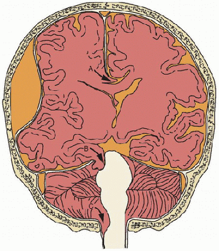
Focal and global brain swelling can act as a mass leading to shifts in brain tissue across neighboring intracranial compartments (Fig. 61.1). These tissue herniation syndromes can exist despite normal global intracranial pressure (ICP) (Table 61.4).


Posttraumatic Ischemia, Tissue Oxygenation, and Metabolism

In adults seen early after injury, cerebral blood flow (CBF) is reduced and secondary insults such as hypotension and hypoxemia have devastating effect. Brain swelling and any accompanying intracranial hypertension can contribute to this early secondary ischemia. Problems in CBF and metabolism are not exclusive to adults and are observed in infants and children. Infants with severe TBI commonly have cerebral hypoperfusion, and a global CBF of <20 mL/100 g/min was associated with poor outcome (32). Following early posttraumatic hypoperfusion, CBF may increase to levels
greater than metabolic demands, producing a state of relative hyperemia (33). Alternatively, a phase of increased cerebral metabolism of glucose may accompany posttraumatic hypoperfusion. In adults, the use of monitoring techniques that reflect the coupling between CBF and metabolism (e.g., using brain microdialysis and oxygen probes) is increasing, and the presence of key phenomena is influencing practice. In children, a recent report indicates a unique pattern of brain tissue oxygen tension with severe TBI: the best threshold for favorable outcome was noted at a level of 30 mm Hg, and high, rather than low, values were observed in some patients with refractory intracranial hypertension and compromised cerebral perfusion, likely reflecting failed uptake rather than failed delivery (34). Such impaired metabolism after severe TBI in children was recently confirmed in an MRI study of 17 children where the authors found variable CBF after injury, but hypoperfusion and low oxygenation predominating (35). Taken together, these findings are consistent with preclinical and adult clinical studies of brain metabolism suggesting mitochondrial dysfunction after TBI.


FIGURE 61.1. Herniation syndromes: (A) subfalcine and cingulate, (B) uncal, (C) foramen magnum (see Table 61.4). Subfalcine herniation occurs when one cerebral hemisphere is displaced under the falx cerebri across the midline (A). Uncal herniation refers to displacement of supratentorial structures inferiorly under the tentorium cerebelli, causing distortion and compression of the blood supply to infratentorial structures (B). Downward herniation of the cerebellum causes compression of the brainstem (C).


TABLE 61.4 TYPES OF BRAIN TISSUE HERNIATION SYNDROMES


Only gold members can continue reading. Log In or Register to continue

Stay updated, free articles. Join our Telegram channel

Jun 4, 2016 | Posted by in CRITICAL CARE | Comments Off on Head and Spinal Cord Trauma

Full access? Get Clinical Tree

Get Clinical Tree app for offline access

SYNDROME


MECHANISM


CLINICAL FEATURES


Foramen magnum


Herniating tissue:


Downward mesial displacement of cerebellar hemispheres


Episodic tonic extension with opisthotonic posturing, leading to quadriparesis


Compression:


Unilateral or bilateral medulla by ventral parafollicular or tonsillae through foramen magnum


Changes in blood pressure, heart rate, and arrhythmias


Ataxic breathing


Small pupils and disturbance of conjugate gaze


Central tentorial


Herniating tissue:


Downward displacement of one or both cerebral hemispheres