Gastrointestinal Bleeding


Gastrointestinal Bleeding


Sabina Mir

Douglas S. Fishman


INTRODUCTION AND EPIDEMIOLOGY

Gastrointestinal (GI) bleeding is a common condition in the pediatric age group with a higher incidence in children admitted in the intensive care unit (ICU). The reported incidence of GI bleeding in the pediatric ICU (PICU) is 6%-25% (1,2,3). Fortunately, the risk of life-threatening bleeding in children admitted in the PICU with upper GI bleeding is only 0.4% (3). Critically ill patients have an increased risk of GI bleeding secondary to multiple comorbidities (high blood pressure, mechanical ventilation (2), disseminated intravascular coagulopathy (DIC), medications, circulatory shock, trauma, and septicemia).

In 2009, 23,383 children aged 11-15 years were admitted in US hospitals with a diagnosis of GI bleeding. The highest mortality rates associated with GI bleeding were in cases with intestinal perforation (8.7%) and esophageal perforation (8.4%). Children with GI bleed had higher rates of comorbidities (12.3% vs. 2.3%; p<.001) (4).

In adult patients, non variceal upper GI bleeding is associated with costs ranging from $3180 to 8890 per admission and a mortality of 3.5% (5). The severity of upper GI bleeding may range from self-resolving benign conditions to progressive and life-threatening bleeding that prompts immediate intervention. This chapter focuses on the initial approach, diagnostic investigation, and management (medical and therapeutic) of GI bleeding in the PICU.


ANATOMICAL LOCATION

GI bleeding is broadly divided into upper and lower GI tract, depending on the anatomical origin. The ligament of Treitz, located at the junction of the end of the duodenum and beginning of the jejunum, provides the division between the upper and lower GI tracts; upper bleeding originates proximal to the ligament, and lower bleeding originates distally. Upper GI bleeding is further characterized into variceal and nonvariceal bleeding. It is essential to exclude other, non-GI, sites where bleeding may originate, such as from the nose or lungs, as different management is required.


DEFINITIONS AND CLINICAL PRESENTATION

Hematemesis is vomiting fresh blood and indicates a rapidly bleeding lesion.

Coffee ground emesis is vomit with a dark brown color due to the effects of gastric acid on the blood.

Melena is a dark-, black-, or tar-colored stool, secondary to bleeding from above the ileocecal valve or, if colonic transit time is slow, from proximal large bowel. A small volume of blood in the stomach can cause melena for 3-5 days and may not indicate ongoing bleeding.

Hematochezia is the passage of bright red blood per rectum or maroon-colored stools, resulting from a colonic source or massive upper GI bleeding.

Obscure GI bleeding (OGIB) is blood loss that occurs with a negative upper endoscopy and colonoscopy or obvious source from basic imaging.


ETIOLOGY

There are numerous causes of GI bleeding in the pediatric population, varying with the age of the child. Tables 99.1, 99.2, 99.3 summarize the etiology of GI bleeding in children (6).


DIAGNOSTIC EVALUATION

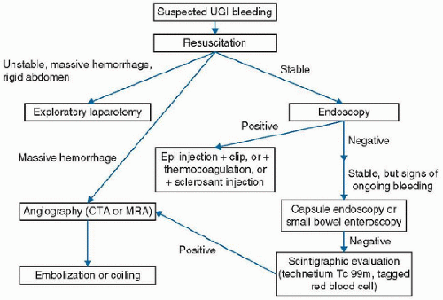
A detailed history, including medications that may potentiate GI bleeding (e.g., anti platelet agents and non steroidal anti-inflammatory drugs [NSAIDS]), and physical examination are important in elucidating the cause and source of bleeding. Physical findings characteristic of underlying disease can help establish the diagnosis (Table 99.4). There are a number of interventional options available for diagnostic evaluation and intervention; they are described in the following section and the next, and summarized in Figure 99.1.


TABLE 99.1 CAUSES OF NON VARICEAL UPPER GASTROINTESTINAL BLEEDING IN CHILDREN


NEWBORN


INFANT


CHILD-ADOLESCENT


Swallowed maternal blood


Esophagitis


Esophagitis


Maternal breast milk irritation


Maternal breast milk irritation


H. pylori (gastritis)


Stress gastritis, ulcers


Duodenitis


Salicylates, NSAIDS


Vitamin K deficiency


Peptic ulcer disease


Mallory-Weiss tear


Vascular malformation


Mallory-Weiss tear


Duodenal Crohn


Milk protein sensitivity


Gastric esophageal duplication


Vascular malformation


Necrotizing enterocolitis


Salicylates, NSAIDS


Dieulafoy lesion


Hemophilia


Foreign body


Coagulopathy (DIC, ITP)


Gastric esophageal duplication


Coagulopathy (DIC, ITP)


Maternal ITP


Vascular malformation


Dieulafoy lesion


DIC, disseminated intravascular coagulopathy; ITP, idiopathic thrombocytopenic purpura; NSAIDS, non steroidal anti-inflammatory drugs.


TABLE 99.2 CAUSES OF VARICEAL GASTROINTESTINAL BLEEDING IN CHILDREN


PREHEPATIC


POSTHEPATIC


INTRAHEPATIC


Portal vein thrombosis


Budd-Chiari syndrome


Biliary atresia


Splenic vein thrombosis


Congestive heart failure


Autoimmune hepatitis


Arteriovenous fistula


Inferior vena cava obstruction


Metabolic liver disease (Wilson disease, hemachromatosis)


Constrictive pericarditis


Toxins


Sinusoidal obstruction syndrome (veno-occlusive disease)


Infectious hepatitis


The Nasogastric Tube

An assessment of active bleeding (and gastric decompression) can be determined by nasogastric (NG) tube placement, although the role of tube placement is controversial. A meta-analysis of adult patients with GI bleeding revealed that blood or coffee ground material in an NG aspirate has a 44% sensitivity, 95% specificity, and positive likelihood ratio of 9.4 for UGI bleeding (7). In a study of 520 patients that underwent NG aspirate prior to endoscopy for UGI bleed, 15% had a clear aspirate but were found to have an upper GI lesion on endoscopy (8). Hence, a clear NG aspirate does not exclude a image bleeding source proximal to the ligament of Treitz. Conversely, the presence of blood in the NG aspirate generally confirms the diagnosis of UGI bleeding in an otherwise consistent clinical context and provided swallowed blood can be ruled out. On balance, we favor the placement of an NG tube in children in the PICU with suspected UGI bleeding to confirm the diagnosis and improve the conditions for endoscopy. Potential
complications of NG tube insertion should be considered in patients with basil skull fracture, severe facial trauma, clotting disorders, esophageal varices, esophageal tumors, or esophageal surgery.


TABLE 99.3 CAUSES OF LOWER GASTROINTESTINAL BLEEDING IN CHILDREN


NEONATE


INFANT


CHILD-ADOLESCENT


Hemorrhagic disease of newborn


Anal fissure


Anal fissure


Coagulopathy


Infectious colitis


Infectious colitis


Milk protein sensitivity


Intussusception


Polyp


Necrotizing enterocolitis


Meckel diverticulum


Vascular malformation


Volvulus


Volvulus


Inflammatory bowel disease


Vascular malformation


Vascular malformation


Vasculitis (Henoch-Schönlein purpura)


Hirschsprung disease enterocolitis


Intestinal duplication


Intestinal duplication


TABLE 99.4 PHYSICAL EXAMINATION


SYSTEM


SIGN


SUSPECTED DIAGNOSIS


Eyes


Jaundice


Cirrhosis


Iritis


IBD


Skin


Hemangiomas, telangiectasia


Vascular anomalies


Petechia, bruises


Cirrhosis, DIC


Jaundice


Cirrhosis


Purpura


HSP


Skin tags, anal fissures, erythema nodosum, pyoderma gangrenosum


IBD


Abdomen


Hepatosplenomegaly, ascites


Cirrhosis


Distension, rebound tenderness


Bowel perforation


Joints


Arthritis


HSP, IBD


DIC, disseminated intravascular coagulopathy; HSP, Henoch-Schönlein purpura; IBD, inflammatory bowel disease.


FIGURE 99.1. Diagnostic algorithm for severe upper GI hemorrhage. Epi, epinephrine.


Laboratory Tests

Hemodynamic changes (e.g., tachycardia, hypotension) are signs of hypovolemic shock and require immediate resuscitation measures prior to further evaluation. Initial laboratory investigations (complete blood count and coagulation profile) should be instituted during resuscitation. Once the child is hemodynamically stable, additional laboratory testing (Table 99.5) to investigate the cause and to direct therapy may include liver panel, metabolic panel, erythrocyte sedimentation rate (ESR), C-reactive protein (CRP), and stool cultures (E. coli O157:H7, Shigella, Salmonella, Yersinia, Campylobacter, and Clostridium image difficile [C. diff] PCR). Fecal and gastric contents can be tested for the presence of hemoglobin using a guaiac-based test to detect occult bleeding. False-positive guaiac reactions occur with intake of red meat, raw fruits or vegetables, and other foods with peroxidase activity. Inflammatory markers (ESR and CRP) are useful when inflammatory bowel disease is considered.


TABLE 99.5 INVESTIGATION


INVESTIGATION


INDICATION


Laboratory


Complete blood count


Assess severity of bleeding (anemia and thrombocytopenia)


PT/INR and PTT


Coagulopathy (liver dysfunction, DIC)


BUN/Cr


High ratio (>30) suggestive of UGI bleed (in adults)


Stool culture


Infectious colitis (Shigella, Salmonella, Yersenia, Campylobacter, and enteropathic E. coli) Stool PCR for C. diff


ESR/CRP


Infectious and inflammatory disorders


Guaiac-based tests


Detection of hemoglobin in gastric or fecal content


Imaging


X-ray


Foreign body, bowel perforation or obstruction


Abdominal ultrasound


Portal hypertension, vascular malformation, intussusception


Radionuclide scanning


TPT (Meckel scan), detection of heterotopic gastric mucosa


CT/MRI abdomen


Mass, vascular malformation, intestinal duplication


Angiography


In massive UGI bleed (bleeding must be at least 0.5 mL/min to be detected


Endoscopy


Esophagogastroduodenoscopy


For assessing acute UGI bleeding requiring transfusion or unexplained recurrent bleeding


Colonoscopy


Polyps, colitis, vascular malformations


Enteroscopy


Small bowel polyps, ulcers, colitis, vascular malformations


Wireless capsule study


Small bowel polyps, ulcers, colitis, vascular malformations


DIC, disseminated intravascular coagulopathy; UGI, upper gastrointestinal; TPT, technetium 99m pertechnetate.


Radiologic and Nuclear Medicine Imaging

Plain x-ray film may be useful in the setting of suspected GI perforation or intestinal obstruction presenting with bleeding. Ultrasonography with Doppler flow is useful for the evaluation of portal hypertension, liver disease, large vascular anomalies (arteriovenous malformations, hemangiomas), and intussusception. Air contrast or barium contrast enema can be used to confirm the diagnosis and treat colonic intussusception. Regular and angiographic CT and MRI are noninvasive and useful in detecting mass lesions and vascular malformations. These modalities are noninvasive but may require sedation. Technetium 99m pertechnetate (TPT) rapidly binds to gastric mucosa and is useful in detecting ectopic gastric mucosa, particularly Meckel diverticulum. The sensitivity of TPT scintigraphy has been reported to range from 50% to 91% (9,10). Pretreatment with pentagastrin, histamine H2 blockers, or glucagon is reported to enhance the sensitivity of the Meckel scan (11

Only gold members can continue reading. Log In or Register to continue

Stay updated, free articles. Join our Telegram channel

Jun 4, 2016 | Posted by in CRITICAL CARE | Comments Off on Gastrointestinal Bleeding

Full access? Get Clinical Tree

Get Clinical Tree app for offline access