Chapter 68 Flail Chest and Pulmonary Contusion
1 What are the most common injuries in patients sustaining blunt chest trauma?
Rib fractures are the most common injury after chest trauma, and multiple rib fractures leading to flail chest occur in 15% to 25% of patients. Pulmonary contusion is the most common intrathoracic injury, occurring in 40% to 60% of patients with blunt chest trauma. Although isolated pulmonary contusion may occur after an explosion injury, most trauma patients have concurrent injury to the chest wall.
2 What are the risk factors for adverse outcomes after blunt thoracic injury?
Thoracic injury and its complications are responsible for up to 25% of blunt trauma mortality. Increasing age and a larger number of rib fractures are most closely linked with increased complications. The greatest risk factors for mortality in patients with blunt chest trauma are age of 65 years or more, sustaining three or more rib fractures, and the presence of medical comorbidities, especially cardiopulmonary disease. Rib fractures cause intense pain and can lead to splinting of the chest with a rapid, shallow breathing pattern, as well as poor secretion clearance, increasing the risk for development of pneumonia. After injury, the development of pneumonia is a significant risk factor for mortality.
3 What is the sensitivity of chest radiograph for diagnosis of rib fractures?
Compared with a computed tomography (CT) scan, chest radiography misses approximately 50% of rib fractures. For stable patients with mild injury, the diagnosis of rib fracture is often clinical, with findings of significant pain and tenderness on examination. However, for patients with more severe injuries, any rib fracture or pulmonary contusion visible on the initial chest radiograph significantly increases the incidence of pulmonary morbidity or mortality.
4 What is a flail chest, and how is it diagnosed?
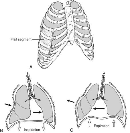
Flail chest is defined as fractures of three or more consecutive ribs or costal cartilages fractured in two or more places (Fig. 68-1). These fractured segments give rise to a free-floating portion of the thorax, which moves paradoxically throughout the respiratory cycle, with inward motion with inspiration and outward motion with exhalation. Although rib fractures may be diagnosed radiographically, flail chest is a clinical diagnosis. Patients often present with chest wall pain, tenderness, bruising, and palpable step-offs of the ribs, but flail chest is distinguished from other chest trauma by noting the paradoxical movement of the chest wall during spontaneous respiration. Patients receiving positive pressure ventilation usually do not demonstrate the classic paradoxical movements. Respiratory dysfunction usually does not arise from the paradoxical chest motion but rather is due to underlying contusions and splinting from pain.
5 What is a pulmonary contusion?
Pulmonary contusion is a bruise of the lung, with alveolar and interstitial hemorrhage and destruction of the pulmonary parenchyma. The subsequent inflammation leads to asymmetric edema, atelectasis, and poor mucous clearance from the airways. These factors lead to progressive ventilation-perfusion mismatch and loss of pulmonary compliance, which may be manifested clinically as progressive respiratory failure develops over the first 6 to 24 hours after the injury. Contusions tend to worsen over the 24 to 48 hours after injury and then slowly resolve within 7 days.
6 What is the role of radiographs in the diagnosis of pulmonary contusion?
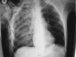
Pulmonary contusions are diagnosed radiographically. Although initial chest radiographs may be unremarkable, a nonsegmental infiltrate typically develops over a 6-hour period. If the contusions are visible on the initial chest radiograph, the injury is likely to be more severe, and enlargement of the contused area on the radiograph over the next 24 hours is a poor prognostic sign. Classic radiograph patterns include irregular consolidations or a diffuse patchy pattern (Fig. 68-2). Even after development of chest radiograph findings, plain radiographs may underestimate the severity of the contusions. CT scan is more sensitive for diagnosis of pulmonary contusions and can quantify the volume of lung involved.
7 What is the relationship among rib fractures, flail chest, and pulmonary contusions?
Pulmonary contusions often occur without flail chest and may even be present in the setting of minimal to no rib fractures. Flail chest, however, indicates that the chest wall sustained a large force, and therefore more than 90% of patients with flail chest have associated intrathoracic injuries, often pulmonary contusions. These patients also frequently have a hemothorax, a pneumothorax, or both. Patients with flail chest are likely to have additional traumatic injuries, including head injury and intraabdominal injuries. The pattern of rib fractures on imaging has been shown to be suggestive of other injuries in a recent series of trauma patients. Lower rib fractures were highly predictive of solid organ injury when compared with upper and midzone rib fractures, and scapular and sternal fractures were more common with upper zone fractures.
8 What is the relationship between pulmonary contusions and acute respiratory distress syndrome (ARDS)?
Patients with a pulmonary contusion are at higher risk than other patients for development of pneumonia and ARDS. The volume of lung parenchyma involved as determined by CT scan has been shown to be a risk factor for the development of ARDS, with patients having contusion volumes of greater than 20% being at the highest risk. Of these patients, ARDS has been shown to develop in approximately 80%.
9 What is the mortality rate and cause of death for patients with flail chest and pulmonary contusions?
The overall mortality rate of patients with blunt chest trauma is 16% with either a pulmonary contusion or flail chest to 42% when patients had both. Although these patients have severe thoracic injury, the most common cause of death in patients with flail chest and pulmonary contusions is brain injury.
10 What are the basic treatment strategies for flail chest or pulmonary contusions?
All trauma patients should be assessed with use of the principles of advanced trauma life support, directed at diagnosing and intervening in life-threatening injuries immediately. Using a primary survey, verifying the ABCs (airway, breathing, and circulation) is paramount. Prompt endotracheal intubation, tube thoracostomy for suspected hemopneumothorax, and mechanical ventilation are warranted in the unstable patient with chest trauma. For the stable patient with chest trauma, management centers around close monitoring of the respiratory status, pain control, aggressive lung physiotherapy, early mobilization, and adequate nutrition.

Stay updated, free articles. Join our Telegram channel

Full access? Get Clinical Tree

