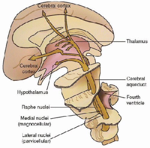
 state, or brain death.
TABLE 57.1 STATES OF ALTERED CONSCIOUSNESS | ||||||||||||||||||||||||||||||
|---|---|---|---|---|---|---|---|---|---|---|---|---|---|---|---|---|---|---|---|---|---|---|---|---|---|---|---|---|---|---|
|
Although such patients may make sounds, facial expressions, or body movements, detailed testing does not demonstrate reproducible purposeful responses to stimulation. Since patients in whom coma is due to head trauma may recover consciousness after a longer period than those in whom coma is nontraumatic, the diagnosis of a persistent vegetative state may be made 3 months following nontraumatic brain injury, but not until 12 months following traumatic brain injury (5,6). Of children in the vegetative state 1 month after head injury, 1-year outcome was death (9%), persistent vegetative state (29%), and recovery of consciousness (62%). However, outcome was worse when the etiology was nontraumatic, with death (22%), persistent vegetative state (65%), and recovery of consciousness (usually with severe cognitive disabilities) in only 13% (7). A patient in a minimally conscious state has severely altered consciousness but has behavioral evidence of self or environmental awareness, such as following simple commands or making simple nonreflexive gestures. Recent functional neuroimaging and electrophysiological studies have indicated that some patients considered to be in the vegetative state demonstrate reproducible physiologic changes in response to tasks, indicating there may be some awareness despite the absence of behavioral signs of responsiveness (8,9,10). These findings have prompted the use of more neutral and descriptive terms (11,12). The vegetative state may be referred to as the unresponsive wakefulness syndrome. The minimally conscious state may be subdivided into the minimally conscious positive state (high-level behavioral responses, such as following commands or intelligible verbalizations) and the minimally conscious negative state (lower level behavioral responses, such as visual pursuit or contextually-appropriate crying or smiling) (11).
causes each accounted for 6% of cases. Approximately onequarter of children with traumatic brain injury present with coma (2). Importantly, multiple interrelated factors crossing these subdivisions may be present in one patient. For example, status epilepticus may occur in the setting of encephalitis; infection inducing a catabolic state may produce decompensation in a child with an inborn error of metabolism; and hyponatremia or other electrolyte dysfunctions may accompany brain injury and contribute to cerebral dysfunction.
TABLE 57.2 ETIOLOGIES OF COMA | ||||||||||||||||||||||||||||||||||||||||||||||||||||||||||||||||||||||||||||||||||||||||||||||||||||||||||||||||||||||||||||||||||||||||||||||||||||||||||||||||||||||||||||||||||||||||||||||||||||||||||||||||||||||||||||||||||||||||||
|---|---|---|---|---|---|---|---|---|---|---|---|---|---|---|---|---|---|---|---|---|---|---|---|---|---|---|---|---|---|---|---|---|---|---|---|---|---|---|---|---|---|---|---|---|---|---|---|---|---|---|---|---|---|---|---|---|---|---|---|---|---|---|---|---|---|---|---|---|---|---|---|---|---|---|---|---|---|---|---|---|---|---|---|---|---|---|---|---|---|---|---|---|---|---|---|---|---|---|---|---|---|---|---|---|---|---|---|---|---|---|---|---|---|---|---|---|---|---|---|---|---|---|---|---|---|---|---|---|---|---|---|---|---|---|---|---|---|---|---|---|---|---|---|---|---|---|---|---|---|---|---|---|---|---|---|---|---|---|---|---|---|---|---|---|---|---|---|---|---|---|---|---|---|---|---|---|---|---|---|---|---|---|---|---|---|---|---|---|---|---|---|---|---|---|---|---|---|---|---|---|---|---|---|---|---|---|---|---|---|---|---|---|---|---|---|---|---|---|---|---|---|---|---|---|---|---|---|---|---|---|---|---|---|---|
| ||||||||||||||||||||||||||||||||||||||||||||||||||||||||||||||||||||||||||||||||||||||||||||||||||||||||||||||||||||||||||||||||||||||||||||||||||||||||||||||||||||||||||||||||||||||||||||||||||||||||||||||||||||||||||||||||||||||||||
 reversible etiologies. Medical stabilization must occur as the coma etiology is being investigated to prevent development of secondary brain injury. An algorithm for initial evaluation of coma is outlined in Table 57.3 and discussed below. The Pediatric Accident and Emergency Research Group of the Royal College of Paediatrics and Child Health and the British Association for Emergency Medicine have published guidelines for practice on the basis of an extensive literature review and expert consensus (www.nottingham.ac.uk/paediatric- guideline) (14). Other literature reviews are also available (15,16,17).
to coma, with particular attention to timing, exposures, and accompanying symptoms. Preceding somnolence or headaches suggests metabolic, toxic or infectious etiologies, hydrocephalus, or expanding mass lesions. Sudden onset of coma without trauma suggests seizure, intracranial hemorrhage, or hypoxicischemic encephalopathy caused by a cardiac event. A slowly progressive loss of consciousness suggests hydrocephalus, an expanding mass lesion, or indolent infection. Fluctuations in mental status may occur with metabolic etiologies, seizure, or subdural hemorrhage. Preceding headache aggravated with positional changes or Valsalva maneuver implies increased intracranial pressure from hydrocephalus or a mass lesion. Headache with neck pain or stiffness suggests meningeal irritation from inflammation, infection, or hemorrhage. Fever suggests infection but its absence does not rule it out, particularly in infants younger than 3 months of age, or immunocompromised children. Recent fevers or illnesses suggest autoimmune processes such as acute disseminated encephalomyelitis (ADEM) or possibly Reye-like illness although this is uncommon. Questions about possible toxic ingestions should include a survey of medications and poisons kept in the places that the child has been.
TABLE 57.3 INITIAL EVALUATION OF COMA | ||||||||||||||||
|---|---|---|---|---|---|---|---|---|---|---|---|---|---|---|---|---|
| ||||||||||||||||


