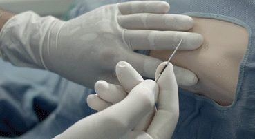
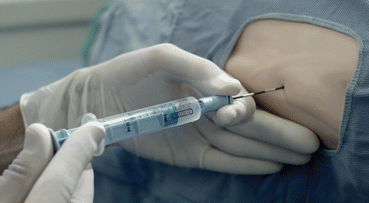
Fig. 5.1
Choice of lumbar interspace
.
A small gauge needle attached to a 5 mL disposable syringe containing the local anesthetic solution (such as 1 or 2% lidocaine) is inserted through the skin to make a wheal over the selected interspace and eventually gently inserted into the underlying tissues until the interspinous ligament, while the local anesthetic solution is slowly injected.
The angle of penetration should be the same as planned for the epidural needle.
Without moving the “landmark fingers,” the epidural Tuohy needle is inserted through the skin wheal previously made exactly in the middle of the interspace.
The epidural needle is held with the palm of the hand resting on the hub, and the shaft of the needle between the fingers of the dominant hand (Fig. 5.2)

.

Fig. 5.2
Introduction of the needle
Once inserted into the skin, the epidural needle should be advanced with the bevel directed cephalad, taking care to remain in the midline. The needle must be advanced very slowly but constantly, without any interruption, in order to be able to recognize the different densities of the underlying tissues (subcutaneous tissue, supraspinous and interspinous ligaments) during its advancement. As soon as it reaches the supraspinous ligament, a resistance is encountered due to the nature of the bevel and the density of the ligament. The needle is then advanced through the loose interspinous ligament which offers much less resistance than the supraspinous ligament (often felt as a “no resistance feeling” in the obstetric patient), until the point of the needle is felt to meet the third, and greater, point of resistance, the ligamentum flavum. This feeling of a greater increase of resistance is often associated with a “crunch” that indicates the initial penetration of the needle in the rear wall of the ligamentum flavum. Instead, if the resistance is absolute the bevel of the needle may be against the bony vertebral arch and any attempt to force the needle may result in pain for the patient due to periostium stimulation. In this case, the needle should be withdrawn, its angle of inclination checked, and the direction changed accordingly.
As soon as the point of the needle has engaged the ligamentum flavum, the advancement of the needle is immediately stopped, and the hands of the operator must change their initial position. The back of the nondominant hand (usually the left hand) rests firmly against the patient’s back to prevent advancement as the needle enters the epidural space with the hub of the needle grasped between the thumb and index fingers. The dominant hand (usually the right hand) removes the stylet and gently attaches to the needle a disposable 10 mL loss of resistance syringe containing a few milliliters (5–7 mL) of sterile saline solution.
Constant, unremitting, pressure is now exerted on the plunger of the syringe by the thumb of the dominant hand and since the content of the syringe (saline) is incompressible, the syringe and needle advance together solely by means of the pressure exerted by the operator on the plunger of the syringe (Fig. 5.3)

.

Fig. 5.3
Loss of resistance
As long as the needle point is in the ligamentum flavum there is a great resistance to injection and the pressure exerted by the thumb on the plunger causes the advancement of the needle.
As the point of the needle emerges from the ligamentum flavum into the epidural space, the resistance suddenly disappears and the advancement of the needle immediately stops, since the driving force exerted on the piston is discharged by the sudden entering of the liquid in the epidural space.
When the epidural needle is positioned in the epidural space, the syringe is removed and the needle observed for the appearance of spinal fluid (a few drops of syringe solution may leak from the needle).
5.1.4 Air or Rebound Test
After negative aspiration for blood or cerebrospinal fluid, proper placement of the needle may be further checked with the air or rebound test. A very small amount of air (1–1.5 mL) is drawn in the loss of resistance syringe and attached to the needle and the plunger of the syringe is tapped sharply. A positive test results when the syringe collapses and does not refill at all, or only refills 0.1–0.2 mL.
This rapid injection of a very few millimeters of air, mixed with the previously injected liquid already in the epidural space, may occasionally result in small air-water bubbles escaping from the hub of the needle. If this occurs, it may be considered another indirect sign of confirmation that the epidural needle is in the right place.
When the plunger rebounds, a partially inserted bevel of the needle in the epidural space could be suspected.
5.1.5 Catheter Placement and Needle Removal
Once the needle is in place, the epidural catheter is advanced through the needle by the dominant hand while the back of the nondominant hand (usually the left hand) keeps resting firmly against the patient’s back with the hub of the needle grasped between the thumb and index fingers.
The parturient should be warned that she might feel an “intense tingle” in her hip or legs when the catheter is advanced a few centimeters beyond the bevel. Such paresthesia may occur when the epidural catheter contacts a spinal nerve root, depending on the type and material of the catheter, on the needle position (midline, paramedian), and on the epidural anatomy of the patient. It may be interpreted as an indirect sign that the catheter is in the epidural space.
Before removing the needle, the catheter should be aspirated with a 2 or 5 mL empty syringe in order to detect blood or cerebrospinal fluid which are, respectively, signs of accidental intravascular or subarachnoid placement of the catheter.
If the aspiration test is negative, the needle is removed. This is an important maneuver since the catheter may be dislodged from the epidural space while removing the needle. The catheter is grasped 1–2 cm distal to the hub of the needle by the thumb and the index finger of the dominant hand while the thumb and the index finger of the other hand pull the needle out of the back of the patient. The dominant hand should attempt to advance the catheter while the nondominant is pulling out the catheter. At the end of the procedure, the catheter distance marks are checked and the catheter properly positioned. Placement of the catheter more than 5 cm in the lumbar epidural space may be associated with a higher incidence of unilateral block and a greater likelihood of the tip entering an epidural blood vessel, while too little catheter length predisposes the catheter to falling out [13].
The catheter is then secured with tape and adhesive dressings, and used for the intended purpose.
Once the catheter is placed, after a test dose, anesthesia for cesarean delivery is achieved by administration of local anesthetics with or without opioids.
Box 1 Dogliotti’s Loss of Resistance to Saline Technique
“When the needle has penetrated the ligamentum interspinosum for a certain distance and before it has gone through the ligamenta subflava into the spinal canal one removes the trocar and attaches a syringe filled with physiological saline. When an attempt is made to inject this fluid a very great resistance is met with since the ligamentum interspinosum and the ligamenta subflava are so dense. If they can be injected at all, it will be only after the employment of considerable force. This resistance is most certain evidence that the needle is still in the posterior fibers of these tissues. The following manoeuvres are then carried out: the syringe is held in one hand the thumb of which applies a continued and uniform pressure to the piston. The other hand slowly advances the needle into the tissues and when it has traversed a few millimetres the hand which is holding it will suddenly note a diminution in the resistance to its passage which has previously been due to the tissues of the ligamenta subflava. At the same instant the injection fluid enters freely. This is certain, practical, and unequivocal evidence that the point of the needle has pierced the ligamenta subflava and is in the peridural space which offers no resistance to the flow of the injected fluid. As soon as this position has been recognized the needle should be left in the position which it now occupies for its point is in the peridural space; any attempt to advance it farther would entail the risk of penetrating the dura.” (Reprinted from [14] with the permission of the Publisher)
5.1.6 Ultrasound Assisted Identification of the Epidural Space
Evidence on ultrasound-guided identification of the epidural space in pregnancy is still limited and this technique is not commonly used as a routine tool to detect the epidural space in obstetrics.
In addition, the visibility of the ligamentum flavum, the dura mater, and of the epidural space decreases significantly during pregnancy [15]. However, preliminary evidence suggests that it is safe and may be helpful in achieving correct placement, especially for teaching purposes and in the obese parturient [16, 17].
Ultrasound can be used in two different ways to ease the performance of an epidural block [18]. One method is to use real-time ultrasound imaging, under sterile conditions, to observe the passage of the needle on the way to the epidural space. In the second method (prepuncture ultrasound), an initial ultrasound scan of the patient’s lumbar area is performed to find the midline and the interspinous space in order to mark on the skin the position of each. The depth of the epidural space may also be determined by using the ultrasound scan. Epidural block is eventually performed in the usual way with the skin markings as an additional guide.
5.2 Epidural Anesthesia
One of the main advantages of epidural anesthesia is that the local anesthetic can be administered in incremental doses and that the total dose can be titrated to the desired sensory level.
This, with the slower onset of anesthesia, allows the maternal cardiovascular system to compensate for the occurrence of sympathetic block reducing the risk of severe hypotension and reduced uteroplacental perfusion.
The use of the epidural catheter, and therefore of a continuous technique, allows the anesthesiologist to give additional local anesthetic to maintain anesthesia, regardless of the duration of surgery and the intensity of surgical stimulation. Usually epidural anesthesia results in less intense motor block than dose spinal anesthesia, especially at the beginning of the block. This may be advantageous for patients in which a high level of motor block may impair ventilation, such as multiple gestation or pulmonary diseases. The epidural catheter may also be used for postoperative analgesia either with exclusive epidural opiods or with an analgesic ultra low concentration solution of local anesthetic and opioids.
5.2.1 Test Dose
The aim of the epidural test dose is to detect the inadvertent intravenous or subarachnoid placement of the epidural catheter in order to avoid, respectively, a too high or a total spinal block or local anesthetic toxicity. The test dose must be formulated to produce a rapid, reliable, and easily detected result when in one of these two situations, without compromising the safety of the mother and the fetus.
In all cases, careful aspiration of the epidural catheter before administering any dose of anesthetic solution is the first extremely important step.
Subarachnoid placement is relatively easy to detect. For practical reasons, the same local anesthetic that is used for producing the anesthetic block is usually chosen. Lidocaine 20–60 mg or bupivacaine (or levobupivacaine or ropivacaine) 7.5–12.5 mg are commonly used. Signs of sensory block in the lower lumbar segments and, most importantly, motor block of the legs should be sought after 3–5 min and this is considered to be specific and sensitive in almost 100% of cases. When the test dose is performed with a relatively “high dose” of local anesthetic, such as 40–60 mg of lidocaine or 12 mg of bupivacaine, in the case of accidental intrathecal injection, a safe but complete sensory and motor block accompanied by maternal hypotension may be observed [19].
Inadvertent intravascular placement of the epidural catheter usually relies on the use of a dose of epinephrine (15 μg) capable of producing detectable changes in heart rate and blood pressure but unfortunately, in obstetrics, intravenous injection of epinephrine has been shown to have a low positive predictive value and may be associated with side effects [20].
Therefore, detection of intravascular multiorifice epidural catheter placement relies on repeated catheter aspiration, observation of gravity-induced fluid efflux within an open-ended catheter, failure of local anesthetic to produce the anticipated effect, and detection of early signs of toxicity by means of slow and incremental injection.
It is therefore vital to aspirate the catheter before giving each dose and fractionate the whole anesthetic dose in small boluses given intermittently, always.
5.2.2 Anesthetic Solution
Approximately 3–5 min after a negative aspiration test and a negative test dose, the therapeutic dose is then administered, in fractionated boluses of 5 mL each.
Although the nerve supply to the uterus extends no higher than the eighth to tenth thoracic nerve roots, it is generally agreed among anesthesiologists that anesthesia for cesarean section should extend to the level of the fourth thoracic dermatome to include afferent fibers running in the greater splanchic nerve. However in some cases, peritoneal stimulation may require a sensory block up to the first thoracic dermatome. An adequate sacral anesthesia level is also required to prevent pain from bladder retraction or uterosacral ligaments traction.
An inadequate sensory assessment prior to surgery or an unrecognized sensory block regression during surgery is a common cause of intraoperative pain. It is therefore most important to check the sensory block with an appropriate and reliable method, such as the loss of sensation to pinprick or to light touch and by using an appropriate evaluation scale [21].
A bilateral, adequately, dense sensory level to T4 is required for cesarean surgery and this could be reached in the majority of cases with 20–25 mL of local anesthetic.
A frequent assessment of the sensory block allows a careful titration of the anesthetic dose at the desired level.
Most anesthesiologists use lidocaine 2%, bupivacaine 0.5%, levobupivacaine 0.5 or 0.75%, or ropivacaine 0.75–1%. 2-chlorprocaine is also used where available (not in Europe).
Epinephrine may be added at the concentration of 1:200,000 or 1:400,000 to decrease vascular absorption of the local anesthetic and to prolong the duration of the block. Due to the well-known pharmacological characteristics of the different local anesthetics, the addition of epinephrine appears to have a rationale only with lidocaine. The addition of sodium bicarbonate to lidocaine hastens the onset of anesthesia and may also improve the quality of analgesia [22].
Opioids are frequently given epidurally to enhance intraoperative analgesia and to provide postoperative pain relief. Fentanyl 50–100 μg or sufentanil 10 μg may be added to the therapeutic dose or given separately at some point during the administration of the epidural boluses without adversely affecting the neonate.
The choice of the drug depends on the desired onset of action and the expected duration of surgery and may vary with the local clinical practice. The most effective solution is 2% lidocaine with epinephrine with a liposoluble opioid, the least is 0.5% plain bupivacaine.
The local anesthetic solution used by the author is a pH adjusted solution of 2% lidocaine with epinephrine 1:400,000 with the addition of 10 μg of sufentanil.
5.2.3 Fluid Preloading and Control of Maternal Hypotension
The incidence and the degree of maternal hypotension after epidural block are dependent on the speed of onset of the sympathetic block, being less with fractioned incremental boluses.
Maternal hypotension may be prevented and/or treated with fluid preloading and vasopressor drugs.
Unfortunately, almost all the recent studies on this topic investigated exclusively spinal rather than epidural anesthesia.
Current literature highlights that prevention of hypotension during spinal anesthesia for cesarean section is mainly based on the use of vasopressor drugs prophylaxis. However, fluid administration remains helpful to further decrease the incidence and severity of maternal hypotension and vasopressor requirement. Hydroxyethyl starch (HES) solution preloading or coloading is the best acknowledged and the more consistent method [23].
With regard to vasopressor use, ephedrine seemed initially to be the logical vasopressor for obstetrics, with both α- and β-sympathomimetic effects, the ideal protection for placental intervillous blood flow. Most likely ephedrine is still the most commonly used vasopressor to prevent and treat maternal hypotension after a spinal block for cesarean section.
Now numerous studies have compared this agent with pure α stimulants, usually phenylephrine, with confusing results, but meta-analysis has shown convincingly that ephedrine is associated with lower pH and BE of the neonate and with a higher risk for fetal acidosis when compared with phenylephrine. Comparing the maternal effects, phenylephrine is associated with an increased risk of maternal bradycardia. Unfortunately, a number of not controlled factors that may also influence fetal blood gases such as the total amount of vasopressor given before delivery, timing of administration, duration, and severity of maternal hypotension and, in addition, a clear definition of hypotension is often not reported in these studies [24, 25].
However, these findings concern spinal rather than epidural anesthesia, and a comparison between vasopressors during epidural anesthesia has not been performed.
Stay updated, free articles. Join our Telegram channel

Full access? Get Clinical Tree


