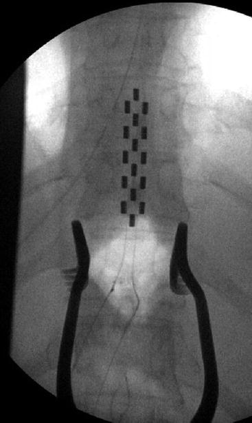
Fig. 20.1
Intraoperative view of thoracic paddle lead implantation. (a) The electrode is behind the body of T9 and T10. Stimulation is right sided with the cathode at the second position and the anode at the third. (b) With right-sided stimulation, there is right-side gastrocnemius activation, which will correlate with an S1 dermatomal paresthesia
Table 20.1
Correlations between EMG activation of specific muscles with postoperative induced paresthesia—thoracic paddle electrode at T9–10 (laminectomy T10–11)
EMG activation, muscle group | Induced paresthesia |
|---|---|
External oblique | Low back |
Tibialis anterior | L4 |
Vastus lateralis | L5 |
Gastrocnemius | S1 |
The general concept of using intraoperative EMG in the placement of the SCS on the PM of the spinal cord is similar with respect to the two- and three-column paddle configuration and differs in terms of whether the “expected” pattern should be symmetrical (the middle column of a three-column array) or “equally asymmetrical” (a two-column array). We have just begun PM evaluation with this technique using the newest five-column array (Penta, St. Jude Medical, Plano, TX).
20.3 Technique of Midline Positioning of the Spinal Cord Stimulator: Tripolar Paddle
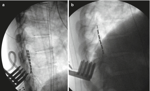
Once the three-column paddle (Fig. 20.2) is placed in the dorsal epidural space, the superior midline contact is stimulated at minimal settings and the EMG trace recording associated with the dermatomal level of stimulation is monitored. The stimulus intensity is gradually increased until MUAPs are seen on EMG. The lowest stimulus intensity needed to elicit a motor response is referred to as the threshold stimulus. MUAPs will be seen bilaterally at the threshold stimulus if the midline contact of the SCS is in line with the PM of the spinal cord. If MUAPs are seen unilaterally, the threshold intensity for that side is recorded and the stimulus is further increased to elicit a response on the other side. A difference in threshold stimulus intensity between the left and the right sides indicates that the SCS is lateral to the PM. Medial/lateral repositioning of the SCS is only necessary, however, if the difference in threshold intensity between the two sides is greater than 2 mA. In this case the paddle may not be perfectly flat on the lateral x-ray (Fig. 20.3a), thus dissection of the lateral recesses or proximal/superior lamina is further performed until the electrode is perfectly aligned with vertebrae on a lateral view (Fig. 20.3b). This ensures optimal electrode column symmetry and programmability relative to the PM. Of course, as a last resort, the laminotomy can be extended to a full laminectomy to allow perfect alignment of the paddle on the PM. In this case tissue must typically be identified to suture the electrode in place to prevent migration. These tenants hold true for all paddle (one-, two-, three- and five-column) array configurations.


Fig. 20.2
Intraoperative image of initial placement of a three-column paddle electrode. Self-retaining retractors are seen providing exposure. The middle column of the paddle lies under the image of the spinous processes, and the array is relatively symmetrical with respect to the pedicles. This is a reasonable starting position to begin physiological mapping

Fig. 20.3
Lateral intraoperative views. (a) Lead placed off the midline. (b) Perfect alignment on the midline
20.4 New Frontiers of Intraoperative EMG Application
There appears to be a correlation between the muscles with objective EMG activation during intraoperative monitoring and the subjective paresthesia obtained postoperatively as described in Table 13.1. Thus this may be explored to generate a precise model of paresthesia coverage and create a functional dermatomal mapping of perceived stimulation threshold after the surgery. Furthermore the EMG activation threshold may be a reliable predictor of the patient’s perceived paresthesia threshold. It has been the author’s experience that EMG activation correlated with pain control at amplitudes lower than the paresthesia threshold (i.e., subthreshold stimulation) and that occasionally persistent EMG activation intra- and postoperatively may be seen lasting as long as 15 min after the stimulation is discontinued. These patients typically respond extremely well to the stimulation therapy. It seems likely that these patients are the occasional patients who use their stimulation only intermittently, often having effective long-term pain relief while using their system only for a portion of each day.
20.5 Conclusions
EMG and SSEP monitoring are well-accepted modalities of neurophysiological testing in traditional spine surgery, but their application relative to implantation of neuromodulatory systems, in particular epidural paddle electrode systems, is comparatively new. Given the limitations associated with other methods of implantation, however, we expect that the techniques described in this chapter will continue to gain acceptance and improve outcomes by objectively localizing neurophysiological stimulation targets.
References
1.
2.
Spielholz NI, Benjamin MV, Engler GL, Ransohoff J. Somatosensory evoked potentials during decompression and stabilization of the spine. Methods and findings. Spine (Phila Pa 1976). 1979;4:500–5.CrossRef
3.
Ryan TP, Britt RH. Spinal and cortical somatosensory evoked potential monitoring during corrective spinal surgery with 108 patients. Spine (Phila Pa 1976) . 1986;11:352–61.CrossRef
4.
Perlik SJ, Van Egeren R, Fisher MA. Somatosensory evoked potential surgical monitoring. Observations during combined isoflurane-nitrous oxide anesthesia. Spine(Phila Pa 1976). 1992;17:273–6.CrossRef
5.
6.
Ben-David B, Haller G, Taylor P. Anterior spinal fusion complicated by paraplegia. A case report of a false negative somatosensory-evoked potential. Spine (Phila Pa 1976). 1987;12:536–9.CrossRef
Stay updated, free articles. Join our Telegram channel

Full access? Get Clinical Tree


