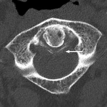
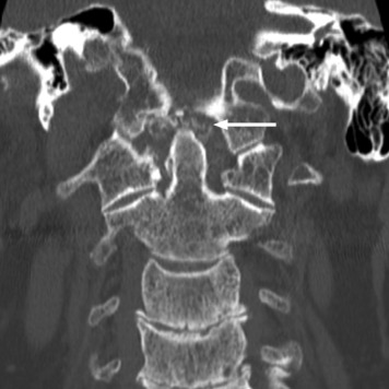
An 82-year-old white man with a medical history of calcium pyrophosphate crystal arthritis (pseudogout) and nephrolithiasis presented to the emergency department with a 2-day history of acute, sharp neck pain, neck stiffness, and low-grade fever. There was no history of trauma. On physical examination, he had neither a septic appearance nor focal neurologic deficits. Range of neck motion was significantly limited, especially to rotation, as well as flexion and extension. Vital signs were normal. Computed tomography (CT) of the cervical spine demonstrated the presence of periodontal calcification ( Figures 1 and 2 ).


Diagnosis
Crowned dens syndrome. Crowned dens syndrome is an inflammatory condition resulting from microcrystalline deposition around the odontoid process, most often calcium pyrophosphate dehydrate crystals. CT focusing on the atlantoaxial joint is the criterion standard and shows crown-shaped calcium deposits surrounding the odontoid process. Risk factors and associated conditions are similar to those of calcium pyrophosphate deposition disease (eg, aging, a history of trauma, osteoarthritis, loop diuretics use; metabolic conditions such as primary hyperparathyroidism, hemochromatosis, hypomagnesemia). Symptoms of crowned dens syndrome include acute neck pain, headache, limited range of motion of the neck, fever, and raised inflammatory markers, and can mimic meningitis, discitis, giant cell arteritis, and polymyalgia rheumatica. Patients often respond to nonsteroidal anti-inflammatory medication. Awareness of this condition may allow rapid diagnosis with CT scan of the cervical spine and avoidance of invasive diagnostic procedures and treatments.
For the diagnosis and teaching points, see page 673.
To view the entire collection of Images in Emergency Medicine, visit www.annemergmed.com .
Stay updated, free articles. Join our Telegram channel

Full access? Get Clinical Tree


