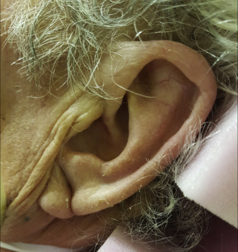
A 69-year-old man with a history of coronary artery disease was admitted to the emergency department for acute chest pain. A few minutes after admission, cardiac arrest occurred. Return of spontaneous circulation was obtained after defibrillation and intravenous amiodarone. Post–cardiac arrest ECG revealed a left bundle branch block. Emergency coronary angiography revealed an acute occlusion of the middle segment of the right coronary artery, with successful recanalization after thrombectomy and angioplasty. On ICU admission, an attentive physical examination showed a bilateral marked diagonal earlobe crease ( Figure ).
Diagnosis
Frank’s sign . Frank’s sign is a profound and permanent wrinkle that goes diagonally from the tragus to the back of each earlobe. First described in 1973, this easily detectable sign is associated with the presence, extent, and severity of coronary artery disease. The absence of the Frank’s sign does not exclude coronary artery disease, but its presence is highly suggestive, with a positive predictive value higher than 80%. Frank’s sign is also associated with carotid intima media thickness and has been reported to be associated with ischemic stroke.
For the diagnosis and teaching points, see page 672.
To view the entire collection of Images in Emergency Medicine, visit www.annemergmed.com .
Related posts:


Stay updated, free articles. Join our Telegram channel

Full access? Get Clinical Tree


