Do Not Use a Negative Focused Assessment With Sonography for Trauma (Fast) Exam to Rule Out Bowel Injury or Injury to the Retroperitoneum or as the Only Test in Penetrating Trauma
Brendan G. Carr MD
Patrick K. Kim MD
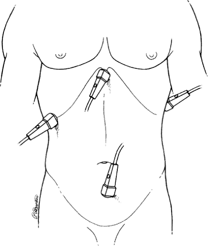
Focused assessment with sonography for trauma (FAST) is a noninvasive ultrasound procedure that is a quick, reliable exam used to detect free intraperitoneal fluid in the injured patient. Initially used in Europe as a diagnostic modality, the ease of use and ability to evaluate the injured patient in the resuscitation area has led to its widespread use. The FAST exam aids in the triage of injured patients directly to the operating room without obtaining further imaging studies. The traditional FAST exam consists of the following four views (one pericardial and three abdominal) (Fig. 279.1):
Subxiphoid view (to evaluate the pericardium)
Right upper quadrant view (hepatorenal recess or Morrison’s pouch)
Left upper quadrant view (splenorenal recess)
Suprapubic view
For decades, the standard for evaluation of the abdomen in unstable blunt trauma was diagnostic peritoneal lavage (DPL). This invasive modality is sensitive but nonspecific and reliance on DPL results in high rates of nontherapeutic laparotomies. Computed tomography (CT) has largely replaced DPL in the evaluation of the stable blunt trauma patient and is highly sensitive and specific. However, CT requires transport of the patient to the radiology suite, with the inevitable delay in obtaining and evaluating the images.
In contrast, ultrasound is available at the bedside, readily learnable by nonradiologists, and very quick to perform (2 to 4 minutes). In the blunt trauma population, reported sensitivity and specificity approximate 83.3% and 99.7%, respectively. More important, in the evaluation of hypotensive trauma patients, the sensitivity and specificity of ultrasound approach 100% in experienced hands.
 FIGURE 279.1. Transducer positions for focused assessment with sonography for trauma (FAST): (1) pericardial area, (2) right and (3) left upper quadrants, and (4) pelvis. (Reprinted with permission from
Get Clinical Tree app for offline access
Rozycki GS, Ballard RB, Feliciano DV, et al. Surgeon-performed ultrasound for the assessment of truncal injuries: lessons learned from 1540 patients. Ann Surg 1998;228(4):557–567.
Stay updated, free articles. Join our Telegram channel
Full access? Get Clinical Tree


|
