Chapter 56 Disseminated Intravascular Coagulation
1 What is disseminated intravascular coagulation (DIC)?
DIC occurs because of aberrant activation of the clotting cascade, leading to fibrin deposition in small vessels, combined with activation of fibrinolytic mechanisms, leading to bleeding. DIC is usually a common final hemostatic disorder caused by other conditions such as sepsis, pancreatitis, or trauma. Because they are consumed by the ongoing prothrombotic and fibrinolytic processes, coagulation proteins and platelets can become depleted, leading to bleeding. Thus, in DIC, hemorrhage and thrombosis can occur simultaneously. DIC can be an acute or a chronic disorder, and the latter is seen mostly in obstetric and oncology patients. Hereafter, the discussion will focus primarily on acute DIC, the form most likely to be encountered in the critical care setting.
2 Why is DIC important?
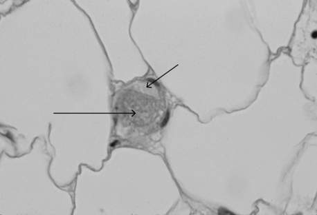
DIC is a common cause of concurrent thrombocytopenia and prolonged clotting times (activated partial thromboplastin time [aPTT] and prothrombin time [PT]) in hospitalized patients. It can also be an independent predictor of mortality. DIC leads to fibrin and platelet deposition in small vessels, which can cause tissue ischemia and result in organ dysfunction (Figure 56-1). The consumptive coagulopathy can also lead to clinically significant bleeding.
3 What is the pathophysiology of acute DIC?
Although the pathophysiology of DIC remains incompletely understood, it is thought to begin at the level of the microvasculature. As a result of widespread endothelial damage due to an underlying illness (i.e., sepsis, trauma, or pancreatitis), tissue factor is released into the circulation, where it combines with factor VIIa to produce thrombin via the extrinsic pathway. The thrombin activates platelets and cleaves fibrinogen such that platelets and fibrin are deposited in the microvasculature. Concurrently, fibrinolytic pathways are activated. As platelets and coagulation proteins are consumed, bleeding can occur as the result of a consumptive coagulopathy. Thus DIC is characterized by simultaneous clotting and hemorrhage.
4 In critical care patients, what conditions are associated with DIC?
Malignancy: adenocarcinoma, acute promyelocytic leukemia (incidence approaches 100%)
Obstetric: hemolysis, elevated liver enzymes, and low platelet count (HELLP) syndrome; retained uterine placental or fetal tissue; placental abruption or previa; amniotic fluid embolus
Vascular: vasculitis, abdominal aortic aneurysm, cavernous hemangiomas
Miscellaneous: burns, anaphylaxis, transfusion reaction, snake bite, acute pancreatitis, transplant rejection, intravenous anti-D immunoglobulin
5 How does DIC present clinically?
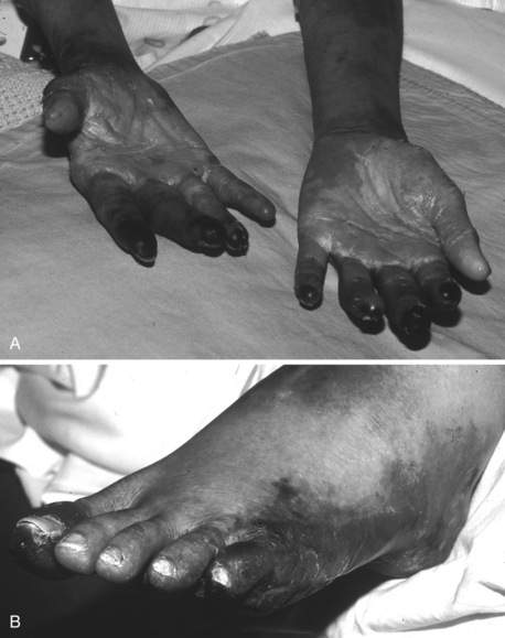
In acutely ill hospitalized patients, DIC usually presents with prolongation of the PT and aPTT along with decreased fibrinogen and platelets; systemic bleeding may or may not be present. Typically, bleeding manifests as ecchymoses, purpura, petechiae; it also occurs at surgical incisions or insertion sites of vascular access catheters. Mucosal and urinary bleeding are common, whereas pulmonary, gastrointestinal, and central nervous system bleeding occur less frequently. Because of widespread intravascular coagulation, tissue ischemia can occur, resulting in cyanosis, delirium, oliguria, hypoxia, and frank tissue necrosis (Figure 56-2).
6 What laboratory abnormalities are typical of acute DIC?
Thrombocytopenia; prolongation of the PT, aPTT, and thrombin time; and hypofibrinogenemia are characteristic. Early in the course of DIC, the platelet count may be in the normal range but is often decreased from baseline. Platelet counts are rarely less than 20,000/mcL. D-dimer and fibrin degradation product (FDP) levels are almost always markedly elevated. The haptoglobin level is variably affected and is not helpful in confirming the diagnosis of DIC.
7 Is the peripheral blood smear useful in the diagnosis of DIC?
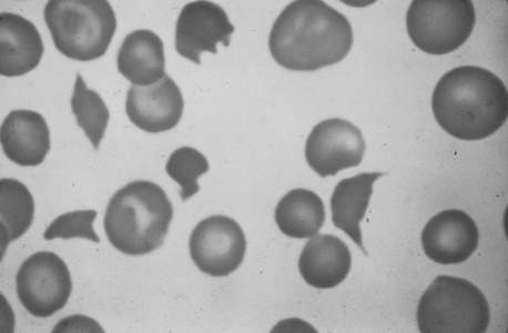
The peripheral blood smear from a patient typically shows mild to moderate thrombocytopenia. The finding of schistocytes (red blood cell fragments created by intravascular hemolysis) (Figure 56-3) is neither sensitive nor specific, and schistocytes are present in only 10% to 50% of cases of acute DIC. If present in DIC, there are usually only one to four per high-power field, in contrast to thrombotic thrombocytopenic purpura where they are much more abundant.

Figure 56-3 Schistocytes in peripheral blood smear of patient with severe acute disseminated intravascular coagulation. Note also the absence of platelets.

Stay updated, free articles. Join our Telegram channel

Full access? Get Clinical Tree

