Dental
6-1 Acute Necrotizing Ulcerative Gingivitis
Matthew Spencer
Clinical Presentation
The three most common features of acute necrotizing ulcerative gingivitis (ANUG) are papillar ulcerations, gingival pain, and gingival bleeding.1,2 It is usually of relatively acute onset, with a grayish-white exudate covering the interdental papilla and accompanying halitosis. Symptoms of severe disease include the systemic manifestations of fever and lymphadenopathy.
Pathophysiology
ANUG is an acute bacterial infection of the gingiva also known as “trench mouth,” Vincent’s disease, or fusospirochetal gingivitis.1,3 ANUG is thought to be the result of gingival infection secondary to overgrowth of oral bacteria. Specifically, anaerobes of Treponema, Selenomonas, Prevotella, Porphyromonas, and Fusobacterium species have been implicated.1,4 Predisposing risk factors include smoking, stress, poor diet, and systemic immune deficiency in the setting of chronic gingival inflammation from poor dental hygiene.1,2,3
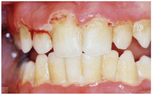 FIGURE 6-1 Generalized severe view of acute necrotizing ulcerative gingivitis (ANUG). (From Mandell, with permission.) |
Clinical Complications
Acute necrotizing periodontitis, which involves the spread of infection to the periodontal ligament and surrounding alveolar bone, may complicate some cases of ANUG.
Management
First-line therapy includes instrumentation and cleaning by a dental professional, antimicrobial mouth rinses, improved personal oral hygiene, and possibly systemic oral antibiotics.2,5 The emergency physician should prescribe penicillin, clindamycin, or metronidazole and refer the patient to a dentist within 24 to 48 hours.
REFERENCES
1. Rowland RW. Necrotizing ulcerative gingivitis. Ann Periodontol 1999;4:65-73.
2. Ahl DR, Hilgeman JL, Snyder JD. Periodontal emergencies. Dent Clin North Am 1986;30:459-472.
3. Hirsch RS, Clarke NG. Infection and periodontal diseases. Rev Infect Dis 1989;11:707-715.
4. Novak MJ. Necrotizing ulcerative periodontitis. Ann Periodontol 1999;4:74-78.
5. Pihlstrom BL, Ammons WF: Position paper: the American Academy of Periodontology. Treatment of gingivitis and periodontitis. J Periodontol 1997;68:1246-1253.
6-2 Dental Abscess
Matthew Spencer
Clinical Presentation
Periapical and periodontal abscesses have similar clinical presentations of local tooth pain, thermal sensitivity, and soft tissue swelling and redness.1,2,3,4 Systemic symptoms of fever, malaise, regional lymphadenopathy, and occasionally trismus are often present as well. Periodontal abscesses generally drain through the deep gingival pocket into the oral cavity, whereas periapical abscesses form fistulas through the bone and develop many different drainage sites, depending on the abscess location and thickness of the surrounding alveolar bone.1
Pathophysiology
Dental abscesses are oral infections that result in localized accumulations of pus; they are classified according to the location of the infection source. An endodontic or periapical abscess develops from an infection of the root canal after pulp necrosis and expands through the apical foramen to form a pocket at the base of the tooth.1,2 A periodontal abscess forms from an infection of the surrounding periodontal tissues: the gingiva, mucosa, periodontal ligament, or bone.1,2 Endodontic and periodontal abscesses are commonly encountered emergency dental infections; they account for between 14% and 25% and between 7% and 14% of oral infections, respectively.2
Periapical abscesses are a result of severe dental caries, trauma, root fracture, or multiple dental procedures resulting in pulp necrosis and subsequent polymicrobial infection with anaerobic oral flora.1,3 Periodontal abscesses form after debris is trapped between the gingiva and the tooth, resulting in inflammation of the periodontal tissues and secondary infection with anaerobic bacteria.1,2,3
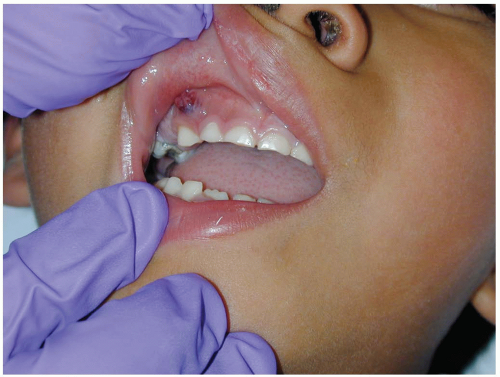 FIGURE 6-2 Dental abscess. (Courtesy of Robert Hendrickson, MD.) |
Diagnosis
The diagnosis of dental abscess is primarily based on clinical examination, with confirmation of periapical abscesses by intraoral periapical or Panorex radiographs if available.
Clinical Complications
The most common complications of these infections are tooth and bone loss, osteomyelitis, and chronic pain. However, if they are left untreated, these infections can spread through the soft tissue fascial planes of the head and neck, leading to life-threatening conditions such as mediastinitis, Ludwig’s angina, and cavernous sinus thrombosis.
Management
Emergency department (ED) management of these abscesses is essentially the same as for any other abscess. After incision and drainage of the purulent collection and irrigation with warm saline, packing material is placed in the wound to ensure adequate drainage.1,2,3 Because of continual recontamination from oral flora, systemic antibiotics should be initiated.1,2,3 The cause of these lesions is usually anaerobic oral flora of the mouth, so penicillin is the drug of choice. In case of penicillin allergy, metronidazole or clindamycin may be substituted. Patients should be treated with antibiotics for 10 to 14 days and referred to a dentist or oral surgeon within 24 hours.
REFERENCES
1. Dahlen G. Microbiology and treatment of dental abscesses and periodontal-endodontic lesions. Periodontology 2000 2002;28:206-239.
2. Herrera D, Roldan S, Sanz A. The periodontal abscess: a review. J Clin Periodontol 2000;27:377-386.
3. Kretzschmar JL, Kretzschmar DP. Common oral conditions. Am Fam Physician 1996;54:225-234.
4. Flynn TR. The swollen face: severe odontogenic infections. Emerg Med Clin North Am 2000;18:481-519.
6-3 Dental Caries
Matthew Spencer
Clinical Presentation
The typical clinical presentation of tooth decay in the emergency department (ED) is tooth pain derived from a carious lesion or fracture of a severely decayed tooth. The classification of carious lesions is based on a scoring system from 0 to 4. Zero (0) involves a slight discoloration of the tooth’s enamel, visible only after drying. One (1) is a moderate white discoloration, and 1a is a brown discoloration of the tooth; both conditions are visible only after drying of the tooth and not when the tooth is wet. Scores of 2 and 2a are, respectively, white and brown discolorations of enamel that are clearly visible on the wet tooth without drying. A score of 3 indicates localized breakdown of enamel resulting in an opaque or grayish color to the tooth. A score of 4 is an actual cavitation exposing the underlying dentin.1 Hidden lesions are found on the occlusal tooth surface, and there may be a large lesion underneath a normal-appearing fissure.1,2 Lesions often are missed on clinical examination. Pain indicates that there is some irritation of the pulp tissues.
Pathophysiology
Dental caries, or tooth decay, is erosion and demineralization of the enamel and other hard tissues of the teeth. According to epidemiologic data, 60% of tooth decay is found in only 20% to 25% of the population, although 70% of individuals 35 to 44 years of age have had at least one tooth extracted because of a carious lesion.2,3 The cause of tooth decay is multifactorial.2 The first step is the formation of dental plaque, which is a thin layer of food debris, bacteria, mucus, and dead epithelial cells that covers the tooth. Plaque forms a hospitable environment for the adherence and growth of the bacteria on the tooth surface. These oral bacteria species produce inorganic acids, leading to demineralization of the enamel and dentin. The entire process is dynamic, with the pulp constantly trying to remineralize the affected area.1,4
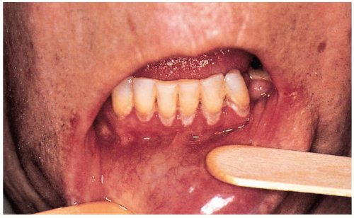 FIGURE 6-3 Dental caries is the gradual decay and disintegration of a tooth with progressive decalcification of the enamel and dentin. Moderate caries in this adult is visible around the roots of the teeth. (From Benjamin, with permission.) |
Diagnosis
The diagnosis is primarily clinical, made by a thorough examination of the involved teeth. Intraoral periapical or bite-wing radiographs may be necessary to diagnose small or hidden lesions in the setting of tooth pain and a normal examination.
Clinical Complications
Complications resulting from dental caries are pain, gingivitis, dental abscess, pulpitis, pulp necrosis, dental fractures, and tooth loss.
Management
The treatment of dental caries is primarily preventive and is aimed at eradication of the plaque through good oral hygiene using a fluoride-containing toothpaste, regular professional cleaning, and a balanced diet. Lesions with a score of 0, 1, or 2 require either no therapy or plaque control alone. Lesions with a score of 3 require restoration with a sealant, whereas a score of 4 requires a temporary restoration and possible removal of all affected dentine.1 Most patients present to the emergency department (ED) with dental pain, and analgesics are the mainstay of therapy for these individuals. Oral nonsteroidal antiinflammatory drugs (NSAIDs), narcotics, or narcotic combinations are the therapy of choice, as well as regional anesthesia with an appropriate nerve block or local alveolar infiltration. Emergency department patients who complain of dental pain usually have advanced disease with pulpal irritation causing pain and should be referred to a dentist within 24 hours. Antibiotics are not necessary unless the ED physician suspects a dental abscess or cellulitis of the surrounding gingival tissues. The antibiotic of choice for these patients is penicillin, metronidazole, or clindamycin, depending on the patient’s allergy profile.
REFERENCES
1. Ekstrand KR, Ricketts DN, Kidd EA. Occlusal caries: pathology, diagnosis, and logical management. Dent Update 2001;28:380-387.
2. Charland R, Voyer R, Cudzinowski L, Salvail P, Abelardo L. Dental caries diagnosis and treatment. N Y State Dent J 2002;68:38-40.
3. Truman BI, Gooch BF, Sulemana I, Gift HC, Horowitz AM, Evans CA, et al. Reviews of evidence on interventions to prevent dental caries, oral and pharyngeal cancers, and sports related craniofacial injuries. Am J Prev Med 2002;23:21-54.
4. Lenander-Lumikari M, Loimaranta V. Saliva and dental caries. Adv Dent Res 2000;14:40-47.
6-4 Postextraction Bleeding
Matthew Spencer
Clinical Presentation
Pathophysiology
Postextraction bleeding involves persistent bleeding after a tooth has been extracted that continues after discharge from the dental office.1,2,3 Tooth extraction creates an open wound in the highly vascular gingival and alveolar bone. Persistent bleeding can be caused by salivary lysis of the blood clot, failure to obey discharge instructions, anticoagulant or antiplatelet medications, alcohol, anticancer agents, or inherited coagulation defects.1,2,3
Clinical Complications
Uncontrolled bleeding that results in anemia, requiring blood product administration or respiratory distress after aspiration of blood, may complicate postextraction bleeding.1,2,3
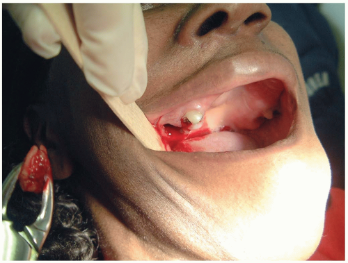 FIGURE 6-4 Bleeding from the socket of a tooth that was removed. (Courtesy of Mark Silverberg, MD.) |
Management
The first step is to insert gauze into the extraction site and have the patient apply direct pressure for 15 to 30 minutes by biting down gently. If direct pressure fails to curtail the bleeding, more aggressive local therapy, consisting of absorbable suture placement and topical hemostatic agents, should be employed. Common topical hemostatic agents include Gelfoam,® Surgicel,® Oxycel,® and bovine collagen products such as Avitene,® CollaTape,® CollaCote,® and CollaPlug.® All can be applied directly to the bleeding socket, and many can be sutured in place. Both Surgicel® and Oxycel® have some bacteriocidal properties and therefore should be chosen in the setting of a concomitant infection. If bleeding still persists, the emergency physician must consider systemic causes of excessive bleeding, such as pharmacologic or inherited coagulation defects. Systemic treatment with fresh-frozen plasma or vitamin K should be used in the setting of an elevated International Normalized Ratio (INR). Factor replacement, desmopressin acetate (DDAVP), cryoprecipitate, or desmopressin should be used for the appropriate factor deficiencies.1,2,3
REFERENCES
1. Matocha DL. Postsurgical complications. Emerg Med Clin North Am 2000;18:549-564.
2. Leonard MS. An approach to some dilemmas and complications of office oral surgery. Aust Dent J 1995;40:159-163.
Related posts:


Stay updated, free articles. Join our Telegram channel

Full access? Get Clinical Tree


