Critical Care
18-1 Acute Respiratory Distress Syndrome
Heatherlee Bailey
Clinical Presentation
Acute respiratory distress syndrome (ARDS) may result from various insults, including trauma, overdose, and infectious processes that promote inflammatory cascades. Patients present with a wide variety of complaints that may include shortness of breath, tachypnea, and hypoxia, followed by rapidly progressive respiratory failure requiring mechanical ventilation. The hypoxemia is often refractory to supplemental oxygen.1,2,3
Pathophysiology
ARDS involves diffuse inflammatory injury to the lungs, with injury to the alveolar-capillary membrane allowing increased permeability. Protein-rich fluid collects in the interstitial and alveolar spaces, impairing gas exchange.2
Diagnosis
The specific diagnostic criteria include acute onset, poor oxygenation as determined by a ratio of 200 or less between the arterial partial pressure of oxygen (Pao2) and the fractional concentration of oxygen in inspired gas (Fio2), and bilateral opacifications on chest radiography. Pulmonary occlusion pressure, or wedge pressure, is less than 18 mm H2O; in other words, there is no evidence of left-sided heart failure1,2,3
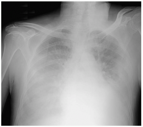 FIGURE 18-1 Bilateral pulmonary infiltrates consistent with, although not diagnostic of, acute respiratory distress syndrome (ARDS). (Courtesy of Mark Silverberg, MD.) |
Clinical Complications
Management
Ventilatory support is critical, with the objectives of improving the oxygenation and not worsening the lung injury by application of lung-protective strategies. High levels of oxygen are commonly used. Increased levels of positive end-expiratory pressure (PEEP) may help to improve gas exchange and lower Fio2 to less than 60%. One of the current approaches is the use of low tidal volumes with high respiratory rates.1 Other therapies include the use of pressure-cycled ventilation, high-frequency oscillatory ventilation,3 and airway pressurerelease ventilation (APRV).
REFERENCES
1. Ventilation with lower tidal volumes as compared with traditional tidal volumes for acute lung injury and the acute respiratory distress syndrome. Acute Respiratory Distress Syndrome Network. N Engl J Med 2000;342:1301-1308.
2. Steinbrook R. How best to ventilate? Trial design and patient safety in studies of the acute respiratory distress syndrome. N Engl J Med 2003;348:1393-1401.
3. Derdak S. High-frequency oscillatory ventilation for acute respiratory distress syndrome in adult patients. Crit Care Med 2003;31[4 Suppl]:S317-S323.
18-2 Disseminated Intravascular Coagulopathy
Heatherlee Bailey
Clinical Presentation
Disseminated intravascular coagulopathy (DIC) leads to a combination of bleeding and thrombosis. Spontaneous oozing at wounds and venipuncture sites are important findings and should prompt laboratory evaluation for DIC. Thrombosis tends to occur in the digits and may lead to gangrene. However, solid organs such as the kidneys may also be affected.1,2
Related posts:


Stay updated, free articles. Join our Telegram channel

Full access? Get Clinical Tree


