A normal ejection fraction does not exclude congestive heart failure (CHF), as CHF can occur secondary to either systolic or diastolic dysfunction.
Nitroglycerin is the initial treatment of choice because it reduces both preload and afterload and rapidly improves patient symptoms.
Consider acute coronary syndrome as the primary precipitant of CHF.
CHF associated with cardiogenic shock maintains a very high mortality rate despite appropriate medical management.
Congestive heart failure (CHF) is the leading cause of hospitalizations in the United States in patients older than 65 years. Once symptomatic, up to 35% of patients will die within 2 years of the diagnosis, and more than 60% will succumb within 6 years. The annual costs of treatment are more than $27 billion and will only increase given the aging population.
Heart failure occurs when the myocardium is unable to provide sufficient cardiac output to meet the metabolic demands of the body. As the myocardium can no longer keep up with the return of venous blood, pulmonary and systemic vascular congestion occurs. Common causes of CHF include myocardial infarction, valvulopathies, cardiomyopathies, and chronic uncontrolled hypertension.
Based on the underlying pathophysiology, heart failure can be divided into systolic and diastolic subtypes. Systolic heart failure develops when a direct myocardial injury impairs normal cardiac contractility causing a secondary decline in ejection fraction (eg, myocardial infarction). Diastolic heart failure develops when impaired cardiac compliance limits ventricular filling (preload) causing a consequent drop in overall cardiac output (eg, left ventricular hypertrophy).
In acute decompensated CHF, the global decrease in cardiac output forces a compensatory increase in systemic vascular resistance (SVR) to maintain vital organ perfusion. This increase in SVR is actually counterproductive and causes a further reduction in cardiac output as the already compromised myocardium now faces an ever higher afterload. The downward spiral continues as myocardial oxygen demand increases because of the increased ventricular workload, resulting in further compromise of the myocardium. Consequent elevations in left atrial and ventricular pressures eventually beget pulmonary edema and respiratory distress.
Decompensated CHF is commonly precipitated by acute coronary syndrome (ACS), rapid atrial fibrillation, acute renal failure, or medication and dietary noncompliance. Other important precipitants to consider are pulmonary embolus, uncontrolled hypertension, profound anemia, thyroid dysfunction, and states of increased metabolic demand such as infection. Cardiotoxic drugs including alcohol, cocaine, and some chemotherapeutic agents should also be considered.
Patients most commonly present with shortness of breath with exertion or at rest with severe exacerbations. Orthopnea, or dyspnea while lying flat, is common as a result of the redistribution of fluid from the lower extremities to the central circulation when the legs are elevated. The increase in central circulation produces a higher pulmonary capillary wedge pressure and secondary pulmonary edema. Attempt to quantify the severity of the orthopnea by asking on how many pillows the patient sleeps and note any changes from baseline. Paroxysmal nocturnal dyspnea occurs when sleeping patients awake suddenly with marked shortness of breath with the need to sit up and hang the legs over the side of the bed or go to a window for air. In certain patients, pulmonary congestion presents rather occultly with a persistent mild nocturnal cough as the only symptom.
Patients may complain of peripheral edema, but this is neither sensitive nor specific for CHF and should prompt an investigation for alternative etiologies. Right upper quadrant pain may occur in patients with hepatic congestion and can be confused with biliary colic.
Always obtain a detailed review of systems to try to identify any possible precipitants of CHF. Specifically, ask patients about antecedent or ongoing chest pain, palpitations, recent illnesses or infections, and medication or dietary changes or noncompliance.
Quickly evaluate patient stability with a careful assessment of vital signs and a focused physical exam. Check the respiratory rate, obtain a pulse oximetry, look for accessory muscle use, and determine whether the patient can speak in complete sentences to assess the severity of respiratory distress. Decreased stroke volume and impaired cardiac output may manifest as tachycardia, a narrowed pulse pressure, or marked peripheral vasoconstriction. Recognize hypotension and/or signs of hypoperfusion immediately and treat as cardiogenic shock.
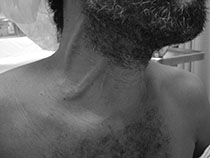
After the initial assessment, focus on signs of total body volume overload. Patients with left ventricular failure typically present with pulmonary signs, including inspiratory crackles, a persistent cough, or a “cardiac wheeze.” Patients with right ventricular failure show signs of systemic congestion. Check for peripheral edema, jugular venous distention, and hepatojugular reflux (an increase in jugular venous pressure with deep palpation of the right upper quadrant) (Figure 15-1). Auscultate the heart for murmurs or gallops. Although often difficult to appreciate in the emergency department (ED), an S3 gallop is highly specific for decompensated heart failure.


