COMPUTED TOMOGRAPHY: BASICS
INTRODUCTION
Computed tomography (CT) has gained widespread acceptance for the evaluation of patients in the emergency setting. Its cross-sectional nature confers significant reliability and accuracy in detecting small anatomic changes, and it has several advantages over traditional two-dimensional radiographs.
CT completely eliminates the superimposition of images outside the area of interest that can occur with plain films. Additionally, its inherent high-contrast resolution allows for distinction between tissues that differ in physical density by less than 1%, permitting much finer detail of anatomic structures. Data from a single CT imaging procedure can be viewed as images in the axial, coronal, or sagittal planes, depending on the diagnostic task. Finally, CT scan was the first technology to marry a computer-based system to a diagnostic imaging technique, enabling a new era of digital imaging. It is a rapid and cost-effective modality that can provide invaluable diagnostic information and characterize the full extent of disease.
CT SCAN IN EMERGENT SCENARIOS
CT scans are completed with the use of a 360-degree x-ray beam and computer production of images. These features allow for cross-sectional views of organs and body structures yielding sharp, focused, and three-dimensional images. CT is most often indicated in patients who may require a form of immediate intervention and is most helpful when the patient has confusing clinical signs and symptoms. In fact, the accuracy of conventional CT has been shown to be as high as 95% in acute abdomen.1 CT is considered the imaging modality of choice in many hospitals for patient triage in the emergency department, and now almost all hospitals have CT scanners on site in the emergency department.
CT technology growth has focused on dramatically increasing the speed of scanning and image reconstruction, both of which are critical in the emergency setting. Rapid speeds have been accomplished by simultaneous data acquisition through more extensive detector arrays, which facilitates the acquisition of multiple “slices” at the same time and, as a result, shortens acquisition time. Currently, CT systems have up to 64 rows of detectors, allowing up to 4 cm to be imaged per revolution (with each revolution being approximately 0.4 seconds).
INDICATIONS FOR CT SCAN
The applications of CT scan in emergencies are numerous, with contributions to diagnoses ranging from trauma to soft tissue infections to pulmonary embolism. The following list outlines a few major indications for CT scan in the acute surgical setting.
1. Imaging in patients with trauma:
a. Significant mechanism of injury and normal clinical examination
b. To localize a hematoma
c. Traumatic aortic injury, as a normal chest film does not exclude aortic injury
d. Nondisplaced subtle fractures
e. To rule out hemorrhage in patients with head trauma
2. Excluding primary intracerebral hemorrhage as a cause of acute stroke (provided it is performed within about a week of onset)
3. Acute abdomen workup
BASIC PRINCIPLES
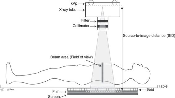
CT involves focused x-ray beams, a detector array, and computerized production of an image. A thin-beam x-ray passes through tissues as an x-ray tube moves around the patient in a continuous arc. The detectors then convert the received beam into an electrical impulse (Figure 4–1). The intensity of this impulse depends on the number of x-rays that have passed through intervening tissue. A computer calculates the amount of beam absorption for each voxel (voxel is a combination of “volume” and “pixel” and represents a value on a regular grid in three-dimensional space) and generates the final CT images. In this way, CT uses computer-processed x-rays to produce tomographic images, or “slices,” of specific areas of the body. Each tomogram is measured in millimeters, which allows for the accurate localization of objects in that section. This is in contrast to conventional radiographs that superimpose all of the structures within a given field of view.
Figure 4–1 Schematic representation of a CT scanner.
Modern scanners generate images at axial or transverse planes, perpendicular to the long axis of the body, and can also reformat the data in various planes or even as volumetric (3D) representations of structures. Three-dimensional images are made using digital geometry processing from a large series of conventional x-ray images that have been taken while the device rotates around a single axis.2,3 This process creates sets of data that can be changed using a process known as “windowing.”
 Window Level and Hounsfield Units
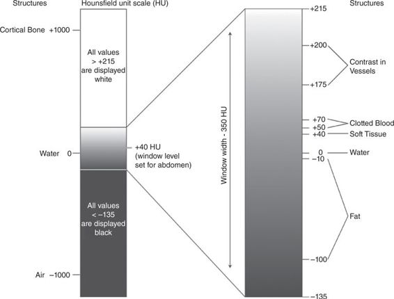
A CT image is composed of a matrix of thousands of pixels, and each of these pixels is assigned a CT number from –1000 to +1000 Hounsfield units (HU). A Hounsfield unit is a measure of the density of a structure on CT4 and depends on how much of the x-ray beam is absorbed (Figure 4–2). A range of densities is called a window, or window-width setting, and the number within that range that is arbitrarily chosen to be the center of the gray scale is called the window level. Windowing allows us to focus on certain tissues within the images to facilitate visualization of different body structures based on their ability to block the x-ray. Tissues of interest can be assigned the full range of blacks and whites, rather than a narrow portion of the gray scale. This technique enables us to identify subtle differences in tissue densities. As a routine setup, most of the reading stations include windows that are optimized for lung, brain, blood, and bone.
Figure 4–2 CT window level and tissue range of Hounsfield units.
 Phases of Acquisition
Phases of acquisition depend on the time to acquire images following the injection of contrast material. This provides additional information in patients for differentiating vascularized structures that need further evaluation. For example, arterial phase acquisitions are performed beginning 20 to 30 seconds following intravenous injection of contrast material, while portal venous phase acquisition begins 70 to 90 seconds after injection. In some cases, there is a need to acquire images during both phases, especially for vascularized structures, e.g., liver or kidney lesions. Delayed images are acquired beginning 4 minutes after injection.5
CONTRAST MATERIAL
Contrast agents have been used for the imaging of anatomic boundaries and to explore normal and abnormal physiologic findings. These agents are administered to increase the differences in density between two tissues. Contrast materials improve the spatial and temporal resolution of multidetector CT, which allows previously highly technically demanding clinical applications such as CT angiography to be practiced on a daily basis.6–8 Accordingly, multidetector CT urography is performed instead of conventional intravenous urography, and functional CT imaging has become a routine clinical protocol.
Radiocontrast agents are typically iodine or barium compounds. As the contrast agents have the potential to meaningfully alter patient physiologic features, produce immunologic reactions, and affect patient outcomes, it is important that all physicians have an appreciation for the indications and selection of these agents. Iodine-based contrast media are often used due to their water solubility and limited side effects and are classified as ionic or nonionic. Ionic agents were developed first and are still in widespread use depending on the requirements, but their use may result in a higher rate of contrast-induced adverse effects. The adverse reactions range from minor dermatologic reactions to severe and life-threatening anaphylaxis or renal failure. Organic (nonionic) agents have fewer side effects, as they do not dissociate into component molecules.
Modern iodinated contrast agents can be used almost anywhere in the body. Most often they are used intravenously, but for various purposes they can also be used intra-arterially, intrathecally, and intra-abdominally, into most body cavities or potential spaces.
Examples of indications for iodinated contrast are:
• Angiography (arterial investigations)
• Venography (venous investigations)
• VCUG (voiding cystourethrography to evaluate the bladder and urethra)
• HSG (hysterosalpingogram to evaluate the uterus and fallopian tubes)
• IVU (intravenous urography to evaluate the ureters)
 Intravascularly Administered Contrast Material
CT scans can be performed with or without the intravenous administration of iodinated contrast material, but in general they yield more diagnostic information when contrast can be used. Intravascular administration of contrast is by far the most common use of iodinated contrast media and can be either intra-arterial or intravenous. The intra-arterial route is the method of choice when diagnostic angiography and catheter-directed arterial intervention, such as percutaneous angioplasty and stent placement, are required. However, intravenous contrast injection for CT scanning is the most common use of iodinated contrast media, secondary to its ease of administration.
The dose and the rate of contrast material administered depend on the working diagnosis. In general, 120 mL of iodinated contrast material injected at a rate of 2 mL per second is adequate. Images are usually obtained during the portal venous phase of enhancement, beginning approximately 70 seconds after injection.
For imaging of specific vascular pathology, for example, aortic aneurysm or dissection, CT angiography should be performed after intravenous bolus administration of 150 mL of contrast material at a rate of 3 to 4 mL per second. Scanning should be initiated within 20 to 30 seconds after initiation of injection.9 Initial precontrast images can be helpful in these scenarios to localize intramural hematoma or impending rupture.
 Orally Administered Contrast Material
Oral contrasts assist in differentiating the gastrointestinal tract from other structures. Usually, 750 to 1000 mL of water-soluble contrast agent containing 3% iodine is administered. The oral contrast is not recommended if high-grade small-bowel obstruction or ureteral colic is suspected, as oral contrast will not reach the site of obstruction, wastes time, adds expense, can induce further patient discomfort, will not add to diagnostic accuracy, and can lead to complications, particularly vomiting and aspiration. In addition, oral contrast material should not be used for CT angiography, because it can interfere with three-dimensional imaging.9
The optimal delay for imaging appendicitis or pelvic disease should be at least 1 hour after oral contrast administration, to allow for optimal opacification of the bowels. Rectal contrast material is not routinely used, although its use has been advocated as an alternative protocol for evaluation of suspected appendicitis or diverticulitis.10
It is important to choose the most appropriate and safest contrast agent based on the clinical scenario. The two most commonly used oral contrast agents are barium and the water-soluble agent gastrografin. The best coating of the membranes, e.g., the intestinal mucosa, is achieved with barium; however, it is not water soluble and can impose certain risks. Accordingly, barium should not be used when any injury to bowel or presence of fistula is suspected. Also, we should avoid barium if abdominal surgery involving opening of bowel is expected. The contrast agent of choice in this setting would be gastrografin, as it can be resorbed by the body.11
SAFETY CONSIDERATIONS
 Contrast Materials
The incidence of adverse reaction is more common after the use of high-osmolarity intravascular contrast agents, with a rate of 15% versus only 3% when using a low-osmolarity agent.10 The etiology of adverse reactions are usually multifactorial, and reactions can be acute or delayed. Acute adverse reactions occur within the first hour after injection. The incidence, severity, and timing of acute reactions vary between different classes of agents. Pain on intravascular injection, nausea and vomiting, rash, and hemodynamic changes are the most common acute reactions.12–14 Severe acute reactions have been reported as pruritus and urticaria, bronchospasm, shortness of breath, hypotension, and cardiovascular collapse. Most of these reactions are probably anaphylactoid in nature, as prior exposure to the contrast agent is not mandatory.15,16
Delayed reactions to contrast agents can manifest with signs and symptoms similar to an acute reaction, such as rash, pruritus, nausea, vomiting, diarrhea, and, occasionally, hypotension. Contrast-induced nephropathy refers to a greater than 25% increase in baseline serum creatinine concentration within 3 days of receiving a contrast agent, after other possible causes of renal failure have been excluded. This process usually has a benign course and is reversible; however, it prolongs hospital stay, increases the need for dialysis, and increases overall mortality.17,18 The risk of contrast-induced nephropathy is closely associated with the dose of iodine used and preexisting renal function.19 Preexisting renal insufficiency is the greatest risk factor, with the probability rising with increasing baseline renal impairment to as high as 50%.20 Adequate hydration, especially with isotonic solution (i.e., 0.9% sodium chloride), and avoidance of dehydration are critical for decreasing the risk of contrast-induced nephropathy.21 The evidence supporting N-acetylcysteine administration, as a free-radical scavenger and local vasodilator after contrast agent–induced nephropathy, is controversial, and a recent meta-analysis demonstrated a beneficial effect of N-acetylcysteine in high-risk patients; however, there was considerable variability in both dosing and efficacy.22–24
 Radiation Dose
The precise risk of malignancy from CT is a topic of ongoing debate. The main concern is the well-established role of radiation in carcinogenesis, even at relatively low doses.25 It is generally accepted that there is meaningful risk of cancer for radiation doses greater than 100 mSv.26 The dose of CT scan that is currently used can be approximately 2 mSv for a head CT to greater than 30 mSv for a multiphase abdominopelvic CT scan; however, this depends on the technique and body site.2,27 The radiation dose for a particular study depends on multiple factors: volume scanned, patient build, number and type of scan sequences, and desired resolution and image quality.28 Given the relatively small risk of CT for a single patient and the relatively high background risk of cancer in the population, there has been no epidemiologic study to yield sufficient power to generate precise risk estimates; however, this will remain an important topic for future research.
The demand for increasingly complex, noninvasive, and rapid diagnostic procedures in emergency situations mandates dose-reduction policies for all patients. To minimize risk, surgeons should ensure that CT is indeed the most appropriate test for a given patient, and radiologists should ensure that each study is justified and the imaging protocols are optimized to answer the clinical question. ALARA (As Low As Reasonably Achievable dose of radiation) principles in CT imaging imply that the right dose for a CT examination takes into account the specific patient attenuation and the specific diagnostic task. For large patients, this indeed means a dose increase consistent with ALARA principles. The low-dose preset protocols are the key to maximizing patient safety.28 For this purpose, radiologists should make every effort to reduce the radiation dose of CT examinations while maintaining diagnostic quality.29,30 An effective strategy is minimizing the scan range of CT examinations as required. Another important consideration is minimizing the number of repeated scanning for multiphase CT protocols; for example, the pre-contrast scanning should be obtained only when diagnostic information on pre-contrast CT images is not obtainable from post-contrast scanning.
IMAGE INTERPRETATION
Image interpretation on CT scan depends on an understanding that tissues absorb x-rays differently based on their tissue density. Accordingly, dense tissues such as bone
Stay updated, free articles. Join our Telegram channel

Full access? Get Clinical Tree


