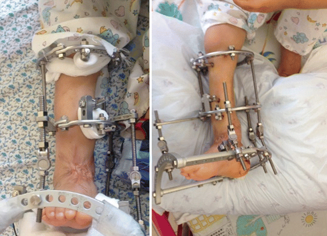
Fig. 15.1
Clinical photos on admission demonstrate severe varus deformity of the right ankle with extensive scarring process around the ankle and distal leg
Radiographs on admission demonstrate subluxation of the ankle joint with loss of distal articular surface of the tibial bone. Reduction of ankle subluxation with correction of deformity was started by a closer manner in the operation room and gradually continued using hinged circular Ilizarov frame. Cancellous bone allograft material was used to fill the distal tibial defect. One week later, the malalignment was gradually restored (Fig. 15.2).


