The pH of the bicarbonate solution falls in proportion to the CO2 concentration, which is measured by the adjacent pH electrode. The Severinghaus electrode has a response time of 2–3 minutes, and needs frequent calibration and temperature maintenance at 37°C. Samples must be free of air bubbles to avoid CO2 diffusing out of solution, and analysis should be undertaken immediately to prevent ongoing red cell metabolic activity increasing the CO2 content.
Cutaneous electrode: A Severinghaus electrode is placed directly upon skin heated to 42–43°C. The heating causes capillary dilatation, enabling CO2 to diffuse out through the skin and into the measuring electrode. Cutaneous electrodes are inaccurate in low cardiac output states and on oedematous skin, and prolonged use can cause burns.
Intravascular electrode: These comprise a needle probe made of CO2-permeable silicon, containing a CO2-sensitive fluorescent dye such as 1-hydroxypyrene-3,6,8-trisulfonate. Fluorescence changes linearly with the CO2 concentration, which is measured by a fibre-optic light source and photo-detector. This gives a real-time measure of CO2 concentration, but is invasive and relatively expensive.
Indirect methods of CO2 analysis include the following.
Calorimetric devices: These comprise an encased pH-sensitive indicator placed onto the endotracheal tube. Expired CO2 creates a visible colour change in the indicator, but no quantitative measure of CO2.
Capnograph: This provides a continuous measurement and display of expired CO2 concentration. It is an essential anaesthetic monitor, which uses infrared spectroscopy. The capnograph underestimates the arterial pCO2 by 0.5–0.8 kPa, although this can be greater in cases of respiratory disease with ventilation perfusion mismatching, or low cardiac output states. They can be either sidestream or mainstream.
Sidestream capnographs continuously aspirate 150 ml·min−1 of gas from a connector at the endotracheal tube. The sample passes through a water trap, then is analysed by an infrared spectrometer before it is returned to the breathing system or scavenged.
Mainstream capnographs incorporate a spectrometer between the endotracheal tube and breathing system. The infrared beam passes directly through expired gas onto a detector on the opposite side. The spectrometer is heated to >37°C to prevent water condensation. 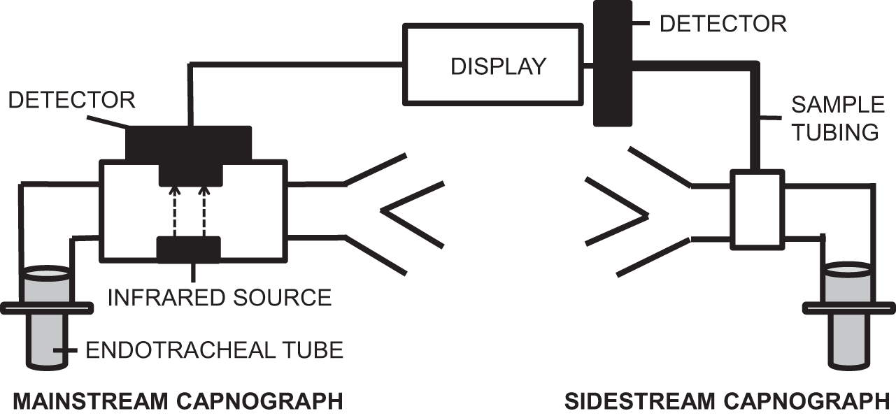
Capnometers: Similar to mainstream capnographs, but they omit the waveform display, displaying only the end tidal carbon dioxide (etCO2) concentration value.
Additionally, mass spectrometry, gas chromatography, Raman spectrometry and the Siggaard–Andersen nomogram can also be used.
Related posts:


Stay updated, free articles. Join our Telegram channel

Full access? Get Clinical Tree


