Chronic Respiratory Failure


Chronic Respiratory Failure


Thomas G. Keens

Sheila S. Kun

Sally L. Davidson Ward


Advancements in pediatric critical care medicine have increased survival from catastrophic childhood illnesses and injuries. However, this improved survival has come at a price. Sometimes, children survive, but with chronic illnesses (1,2,3,4). Children treated in the PICU for acute respiratory failure may emerge with chronic lung disease. A smaller proportion of survivors develop chronic respiratory failure (CRF), and may require long-term ventilatory support (2,5,6,7). Many children already had significant chronic disease when they were admitted to the PICU (5). Thus, their reserve was likely already compromised, making it more likely that these patients may develop CRF (5). Rather than being maintained as long-term residents in the PICU, most of these children can now be cared for in the home, even if ventilatory support is required. Home mechanical ventilation (HMV) is often performed with a relatively good quality of life for many children (8,9,10,11,12). However, children requiring ventilatory support for CRF are, by definition, fragile, have little reserve, and face a 20% mortality rate in the first 5 years after discharge (13). This chapter reviews CRF, including what can be done to attempt to wean patients from long-term assisted ventilation in the PICU, and how to care for the child who requires HMV.

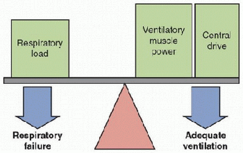
The ability to sustain spontaneous ventilation requires adequate mechanisms that control ventilation, ventilatory muscle function, and lung mechanics. Significant dysfunction of any of these three components of the respiratory system may impair a child’s ability to breathe spontaneously. Respiratory failure occurs when central respiratory drive and/or ventilatory muscle power are inadequate to overcome the respiratory load (Fig. 50.1). For some children, severe lung disease alone (i.e., acute respiratory distress syndrome [ARDS]) may impose a respiratory load so high that it cannot be overcome by normal ventilatory muscles and central respiratory drive. For others, decreased ventilatory muscle power (i.e., neuromuscular diseases) and/or central respiratory drive (i.e., central hypoventilation syndromes or pharmacologic central nervous system depression) may be inadequate to overcome even a normal respiratory load. CRF occurs if the cause of this imbalance is not reversible, and chronic ventilatory support will be required. Once the decision has been made to institute long-term mechanical ventilation in an infant or child with a stable or progressive disorder, a disposition plan is required since the child cannot reside indefinitely in a PICU (8,9,10,11,12,14).
Thus, the approach to managing CRF must be appropriate for home or chronic care facility settings. Chronic ventilatory support in the home is, for many patients, a safe and relatively inexpensive alternative.


FIGURE 50.1. The respiratory balance. In normal individuals, ventilatory muscle power and central drive are more than adequate to overcome the respiratory load, tipping the balance to the right, which results in adequate ventilation. However, when ventilatory muscle power and/or central drive are decreased and/or the respiratory load is increased, or some combination thereof, ventilatory muscle power and central drive may not be sufficient to overcome the respiratory load. The balance will tip to the left, and respiratory failure will result.

The goals of ventilatory support in the home for children with CRF are quite different from the goals of assisted ventilation for children with acute respiratory failure in the PICU (9,15,16,17). The support at home goals include (a) ensure medical safety of the child, (b) prevent or minimize complications, (c) optimize the child’s quality of life, (d) maximize rehabilitative potential, and (e) reintegrate with the family. Medical safety is ensured by attempting to normalize respiratory function. The child’s quality of life is optimized by rehabilitation to as normal a lifestyle as possible and by reintegration into the family. To achieve these goals, it is necessary to adopt a different approach from that employed in the care of children with acute respiratory failure.


RESPIRATORY SYSTEM IN CHILDREN

image Neurologic control of breathing must ensure adequate ventilation to meet the metabolic needs of the body during sleep, rest, and exercise. Ventilation varies with the state of the individual. It becomes less adequate during sleep, and it is less responsive to modulation by chemoreceptor input during rapid eye movement (REM) or active sleep. It is not surprising that sleep is the most vulnerable period for the development of inadequate ventilation in disorders of respiratory control (15,17,18). Immaturity of the respiratory control systems in the infant and young child predispose to apnea and hypoventilation. Furthermore, the infant spends ˜50% of sleep time in REM sleep (in contrast to 15%-20% spent by the adult), and an infant sleeps for a longer portion of the day. Active sleep is associated with greater variation in respiratory timing and amplitude, resulting in periods of inadequate gas exchange. Thus, CRF always includes inadequate gas exchange during sleep.

The diaphragm must perform the work of breathing. Ventilatory muscles of normal strength and endurance may fatigue in the face of increased respiratory loads (19,20). On the other hand, weak ventilatory muscles may successfully perform the work of breathing when pulmonary mechanics are optimal, yet fail when mechanics worsen, as during respiratory infections (21). The factors that predispose to ventilatory muscle fatigue include hypoxia, hypercapnia, acidosis, malnutrition, hyperinflation, changes in pulmonary mechanics that cause increased work of breathing, and disuse. The infant diaphragm also has a significantly smaller proportion of fatigue-resistant muscle fibers and is weaker than the diaphragm of older children and adults (22,23). Consequently, the infant is predisposed to ventilatory muscle fatigue. Underlying muscle pathology will further limit ventilatory muscle endurance. Therefore, infants and children are predisposed to respiratory failure compared with adults, because of differences in the control of sleep and breathing, and decreased ventilatory muscle strength and endurance.


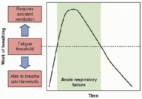
FIGURE 50.2. Acute respiratory failure. Work of breathing (Y axis) increases with worsening lung disease until it exceeds the fatigue threshold, at which point, mechanically assisted ventilation is required (shaded area) until the lung disease improves to the point at which work of breathing falls below the fatigue threshold. Then, the child is able to perform the work of breathing required to breathe spontaneously and he/she can be weaned from mechanical ventilation.


ACUTE RESPIRATORY FAILURE

The clinical picture, etiology, pathophysiology, medical management, and ventilator management of acute respiratory failure are discussed in detail in other chapters of this text. However, aspects of CRF can be contrasted with acute respiratory failure as follows. Acute respiratory failure is most commonly seen in children who experience the abrupt onset of a severe respiratory disorder, such as severe pneumonia or ARDS. This is accompanied by an increase in the respiratory load, which exceeds the ability of the child’s physiology to continue performing that level of work. The ventilatory muscles fatigue while attempting to exert increased effort for breathing (24,25). Thus, the child breathes inadequately, resulting in hypoxia, hypercapnia, and acidosis. If work of breathing is plotted on the ordinate, the fatigue threshold indicates the highest level of work that a subject can sustain indefinitely without fatigue (Fig. 50.2). In adults, this limit for diaphragmatic work is at about 40% of the maximal diaphragm strength, and 60%-70% of combined inspiratory muscle strength (24,25). If the work of breathing that is associated with a respiratory illness remains below the fatigue threshold, a child can continue to breathe spontaneously and acute respiratory failure may not occur. In a typical episode of acute respiratory failure, the work of breathing rapidly exceeds the child’s fatigue threshold.

Most children with acute respiratory failure can be weaned from mechanically assisted ventilation when the work of breathing decreases with recovery from the lung disease. Usually, in the PICU, ventilator settings for children with acute respiratory failure are adjusted to provide the minimal level of support necessary to achieve adequate gas exchange. Weaning is performed in such a way that the patient assumes increasing proportions of the work of breathing. As the primary objective in a child with acute respiratory failure is to wean from assisted ventilation, settings are adjusted so that the child expends all available energy as work of breathing, leaving little or no reserve for other activities. However, this strategy is successful only if the underlying cause of respiratory failure is subsiding.


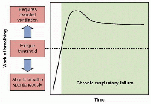
FIGURE 50.3. Chronic respiratory failure. Work of breathing (Y axis) increases with worsening lung disease until it exceeds the fatigue threshold. Mechanically assisted ventilation is required at this point (shaded area). However, the lung disease does not improve to the point that work of breathing falls below the fatigue threshold. Thus, the child is not able to perform the work of breathing required to breathe spontaneously and remains ventilator dependent.


CHRONIC RESPIRATORY FAILURE

CRF implies that a chronic, perhaps irreversible, underlying respiratory disorder is causing respiratory insufficiency that results in inadequate ventilation or hypoxia (16). The diagnosis of CRF is traditionally made once repeated attempts to wean from assisted ventilation have failed for at least 1 month in a patient without superimposed acute respiratory disease or a patient who has a diagnosis with no prospect of being weaned from the ventilator, such as high spinal cord injury (16) (Fig. 50.3).

For purposes of this discussion, the term “prolonged respiratory failure” will be used for children who are difficult to wean from mechanically assisted ventilation but have not yet satisfied the time criteria for the diagnosis of “CRF,” defined as at least 1 month of ventilator dependence. Some patients with prolonged respiratory failure will develop CRF if appropriate therapeutic interventions fail, but others may be able to be weaned from mechanically assisted ventilation. The proper approach to a patient with prolonged respiratory failure must include addressing all barriers to weaning. Keeping the respiratory system balance in mind (Fig. 50.1), therapy should be directed toward reducing the respiratory load, improving ventilatory muscle power, and increasing central respiratory drive as much as possible.


Reduce the Respiratory Load

Reducing the respiratory load requires that pulmonary mechanics be optimized (15). Infection should be treated vigorously with appropriate antibiotics. Aggressive chest physiotherapy, along with inhaled bronchodilators and anti-inflammatory agents, reduces atelectasis and airway resistance by enhancing mucociliary activity and clearing secretions. In infants and young children, respiratory failure is often complicated by increased lung fluid, either interstitial or alveolar edema and diuretics may be helpful. Careful attention to electrolyte balance is required whenever diuretics are used, especially the development of metabolic alkalosis associated with potassium loss.


Increase Ventilatory Muscle Power

Ventilatory muscle power is adversely affected by many conditions commonly present in children with chronic lung disease. The work output of the ventilatory muscles is measured as the generated pleural, airway, or transdiaphragmatic pressures (21). Fatigue of the ventilatory muscles occurs when muscle energy production is hindered (22,24,25,26). Ventilatory muscles cannot perform work if they cannot produce energy. Hypoxia, hypercapnia, and acidosis all decrease the efficiency of muscle energy production, predisposing the muscle to fatigue. Malnutrition decreases oxidative energy-producing enzymes in muscle. Hyperinflation places the diaphragm at a mechanical disadvantage, so that the same amount of muscle tension develops less pressure. Infants have decreased strength and endurance of the ventilatory muscles compared with adults or older children (22,23). If the child has received assisted ventilation for some time, muscle changes may occur from disuse (26). Thus, even a child who does not have a diagnosis of a neuromuscular disorder may have ventilatory muscle dysfunction or fatigue, contributing to respiratory failure. Bronchopulmonary dysplasia, for example, is a primary lung disease associated with hypoxia, hypercapnia, hyperinflation, malnutrition, and infancy, all of which decrease ventilatory muscle endurance. Thus, therapy should be directed toward adequate oxygenation and ventilation, removal of airway obstruction and hyperinflation, adequate nutrition, and ventilatory muscle training (26). Pharmacologic neuromuscular blockade, sedation, and pain medications also decrease ventilatory muscle function. When possible, these medications should be weaned as tolerated. Attention to the optimization of ventilatory muscle function is an important adjunct to the treatment of any child with prolonged respiratory failure.

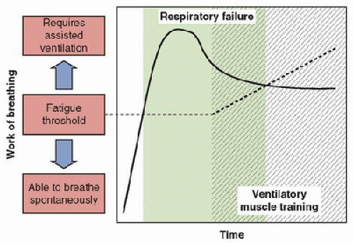
The approach to weaning from the ventilator should be designed to improve ventilatory muscle power in an attempt to raise the child’s fatigue threshold (Fig. 50.4). The desired approach is similar to athletic training of any other skeletal muscle (26). Athletes train for performance by bursts of muscle activity (training stress) followed by rest periods. “Sprint weaning” is analogous to this form of athletic training, and ventilatory muscle training may result. Intermittent mandatory ventilation (IMV) weaning imposes a gradually increasing functional demand on the ventilatory muscles, but it does not provide the alternating stress and rest training pattern. In our experience, some children who have not been weaned from mechanically assisted ventilation by traditional IMV weaning approaches were able to be weaned by sprint weaning, though this may take several weeks.

In a child with prolonged respiratory failure, sprint weaning, or sprinting, is instituted in the following way. Ventilator settings are adjusted to completely meet the child’s ventilatory demands by the use of a physiologic ventilator rate for age and the attainment of normal noninvasive monitoring of gas
exchange (SpO2 ≥95% and end-tidal PcO2 [ETCO2] of 30-40 torr). The goal is to provide total ventilatory muscle rest. The patient is then removed from the ventilator for short periods of time during wakefulness approximately two to four times per day. In some cases, these initial sprints may last only 1-2 minutes. The child is carefully monitored noninvasively during sprints to identify hypoxia or hypercapnia, using pulse oximetry and ETCO2 monitoring. Increased supplemental oxygen, above that required on the ventilator, may be required during sprinting. Guidelines for terminating sprints, such as SpO2 <95% or ETCO2 >45-50 torr, should be provided as written orders. In addition, if the child develops signs of distress such as tachypnea, retractions, diaphoresis, tachycardia, hypoxia, or hypercapnia, the sprint should be stopped. Note that the child with a respiratory control disorder may not exhibit these signs of distress. The length of each sprint is increased daily as tolerated. The physicians should avoid the temptation to increase the sprint length too rapidly, as this often hinders the progress of weaning. Initially, sprinting should be performed only during wakefulness, as ventilatory muscle function and central respiratory drive are more intact during this period than during sleep. Usually a child is weaned off the ventilator completely during wakefulness, before attempting to reduce sleeping ventilatory support. It is important to remember that sprint weaning requires that the child receives complete ventilatory support during rests (27). Because sprint weaning simulates athletic training, better success has been observed with this form of ventilator weaning in prolonged respiratory failure when ventilatory muscle fatigue is thought to be a component. In effect, this technique raises the fatigue threshold, so that a child can perform an increased level of work of breathing, and sustain adequate spontaneous ventilation (26) (Fig. 50.4).


FIGURE 50.4. Ventilatory muscle training. Work of breathing (Y axis) increases with worsening lung disease until is exceeds the fatigue threshold. Mechanically assisted ventilation is required at this point (shaded area). The lung disease does not improve to the point that work of breathing falls below the fatigue threshold. However, ventilatory muscle training may increase the work of breathing that the patient can perform (hatched area), raising the fatigue threshold until it exceeds the work of breathing required. Respiratory failure overlaps ventilatory muscle training until the fatigue threshold exceeds the required work of breathing. Then, the child is able to perform the work of breathing required to breathe spontaneously and can be weaned.


Improve Central Respiratory drive

Central respiratory drive can be inhibited by metabolic imbalance (15,17,18,28). Chronic metabolic alkalosis, for example, decreases central respiratory drive. Thus, electrolyte balance should be maintained, with careful attention to maintaining serum chloride concentrations >95 mEq/dL and avoiding alkalosis. Chronic hypoxia and/or hypercapnia may cause habituation of chemoreceptors, leading to a decrease in respiratory center stimulation, and decreased central respiratory drive. Although methylxanthines have been used to stimulate drive by some clinicians, Swaminathan et al. (29) demonstrated no effect of theophylline on ventilatory responses to hypercapnia or hypoxia in normal subjects. Furthermore, children with central hypoventilation syndrome have chemoreceptor dysfunction, which does not respond to pharmacologic stimulation (15,17,18,28). In general, pharmacologic respiratory stimulants have not been shown to be effective in the treatment of prolonged respiratory failure (29).

This three-pronged approach to children with prolonged respiratory failure may result in successful weaning from mechanically assisted ventilation (summarized in Table 50.1). However, when children remain ventilator dependent for at least 1 month despite appropriate use of the above techniques and the respiratory load has been reduced, ventilatory muscle power has been improved, and central respiratory drive has been increased as much as possible, the cause of respiratory failure may be irreversible, or weaning the child from assisted ventilation may take several months to years. In either case, the diagnosis of “CRF” is made, and chronic ventilatory support will be required (16).


DECISION TO INITIATE CHRONIC VENTILATORY SUPPORT

The decision to initiate chronic ventilatory support may be made electively or nonelectively (30,31,32). In the past, most decisions to begin chronic ventilatory support were made nonelectively. Typically, a child with a preexisting respiratory disorder developed acute respiratory failure from a respiratory infection or pneumonia and progression of underlying disease. The child was intubated, and mechanically assisted ventilation was initiated as a life-saving measure. Subsequently, it was not possible to wean the child from mechanical ventilation. Because it is often emotionally difficult to abruptly stop this therapy in an alert child who might experience severe distress without it, the transition to chronic ventilatory support was made. In this setting, the child and family often did not have the opportunity to discuss this therapeutic option in advance. Thus, the child and family were not really given an informed choice about whether or not to initiate chronic ventilatory support (30,32).

Increasingly, the decision to initiate chronic ventilatory support is being made electively to preserve physiologic function and improve the quality of life. Using this decisionmaking approach, the health care team begins the discussion of options for long-term care, including the varying approaches to chronic ventilatory support, in patients who
can be expected to develop CRF (30,31,32). The most obvious example is the child with a progressive neuromuscular disorder, such as spinal muscular atrophy or muscular dystrophy (33,34). Discussion begins long enough before the anticipated need to allow the child and family to thoroughly evaluate the options, discuss their feelings, and reach a decision. Families need to be informed that a home ventilator is not a guarantee that the child will survive. We found that children receiving HMV via tracheostomy face a 20% mortality rate in the first 5 years after discharge home (13). If the family opts for chronic ventilatory support, then noninvasive ventilatory support is usually initiated first, or a tracheostomy is performed and positive-pressure ventilation (PPV) is started electively before the patient develops major complications of CRF (27,33,34,35). Since hypoventilation is more severe during sleep than during wakefulness, nocturnal assisted ventilation often prevents the development of pulmonary hypertension and other complications of chronic intermittent hypoxia (35). Nocturnal ventilation allows ventilatory muscle rest and improves endurance for spontaneous breathing while awake; therefore, it is actually associated with an enhanced quality of life (27,35). Further, with this approach, the child and family do have the opportunity to make a truly informed decision about whether or not to initiate chronic ventilatory support (30,32). Preemptive initiation of assisted ventilation prior to sleep related alveolar hypoventilation has not been shown to be helpful in Duchene muscular dystrophy, and is not indicated.


TABLE 50.1 APPROACHES TO WEANING CHILDREN WITH PROLONGED RESPIRATORY FAILURE


REDUCE THE RESPIRATORY LOAD


Relieve bronchospasm


Aerosolized bronchodilator


Aerosolized corticosteroids or other anti-inflammatory agents


Remove excessive pulmonary secretions


Chest physiotherapy


If ventilatory muscle weakness, cough assist device


Reduce lung fluid and pulmonary edema Treat pulmonary infections


Diuretics with careful attention to electrolyte balance Antibiotics


Consider aerosolized antibiotics for chronically colonized patients


INCREASE VENTILATORY MUSCLE POWER


Increase ventilatory muscle strength


Eliminate or reduce hyperinflation


Increase ventilatory muscle endurance


Adequate oxygenation


Avoid hypercapnia


Avoid acidosis


Achieve optimal nutrition


Reduce respiratory load (as above)


Train ventilatory muscles to improve strength and endurance


Sprint weaning


IMPROVE CENTRAL RESPIRATORY DRIVE


Avoid hypochloremic alkalosis


Maintain serum [Cl-] ≥95 mEq/dL


Avoid alkalosis


Reset chemoreceptors


Ventilate to adequate oxygenation (SpO2 ≥95%) and ventilation (ETCO2 ≤40 torr)


Avoid respiratory depression


Reduce or avoid central nervous system-depressant medications


Consensus has not been reached regarding the length of time that a child can remain intubated before airway damage from prolonged intubation occurs. Therefore, an “outside limit” has not been reached on the length of time intubation is permitted before performing a tracheostomy. In practice, if a child requires PPV via tracheostomy, a tracheostomy should be surgically placed when the diagnosis of CRF is made.

Jun 4, 2016 | Posted by in CRITICAL CARE | Comments Off on Chronic Respiratory Failure

Full access? Get Clinical Tree

Get Clinical Tree app for offline access