Chapter 33 Childhood emergencies
The child in the emergency department presents a challenge to the busy emergency physician, particularly in a setting where both adults and children are being treated and the general culture is not a paediatric one. Children are different in that they are dependent, developing and growing rapidly (see Table 33.1). They also differ in their spectrum of disease and response to illness.
Table 33.1 Normal respiratory rate and cardiovascular values
Age | Normal respiratory values (breaths/min)∗ | Normal cardiovascular values (beats/min)∗∗ |
---|---|---|
Infants | 40 | 160 |
Preschool | 30 | 140 |
School age | 20 | 120 |
∗ Endotracheal tube size = (age in years ÷ 4) + 4
∗∗ Blood volume = 80 mL/kg; systolic blood pressure = 80 mmHg + (age in years x 2)
Keep in mind that you are managing both the child and the family. Parents may often perceive their children to be sicker than staff assess them to be. In many instances they may prove to be right! It is crucial for a successful consultation to listen to the parents and get a clear understanding of their concerns. At the same time, do not be dismissive of the child; involve him/her in your history taking as a prelude to examination. Parental anxiety and coping skills also need to be assessed and any social disadvantage noted.
In all childhood emergencies, take a careful history and examine the whole child. A child can deteriorate rapidly. This must be anticipated. If there is any doubt about a child’s condition, a paediatrician should be involved and transfer to a paediatric hospital considered.
PAEDIATRIC PARAMETERS
In dealing with a sick child it is important to recognise how physiological parameters change with age and the impact that this may have on the interpretation of observations and management.
IMMUNISATION IN CHILDHOOD
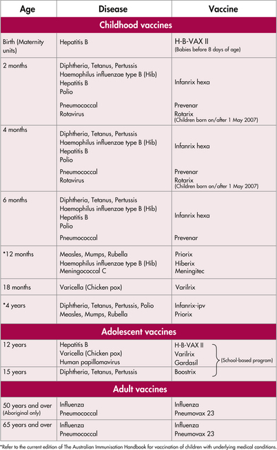
The emergency department offers an excellent chance to give catch-up immunisations. The recommended schedule changes frequently and is listed in the current Australian Immunisation Handbook (readily available from the Commonwealth Department of Health and Aged Care, tel. 1800 671 811, or from the website, see ‘Recommended reading’). There are few contraindications.
RESUSCITATION
In the emergency situation, a child’s condition can deteriorate very rapidly. This is due to:
There may also be a greater risk of acute deterioration in the following cases:
The key to success in managing seriously ill children is early recognition. The outcome from paediatric cardiac arrest is extremely poor, as most children who arrest do so from progressive unrecognised hypoxia or through inadequate or inappropriate resuscitation.
When assessing children, attention should focus on the three major systems—respiratory, cardiovascular and central nervous system (CNS)—to identify the very sick child early.
Respiratory
Increased breathing effort is a sign of increasing respiratory insufficiency and is characterised by: tachypnoea, use of accessory muscles, expiratory grunting, stridor, wheezing, nasal flare, dyspnoea and cyanosis. Other important but often forgotten signs of impending respiratory embarrassment in children are exhaustion and apnoea.
Table 33.2 NSW Immunisation schedule from 1 July 2007
![]() |
Cardiovascular
A child’s haemodynamic status is assessed by examining pulses, capillary refill, blood pressure, mental status and urine output. It must be remembered that in children the ability to compensate through vasoconstriction is strong, hence blood pressure is always one of the last indicators of a decompensated haemodynamic state.
CNS
The level of consciousness in children may be impaired for many reasons. However, invariably in the seriously ill child it is due to hypoxia. Mental state change is one of the most consistent features of shock and generally occurs when cerebral perfusion is being compromised. Neurological assessment therefore involves:
The principles of resuscitation
These are similar in children and adults.
A—airway
In children the goals are to recognise and relieve obstruction, to prevent aspiration and to allow adequate oxygenation.
B—breathing
If breathing is laboured or poor in children, it should be supported with either a bag-valve-mask device or through intubation with mechanical ventilation.
C—circulation
The rate-limiting step in many resuscitative efforts in children is achieving IV access. If unsuccessful after an initial attempt, an intraosseous line should be promptly attempted. An initial fluid bolus to resuscitate a shocked child is 20 mL/kg of normal saline or Hartmann’s solution. A colloid such as 5% albumin can also be used.
D—drugs
When using drugs in resuscitation in children, it is important to be aware that their effect depends on:
IDENTIFYING THE SICK CHILD
Identifying the sick child can be difficult even for experienced staff. The younger the child, the more difficult it can be, particularly when trying to exclude focal bacterial infection. The following simple guidelines will help screen out those children with a greater likelihood of serious illness. The following guidelines provide a useful approach to detecting serious illness in the child under 36 months of age. Combinations of symptoms are even more concerning. No child should be sent home from the emergency department without having had a thorough assessment, appropriate investigations and preferably a period of observation.
Other features of concern include fever, apnoea, convulsions, a petechial rash, cyanosis and the rapid onset of symptoms. Antibiotic use, prolonged symptoms and chronic illness are important features to note. Do not ignore the signs of bile-stained vomiting or blood in the stools alone or in the presence of abdominal pain.
AIRWAY EMERGENCIES
Croup
Croup is viral laryngotracheobronchitis characterised by a barking cough. Often several days of upper respiratory tract symptoms precede the cough. The associated respiratory distress is worse at night and with anxiety in child or parent. Spasmodic croup occurs without accompanying infective symptoms and is often recurrent. Stridor at rest is an indication of severity, while cyanosis is a pre-arrest state. Lateral airways X-rays are not needed and are dangerous as the child is unstable.
A calming environment with the child on the mother’s lap is therapeutic.
If there is moderate or severe airway obstruction, nebulised adrenaline (0.5 mL/kg of 1:1000, maximum 5 mL) can be used as a temporising measure, but rebound after 20 minutes has been well described. Oral dexamethasone (0.15 mg/kg) or prednisolone (1 mg/kg) reduce the risk of intubation and in moderate cases may allow the child to be discharged with review arranged. If this is not tolerated, IV dexamethasone or nebulised budesonide 2 mg can be given. Attention to hydration of the child is important. Humidified air has not been shown to help, and numerous burns occur from steam at home.
Epiglottitis
Epiglottitis is a severe bacterial infection of the epiglottis less commonly seen since Haemophilus influenzae immunisation. The disease is usually rapidly progressive over hours and the child drools, is unable to phonate, does not cough and assumes the ‘sniffing the air’ position to maximise the airway calibre. Cyanosis is a late, pre-arrest state. Interventions should be avoided unless the child is pre-arrest. The initial treatment is rapid transfer to the operating suite for administration of an inhalational anaesthetic and intubation. If there is doubt about the diagnosis, this can be diagnostic. If a respiratory arrest occurs before an inhalational anaesthetic is available, bag-and-mask ventilation, intubation with sedation only or needle cricothyroidotomy is needed. Muscle relaxants must not be used with the anaesthetic as the airway will be lost. Once the airway is controlled, take a swab and commence IV third-generation cephalosporins.
Bacterial tracheitis
This is a rare infection presenting similarly, with acute upper airway compromise. Usually the child appears toxic.
Foreign body inhalation
This causes an acute onset of respiratory distress, often in a child between 6 months and 2 years. If the object is in the upper airway, complete or partial obstruction may be present. If the child is moving air or coughing, removal under inhalational anaesthetic by skilled personnel is advisable. If the airway is completely obstructed, rapid back blows in an infant or the Heimlich manoeuvre in an older child should be performed. Direct visualisation and removal of the object with Magill’s forceps may be possible. If this is not successful, a needle cricothyroidotomy should be performed. Cricothyroidotomy using a scalpel is not recommended for children under 5 years.
RESPIRATORY EMERGENCIES
Asthma
Asthma in childhood is a common condition. Signs of respiratory distress indicating severity include tachypnoea, intercostal recession, use of accessory muscles, prolonged expiration, cyanosis and altered level of consciousness. Exacerbations are treated with salbutamol, initially with spacers or nebulised if moderately severe bronchospasm prevents adequate inspiration. In severe cases, IV salbutamol initially as a bolus, or an infusion if improvement is not maintained, speeds recovery. Except in mild cases nebulised ipratropium bromide is also given. A short course of steroids (prednisolone 1 mg/kg) for 3–5 days is given, by the IV route in severely ill patients. Ventilatory support with either continuous positive airway pressure (CPAP) or intubation, often with assistance with expiration, is needed with apnoea or altered level of consciousness. There is a high attendant risk of barotrauma. Inhalational anaesthetics can also be used as bronchodilators. Long-term preventative therapy aims to reduce the incidence and severity of exacerbations: inhaled sodium cromoglycate and, if needed, inhaled steroids.
Pneumonia
This also presents with respiratory distress. Auscultatory signs may be subtle, especially in the younger child, and X-ray is needed to confirm the diagnosis. Treatment is with antibiotics, either oral or IV.
Organisms causing pneumonia include:
Bronchiolitis
Bronchiolitis occurs particularly in 2–6-month-olds, and is caused by RSV in 75% of cases. After two days of coryza, increasing respiratory distress with tachypnoea, nasal flaring, wheezing and fever is seen and lasts several days. There may be apnoea or difficulty feeding, requiring IV hydration. Chest X-ray is normal or shows hyperinflated lungs with peribronchial cuffing in approximately 50% of cases. Treatment involves oxygen, if required, via nasal prongs, headbox or intubation and attention to hydration. Bronchodilators and steroids are of no proven benefit but a trial of brochodilators may be warranted in the older infant. Patients who are discharged need early review by their local doctor.
Cystic fibrosis patients commonly present with respiratory decompensation related to infection. They have a chronic cough and purulent sputum, and benefit from chest physiotherapy and IV antibiotics.
Chronic lung disease occurs in premature infants and some develop respiratory failure with subsequent upper respiratory tract infection.
Pertussis is associated with spasms of coughing, an inspiratory whoop in older children and then frequently a vomit. Apnoea may be the only symptom in the infant less than 3 months old. Between coughing paroxysms the child is often asymptomatic. There is a leucocytosis of 20,000–50,000 with a predominance of lymphocytes. X-rays are usually normal. Treat with oxygen during spasms, IV rehydration if necessary, and erythromycin if the patient is in the early phase or for prophylaxis of contacts.
THE UNCONSCIOUS CHILD
Remember that the brain is more commonly the target of insult than the primary cause. Always consider and treat those conditions that are correctable, such as hypoglycaemia and hypoxia.
Always examine the whole child in the light of a thorough history.
Immediately
THE FEBRILE CHILD
If the cause is still not evident consider intussusception (may have a ‘cerebral’ presentation). Fever is one of the most common causes of presentation to an emergency department. Most fevers are due to viral infections, but care must be taken to exclude a bacterial infection. Diagnosis can often be difficult, particularly in the younger child where caution is advised. The child without a clear focus presents a real challenge, with pneumococcal and meningococcal infections being the most common infective condition encountered. Various approaches are advised in the literature, ranging from cautious assessment and observation through to aggressive management (see Figure 33.1).
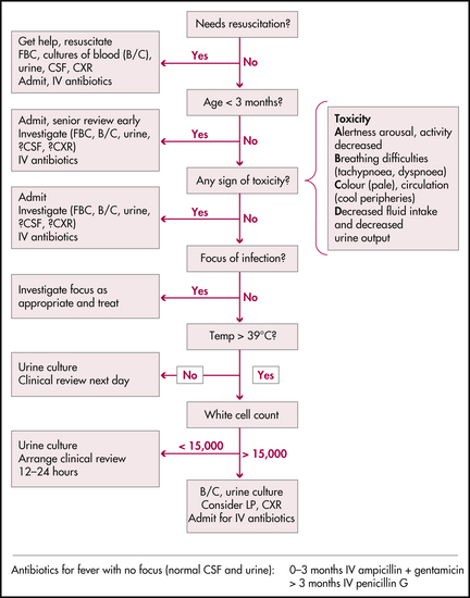
Figure 33.1 Flowchart for an infant with fever
NSW Health, Acute management of infants with fever. Assessment and management: flowchart for child < 3 years old with fever (> 38°C) axillary, p 4
Factors to consider when assessing a febrile child:
A thorough clinical assessment is recommended. Appropriate investigations include a full blood count, blood culture, chest X-ray, urine microscopy and culture, and lumbar puncture in the younger child, where there are associated convulsions, meningism or where the child is on antibiotics. Ensure appropriate observation, review and, if in doubt, seek further consultation. The dilemma of when and where to treat and which antibiotic to use depends upon the individual and the local environment. It is better to err on the side of overinvestigating and treating.
In the acute situation the age of the child, the highest recorded temperature (>39.5°C) and an elevated white cell count with a shift to the left are the most useful guides to underlying serious bacterial infection, in combination with the severity of illness.
Is the child toxic?
Toxicity is determined by the ABCD method. Use this simple system to work out how sick a child appears to be:
A is for arousal, alertness and activity.
B is for breathing difficulties.
C is for poor colour (pale) and poor circulation (cold peripheries).
D is for decreased fluid intake (less than half normal) and decreased urine output (fewer than four wet nappies a day).
Abnormality of any of these signs places the child at high risk of serious illness. The presence of more than one sign increases the risk.
Although temperatures in febrile children fluctuate and may be modified by antipyretics and the technique of measurement, children with high temperatures are more likely to have a serious focus. The risk of acute occult pneumococcal bacteraemia increases from 1.2% for children with temperatures of 39.0–39.4°C to 2.5% for temperatures of 39.5–39.9°C, to 3.2% for temperatures of 40.0–40.4°C and 4.4% for temperatures over 40.5°C. A threshold of 39°C is a reasonable balance between sensitivity and specificity.

Stay updated, free articles. Join our Telegram channel

Full access? Get Clinical Tree