CHEST TRAUMA
CASE SCENARIO
A 22-year-old man complains of shortness of breath and chest pain after a motorcycle crash. He is breathing spontaneously but has decreased breath sounds on the left side. His respiratory rate is 40; his heart rate is 115 beats per minute (bpm); and he has an oxygen saturation of 82% on a non-rebreather mask.
EPIDEMIOLOGY
Motor vehicle crash is the most common etiology of thoracic trauma. Rib fractures occur in about 10% of blunt trauma patients and can indicate injuries to the underlying lung parenchyma, pleura, or abdominal viscera. Among patients with rib fractures, flail chest occurs in about 5% of all trauma patients and confers significant morbidity secondary to the underlying pulmonary contusions. Rib fractures can also be seen after episodes of severe coughing, in repetitive sports (most commonly in rowing and golf), and in the setting of metastatic malignancy. Most mortality in rib fracture patients is actually related to extra-thoracic injuries, including head, craniofacial, and intra-abdominal injuries.1
Pneumothorax is a common problem in acute care surgery, occurring in up to 40% of blunt trauma patients.2 In primary pneumothorax, air can gain entry into the pleural space spontaneously from subpleural blebs, or as a result of diffuse pulmonary disease. Secondary causes of pneumothorax are numerous and include penetrating or blunt trauma, barotrauma from mechanical ventilation, thoracic procedures or instrumentation, and central venous catheter placement. Rare causes include esophageal perforation and catamenial pneumothorax secondary to pleural endometriosis.
Hemothorax occurs in up to 50% of blunt trauma cases,2 but can also result from malignancy, be iatrogenically induced, or develop spontaneously. A list of causes and examples of hemothorax is given in Table 21–1.

PATHOPHYSIOLOGY
Rib fractures are usually uncomplicated and require no specific treatment, but they can also be associated with significant injuries, including pneumothorax, hemothorax, and pulmonary contusion. Fractures of the lower ribs can signal intra-abdominal injuries, notably of the liver and spleen. Despite traditional teaching, there is no evidence of an association between aortic injuries and first and second rib fractures.3
Flail chest refers to a free-floating segment of the chest wall secondary to consecutive rib fractures. The mobile segment moves paradoxically during respiration (e.g., inward during spontaneous inspiration) and is associated with contusions of the underlying pulmonary parenchyma that confer significant morbidity and mortality. Pulmonary contusions are essentially a bruise of the lung, with alveolar hemorrhage and edema related to capillary leak. Direct disruption of capillary and alveolar walls leads to transudation of fluid and blood into the alveolar space and interstitium, with associated impaired oxygen exchange. In addition, there is a change in vascular resistance, leading to decreased blood flow to healthy lung, decreased surfactant, and resultant atelectasis.
Air that accumulates in the pleural space compresses lung tissue, reduces pulmonary compliance, and compromises ventilation and diffusion of oxygen. If air enters the pleural space without the ability to escape, pressure increases, leading to a tension pneumothorax with compression and shifting of the mediastinum, decreased venous return to the heart, and obstructive shock with cardiorespiratory collapse.
Blunt or penetrating injury can result in bleeding from any structure in the chest, including major and minor vascular structures, the chest wall, the lung, or the heart itself, with resultant hemothorax. Bleeding from tumor is usually related to tumor implants on the pleural surface. Rarely, aneurysmal diseases of the aorta or congenital abnormalities, including sequestrations, can present with hemothorax. Hemothorax can also result from intra-abdominal bleeding via a defect in the diaphragm.
The hemodynamic consequences of hemothorax are related to the severity and speed of blood loss, with hemorrhagic shock possible if the bleeding is brisk, especially if injury to a major cardiovascular structure is the culprit. The respiratory consequences of hemothorax, as with pneumothorax, relate to the space-occupying effect of accumulation of blood in the pleural space, with resultant lung collapse, decrease in ventilation, and decreased compliance. Clotting of blood that enters the pleural space is generally incomplete due to the continuous motion of respiration. Clot lysis begins quickly, which increases oncotic pressure, induces transudation of fluid into the space, and enlarges the effusion within a few days, even without ongoing bleeding. Empyma can result if bacterial contamination of the hemothorax occurs. If a hemothorax is not immediately evacuated, fibrin deposition at the pleural surfaces can prevent a portion of the lung from expanding. This “trapped lung” creates permanent atelectasis and persistent decreased ventilation of a portion of the lung.
CLINICAL PRESENTATION
Rib fractures often present with pain that worsens with deep breathing. Crepitance and ecchymosis can be seen as well. A click can sometimes be heard. Decreased breath sounds may reflect splinting, pneumothorax, hemothorax, or pulmonary contusion, with crackles sometimes heard with contusion. Flail chest is, by definition, a freely mobile segment of the chest wall, and can be seen as a portion of the chest wall that moves independently and opposite from the uninjured portion of the chest wall.
The presentation of pneumothorax depends on the amount of air in the pleural space as well as the underlying condition of the lung tissue. Patients with secondary spontaneous pneumothorax often have worse symptoms because of decreased lung function. Most patients with small pneumothoraces present with ipsilateral chest pain, usually sharp and pleuritic, as well as labored tachypnea. Patients may complain of dyspnea, a nonproductive cough, or orthopnea. Tension pneumothorax, by contrast, presents with severe symptoms, respiratory embarrassment, and hemodynamic collapse. Physical examination reveals hyperresonance on percussion, decreased chest wall excursion, and diminished breath sounds on auscultation, and occasionally absent fremitus. Accessory muscles are employed to aid breathing. Distended neck veins can be seen. Rarely, subcutaneous emphysema can be palpated or seen, and mediastinal air can be heard on auscultation (Hamman’s sign). Tracheal deviation away from the side of a tension pneumothorax is a rare and late sign. Arterial oxygen tension can be low, and carbon dioxide tension can be elevated.
Patients with hemothorax present in much the same way as patients with pneumothorax. Indeed, most trauma patients with hemothorax will also have associated pneumothorax. Tachypnea, dyspnea, decreased ipsilateral breath sounds, and tachycardia are common, the latter sign often from blood loss. In contrast to pneumothorax, patients with hemothorax demonstrate dullness to percussion on the affected side, and their jugular veins may be flat given hemorrhagic shock.
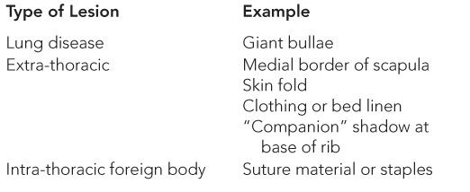
DIFFERENTIAL DIAGNOSIS
 Rib Fractures
Rib fractures are not often confused with other injuries but can easily be missed on initial imaging. Pulmonary contusion can be confused with intrinsic pulmonary lesions such as pneumonia or aspiration.
 Pneumothorax
See Table 21–2.
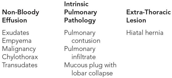
 Hemothorax
See Table 21–3.
WORKUP AND CHOICE OF IMAGING
Chest x-ray is the most commonly employed test in the diagnosis of chest pathology after trauma and a standard part of the trauma evaluation. In fact, along with the focused assessment with sonography for trauma (FAST) examination, plain film of the chest is an adjunct to the primary survey. Plain films allow visualization of large hemothoraces and pneumothoraces, which are important to exclude early in the trauma evaluation.
The radiologic diagnosis of pneumothorax is usually not difficult. Chest x-ray demonstrates a visceral pleural line without distal lung markings. Lateral and decubitus views can be used in equivocal cases. Many clinicians still use expiratory views to detect small pneumothoraces, though their usefulness is controversial. Two centimeters of visible pneumothorax on plain film is equivalent to about 50% of the hemithorax.4 The deep sulcus sign can be seen in cases of tension or in large simple pneumothoraces, especially in supine patients. If the pleura becomes partially adherent or scarred to the chest wall, loculation of the pneumothorax can occur.
Typically, fluid collects in the most dependent portion of the pleural space. Fifty milliliters of blood can be seen on upright chest x-ray in the posterior costophrenic angle, whereas a lateral decubitus film can demonstrate as little as 5 mL.5
With regard to rib fractures, these can often be missed on plain films, with a sensitivity in the anterior-posterior view of as low as 15%.6 Dedicated rib films with oblique views, while more specific, are often not indicated, as detection of additional fractures rarely alters management. Instead, computed tomography (CT) is more useful to rule out additional injuries, especially pulmonary contusions, which plain film fails to detect in up to one third of cases.7,8
Chest CT is the most accurate radiographic tool available to diagnose chest pathology in the stable trauma patient. In pneumothorax, chest CT is the gold standard to which other modalities are compared. Very small collections of gas can be detected, and loculated pneumothoraces are easily and safely evacuated by CT guidance. In addition, any underlying disease of the lung can be assessed and described by CT scan.
For possible hemothorax, CT can detect as little as 2 mL of fluid in the pleural space, and the density of pleural fluid on CT can suggest its origin and acuity. CT can characterize rib fractures and can indicate active bleeding and associated injuries that may require additional attention. In addition, CT is the gold standard for describing pulmonary contusion, seen as parenchymal densities in non-lobar distribution that vary from faint, ill-defined areas to extensive opacification, depending on injury severity. These lesions are typically peripherally located and can be contrecoup from the applied force. Contusion often is not seen on initial imaging and is maximal at 48 hours. If patchy consolidations persist beyond 6 to 10 days, consider wrong diagnosis or superimposed pneumonia, atelectasis, aspiration, or adult respiratory distress syndrome.
Ultrasound (US) is being increasingly used to quickly evaluate patients with acute conditions. A high-frequency linear transducer is employed to scan the anterior chest. In one study, US was equally specific and more sensitive than standard chest x-ray (CXR) in the diagnosis of pneumothorax after trauma.9 Newer studies confirm the utility of US in the diagnosis of rib fractures10 and in differentiating between pathologic and traumatic fractures.11 Hemothorax is echoic on ultrasound and can detect as little as 20 mL of fluid. US performed in the emergency department has a sensitivity of 12.5% to 96.2% and specificity of 98.4% to 100% for all but very small hemothoraces compared to CT.12–14 A scan of the chest can be easily incorporated into the FAST examination as part of the trauma evaluation to evaluate for hemothorax or pneumothorax as a potential cause of shock. However, while US can reliably predict the presence of and even the amount of hemothorax, it is not superior to chest CT and does not provide information about chest wall and mediastinal structures. For these reasons, US should not be considered a definitive test unless CT is unavailable or contraindicated.
Historically, angiography was suggested for first and second rib fractures, out of concern for concurrent aortic injury. Modern evidence suggests, however, that in the absence of a widened mediastinum or other evidence of vascular injury, angiography is not indicated.15
Other imaging modalities are less helpful for chest trauma in the acute setting. Bone scan can play a role in stress fractures related to sports or repetitive injuries, which are often not visible on plain x-ray imaging; however, such a study in the emergent patient is impractical. Magnetic resonance imaging (MRI) is difficult to interpret and often not a reasonable option in acute trauma but can be useful in stress fractures, though even less so than bone scan.16
Table 21–4 compares the sensitivity and specificity of various radiological modalities in the diagnosis of pneumothorax.

Abbreviations: CT, computed tomography; CXR, chest x-ray; US, ultrasound.
IMAGING FINDINGS
 Plain Film
See Table 21–5.
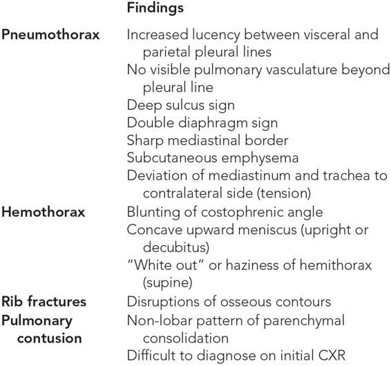
Abbreviation: CXR, chest x-ray.
Normal Chest Film
In a normal chest film, the pleural line is flush with the chest wall, with visible vessels seen extending to the margin of the pleura. As detailed in the following, upright chest films are more helpful than supine films in detailing pathology (Figure 21–1).
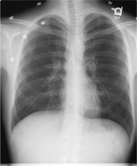
Figure 21–1 Normal postero-anterior chest x-ray. Note lung markings visible to the periphery, and the absence of a clear pleural line.
Pneumothorax
The most important finding in pneumothorax on radiography is the visible white visceral pleural line separated from the parietal pleura by gas that appears black on x-ray. There will be no visible pulmonary vessels beyond the pleural line. As little as 50 mL of air may be visible on upright CXR, while up to 500 mL is required to be seen on supine films. A lateral decubitus film can detect as little as 5 mL of gas.18 Often, the border of the mediastinum is sharply defined, and the “double diaphragm” sign can be seen as a lucent stripe above the ipsilateral diaphragm. Secondary signs seen on CXR that should raise suspicion for pneumothorax include pneumomediastinum and subcutaneous emphysema. In a tension pneumothorax, deviation of the trachea to the contralateral side is seen (Figures 21–2 through 21–5).

Figure 21–2 Upright chest x-ray demonstrating right pneumothorax. Note the clear visceral pleural line (white arrows) and the lucency in the right chest with absent lung markings (asterisk). A deep sulcus sign is also visible (black arrow), with the right hemidiaphragm depressed and sharply demarcated.
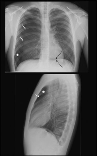
Figure 21–3 Postero-anterior and lateral chest x-ray with right pneumothorax. Note the pleural line (white arrows), lucent hemithorax (asterisk), and double diaphragm sign, identifiable as a lucency in the contralateral hemithorax (black arrow). In the lateral view, the pneumothorax is best visualized as a pleural line and lucency anteriorly.
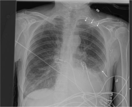
Figure 21–4 Upright chest film revealing a left hydropneumothorax. Note the pleural line at the left apex and base (arrows). There is haziness outside this line, rather than the typical black lucency, because the patient has a concurrent pleural effusion.

Figure 21–5 Chest film showing a small loculated left pneumothorax (arrow) in the setting of pleural scarring.
Hemothorax
Fluid collects in dependent regions and can be seen on upright CXR as a blunting of the normally sharp costophrenic angle with a characteristic concave upward meniscus. The amount of pleural effusion can be estimated based on standard frontal and lateral radiographs. Though rarely an option in trauma, as little as 5 mL can be seen on decubitus films, while 75 mL are needed to obscure the posterior costophrenic sulcus on lateral film, and 175 mL is necessary to obliterate the lateral costophrenic sulcus on an upright film.19 As little as 175 mL of fluid can be seen on supine films, sometimes forming apical caps that disappear in the upright position, but it is not uncommon to miss as much as 500 mL on supine films.20 More often, supine films demonstrate a diffuse haziness caused by layering of blood in the hemithorax. In general, upright and decubitus films are more helpful than supine films in evaluating hemithorax (Figures 21–6 through 21–9).
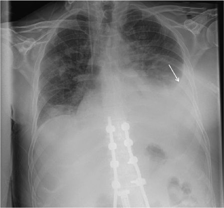
Figure 21–6 Upright chest film in a patient with a left hemothorax. Note the meniscus in the left chest (arrow).
Figure 21–7 Supine chest film in a patient with left hemothorax. Note that in contrast to the upright film in Figure 21–6, no meniscus is visible. Instead, hemothorax is recognizable only as a diffuse haziness of the left hemithorax.
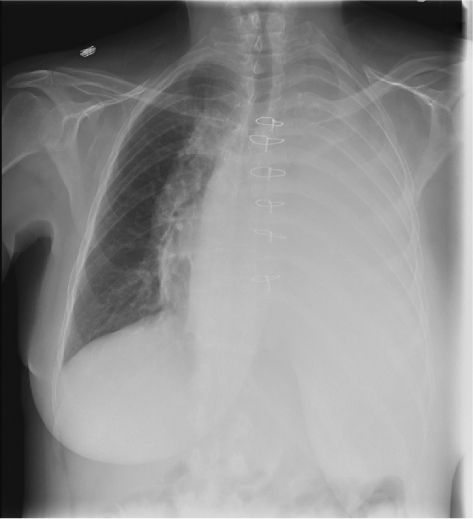
Figure 21–8 Complete “white out” of the left chest in the setting of a large left hemothorax. White out secondary to effusion or hemothorax can be difficult to distinguish from obstructive lung collapse. The key to differentiation is in the position of the trachea. Note that in this film, with hemothorax, the trachea is still midline, with slight contralateral deviation at the level of the carina. In lobar collapse, the trachea is deviated toward the side of the white out.
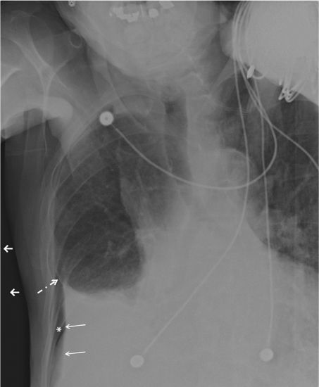
Figure 21–9 Left lateral decubitus film revealing a right hemopneumothorax. Notice the fluid layering in the right base (yellow arrows), as well as lucency (asterisk) and a pleural line (dashed arrow) indicative of concurrent pneumothorax. Decubitus and upright films are optimal for elucidating the presence of fluid in the chest.
Rib fractures are seen as disruptions of the normally smooth contour of the osseous borders but can easily be missed if extremely lateral or subtly displaced (Figures 21–10 through 21–12).
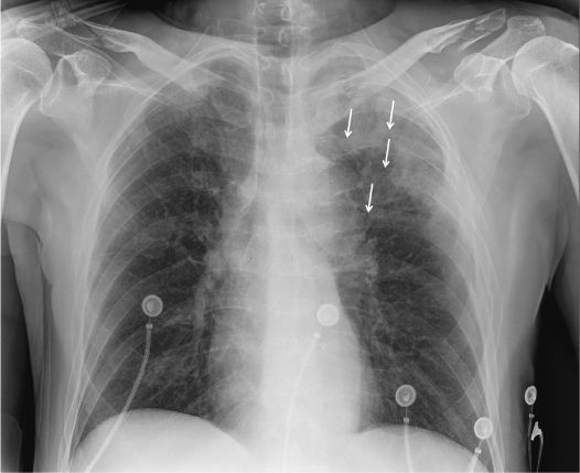
Figure 21–10 Plain film with multiple left-sided rib fractures (arrows). Fractures are identified by a disruption in the normally smooth contour of the rib, with interruption of the cortical line. Note the haziness in the area of the fractures, consistent with the underlying pulmonary contusion.
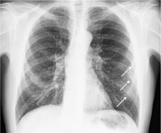
Figure 21–11 Subtle finding of left rib fractures, identifiable by the abnormal contour of the ribs (arrow).
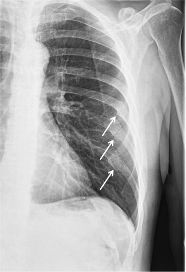
Figure 21–12 Repeat chest film in the patient from Figure 21–11, months later. The fractures are much more obvious after healing (arrows).
Pulmonary Contusion
Pulmonary contusion is difficult to diagnose on initial CXR, as 24 to 48 hours are usually required to develop visible opacities. When seen, contusion is represented by a non-lobar pattern of increased density of varying degrees, depending on the severity of injury (Figure 21–13).
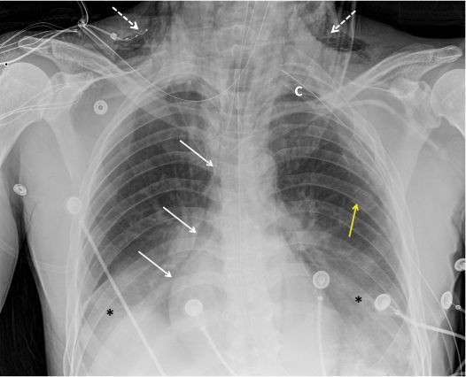
Figure 21–13 Multiple plain film findings in a patient with chest trauma. Note the rib fracture (yellow arrow) and haziness at the pleural bases, consistent with bibasilar pulmonary contusions (asterisks). Subcutaneous emphysema (dashed arrows) is seen, which is a frequent finding in pneumothorax. There is also pneumomediastinum (solid arrows), identified by lucency along the heart border extending cephalad, also consistent with a history of pneumothorax. A left chest tube has been placed (C), with good expansion of the lung.
 CT Scan
See Table 21–6.


