(1)
University of Texas Medical Center, Houston, TX, USA
Keywords
Penetrating neck vascular injuries (PNVI)Blunt neck vascular injuries (BNVI)Gunshot wound (GSW)Stab wound (SW)Carotid injuriesVertebral arteriesInternal jugular veinsTracheaEsophagusPharynxSeatbelt signIntroduction of the Problem
The complex anatomical relationships within a small area make the diagnosis and management of both penetrating (PNVI) and blunt neck vascular injuries (BNVI) challenging. Radiographic evaluation continues to evolve, with a shift from invasive to noninvasive diagnostics. Despite advances in both diagnosis and therapeutics, the optimal management of neck injuries remains a matter of active investigation.
The epidemiology of penetrating and blunt vascular injuries to the neck is distinctly different. Among penetrating injuries, firearms are responsible for about 43 %, stab wounds for about 40 %, shotguns for about 4 %, and other weapons for about 12 % [1]. Overall, about 35 % of all gunshot wounds (GSWs) and 20 % of stab wounds (SWs) to the neck cause significant injuries, but only 16 % of GSWs and 10 % SWs require surgical therapy. Even though transcervical GSWs cause significant injuries in 73 % of victims, only 21 % require surgery [2].
Blunt vascular injury to the neck follows a distinctly different epidemiology. Although BNVI is common, when cervical spine injuries are excluded, injuries to the remaining structures are rare. Though uncommon, blunt cerebrovascular injuries to the vertebral and carotid arteries can be associated with significant lethality. With increased appreciation and availability of noninvasive diagnostics, the rates of these injuries are now between 1.0 and 2.0 % [3–10].
History of Care
Historically, open surgical techniques were utilized for both diagnosis and treatment of cervical vascular injuries. In this context, the division of the cervical region into three anatomical zones facilitated algorithms for evaluation and operative planning (Fig. 17.1). Zone I comprises the area between the clavicles and the cricoid cartilage. Critical structures include the innominate vessels, the origin of the common carotid artery, the subclavian vessels and the vertebral artery, the brachial plexus, the trachea, the esophagus, the apex of the lung, and the thoracic duct. Surgical exposure in zone I can be difficult because of the presence of the clavicle and bony structures of the thoracic inlet. Zone II comprises the area between the cricoid cartilage and the angle of the mandible and contains the carotid and vertebral arteries, internal jugular veins, trachea, and esophagus. This zone is more accessible to clinical exam and surgical exploration than the other zones. Zone III extends between the angle of the mandible and the base of the skull and includes the distal carotid and vertebral arteries and the pharynx. The proximity to the skull base makes zone III structures less amenable to physical exam and difficult to explore. Overall, zone II is the most commonly injured area (47 %) after PNI, followed by zone III (19 %) and I (18 %) [1]. In 16 %, injuries will involve more than one zone [1].
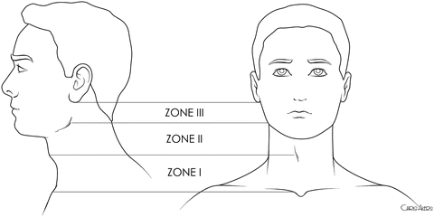
Fig. 17.1
Surgical zones of the neck: zone I is between the clavicle and the cricoid, zone II is between the cricoid and the angle of the mandible, and zone III is between the angle of the mandible and the base of the skull.
Using these zone categorizations to guide operative planning, for many years mandatory operation for all patients with penetrating injuries of the neck that violated the platysma was standard. The rationale was that clinical examination was not reliable. In addition, it has been suggested that routine operation avoided expensive investigations and does not prolong hospital stay [11]. Routine surgical exploration is associated with an unacceptably high incidence of unnecessary operations, however, ranging from 30 to 89 % [11, 12]. Improved appreciation of the reliability of physical exam and “hard” and “soft” (Table 17.1) signs of vascular injury, combined with noninvasive diagnostic capabilities that can be utilized to investigate patients with “soft” signs of injury, has resulted in the use of selective nonoperative management at most centers [1, 13, 14].
Table 17.1
Hard and soft signs of injury after penetrating neck trauma.
Hard signs of injury | Soft signs of injury |
|---|---|
Active arterial hemorrhage | Stable hematoma |
Absent peripheral pulse on affected side | Trajectory |
Expanding hematoma | Dysphagia |
Air or saliva from wound | Pulse abnormality on affected side |
Bruit | Nerve deficit |
Hemoptysis |
GSWs are associated with a higher incidence of significant injuries requiring operation than SWs. However, more than 80 % of GSWs to the neck do not require an operation, and there is strong evidence that these patients can be identified and spared an unnecessary operation [1, 13–16].
Transcervical GSWs are associated with a much higher incidence of significant injuries than GSWs that have not crossed the midline (73 % vs. 31 %) [17]. It has been suggested that all such patients undergo exploration, irrespective of clinical exam [18]. However, many of these injuries, such as spinal cord or nerve injuries, do not require operation. In one prospective study of transcervical GSWs, 73 % of patients had injuries to vital structures, but only 21 % required operation [14, 17]. Several studies have demonstrated that CT angio with thin cuts can reliably identify those patients who do not need further investigation or those who might benefit from specific studies [18–23].
Technique with Personal Tips
Operative Management: Carotid Injuries
The patient is placed in slight Trendelenburg with neck extended and the head rotated away from the side of injury. The patient should be prepped from the chin down to the knees in anticipating the need for a thoracic incision or saphenous vein harvest. The most common incision for exposure of the unilateral carotid artery is a vertical oblique incision made over the anterior border of the sternocleidomastoid muscle (SCM), from the angle of the mandible to the sternoclavicular joint. Retracting the SCM laterally will expose the internal jugular vein, with the carotid artery lying medial and deep to the vein. The vagus nerve is located in the posterior carotid sheath. Division of the facial vein exposes the carotid bifurcation and allows mobilization and control of the internal and external carotids. Simple lacerations of the internal jugular vein or external carotid artery may be repaired, but in most cases veins can be ligated without sequela.
Some zone I injuries may be controlled and repaired through a cervical incision, but proximal zone I injuries may require extension inferiorly into a median sternotomy. Mobilization and superior retraction of the brachiocephalic veins will expose the aortic arch, brachiocephalic artery, and proximal common carotid arteries. Care should be taken to avoid the recurrent laryngeal nerves ascending posterior to the vessels.
Zone III carotid injuries are the most difficult to expose and get distal control. The cervical incision should be extended superiorly into the posterior auricular area and the digastric muscle divided, avoiding injury to the hypoglossal, glossopharyngeal, and facial nerves. Anterior subluxation of the mandible, and further improved by mandibular osteotomy, excision of the styloid process, and removal of the anterior clinoid process improve exposure (Fig. 17.2). Temporary control of uncontrolled zone I or III hemorrhage may be obtained by insertion of an embolectomy catheter through the arterial defect or an arteriotomy and inflation of the balloon.
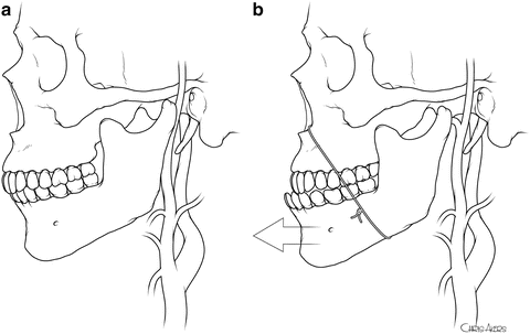
Fig. 17.2
Exposure of zone III carotid injuries.
Most external carotid injuries may be ligated without consequence. Ligation of the common or internal carotid artery can result in devastating neurologic sequelae if collaterals are inadequate. Carotid ligation should be reserved for patients in whom repair is not technically possible, such as injuries at the base of the skull or patients with an established anemic cerebral infarction. In unstable patients, placement of a temporary intraluminal shunt and delayed reconstruction is an option.
Intravenous heparin should be administered if there are no other sites of hemorrhage or intracranial injury, preferably before clamping the artery. Alternatively, local administration of heparin at the site of injury may be used. Adequate collateral flow may not be present. Use of an intraluminal shunt to provide antegrade flow in complex repairs requiring a graft may be wise. Small lacerations may be primarily repaired using an interrupted or running suture after adequate debridement of wound edges. If primary repair is not possible, then a vein or prosthetic patch plasty of the defect is performed. Clean transections, such as stab wounds, may be repaired by mobilization of the proximal and distal artery and primary end-to-end anastomosis if this can be achieved without stenosis or tension.
Many carotid injuries, particularly from GSWs, are not amenable to primary repair or anastomosis after debridement. Reconstruction with either a vein or prosthetic interposition graft is needed. Saphenous vein is preferred for internal carotid artery reconstruction, with some evidence of improved patency and lower infection rates compared to prosthetic graft [24, 25]. Alternatively, reconstruction of the proximal internal carotid may be performed by transecting the proximal external carotid artery and transposing it to the distal transected internal carotid (Fig. 17.3).
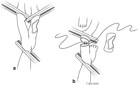
Fig. 17.3
Reconstruction of the proximal internal carotid by transecting the proximal external carotid artery and transposing it to the distal transected internal carotid.
Common carotid artery injuries are best repaired using a thin-walled polytetrafluoroethylene graft, which has a better size match with the native artery and excellent long-term patency. An intraluminal shunt may be used here as well. If associated injuries to the aerodigestive tract have been repaired, well-vascularized tissue such as a sternocleidomastoid muscle flap should be placed between the repairs [26].
If the injury or dissection extends into the distal internal carotid artery (zone III), exposure and repair are significantly more difficult. Ligation or catheter-assisted thrombosis of the injured vessel should be considered in the asymptomatic patient or if the appropriate expertise is not available to perform distal revascularization. Extracranial to intracranial carotid bypass may be performed but requires significant exposure of the intracranial carotid artery. Alternatively, saphenous vein bypass from the proximal internal carotid to the petrous carotid artery or middle cerebral artery has been reported. This technique avoids intracranial dissection of the carotid artery and has been associated with excellent associated long-term outcome and graft patency [24, 27].
Operative Management: Vertebral Arteries
Operative management is almost always necessary when there is severe active bleeding from the vertebral artery. The head is turned away from the injured site and the neck slightly extended. A generous incision is made on the anterior border of the SCM. The fascia is incised and the SCM retracted laterally. The omohyoid muscle is divided, and the carotid sheath is exposed and retracted, while the midline structures are retracted medially. A tissue plane anterior to the prevertebral muscles is opened, taking care to avoid the ganglia of the cervical sympathetic chain. Next the anterior longitudinal ligament is incised longitudinally. The transverse processes are palpated, and the overlying longus coli and the longissimus capitis muscle should be mobilized laterally with a periosteal elevator. The anterior aspect of the vertebral foramen is then best removed with rongeurs to expose the underlying vertebral artery. The artery can then be ligated. The cervical roots are just behind the artery, and care should be taken not to injure them. Blind clamping or clipping should be avoided. Although the artery can be identified between the transverse processes, this is technically challenging. In addition, the venous plexuses can be troublesome.
Another option for rapid control of the proximal vertebral artery is to approach it at the base of the neck where it comes off the subclavian artery. One method is to extend the incision towards the clavicle and transect the SCM off the clavicle, retract the subclavian vein caudal, and transect or retract the anterior scalene muscle laterally. The first portion of the subclavian artery is medial, and it gives off the vertebral artery, the thyrocervical trunk, and the internal mammary muscle. The vertebral artery comes off the superior dorsal aspect of the ascending subclavian artery. When approaching the left vertebral artery, care should be taken not to injure the thoracic duct. The second method is to cut down directly on the clavicle and open the periosteum. The clavicle can be disarticulated at the sternal boarder and resected with towel clamps as a handle. This can be a rapid way of identifying the artery. Repair of the vertebral artery is extremely difficult and is not usually attempted. The collaterals are usually sufficient to not cause an ischemic stroke. When dealing with an active bleeding vertebral artery and obtaining vascular control is difficult, packing is an option if bleeding can be controlled in this manner [28].
Related posts:


Stay updated, free articles. Join our Telegram channel

Full access? Get Clinical Tree


