Cerebrovascular Disease and Stroke


Cerebrovascular Disease and Stroke


John Pappachan

Robert C. Tasker

Fenella Kirkham


Stroke and cerebrovascular disorders (CVD) are important causes of morbidity and mortality in children (1,2). Approximately 3200 cases of stroke occur per year in the population aged between 30 days and 18 years in the US alone. Although outcomes for stroke in children are significantly better than in adults, 20% of children who suffer a stroke die and 50%-80% are left with significant disability. The majority of children with acute stroke syndromes are now admitted to the pediatric intensive care unit (PICU) for endotracheal intubation, mechanical ventilation, and other emergency interventions (3). This chapter concentrates on presentation, pathophysiology, investigation, and treatment. The most recent epidemiologic data are reviewed, and the pathogenesis of hemorrhagic and ischemic stroke syndromes, their risk factors, and conditions that predispose to stroke are described.


PRESENTATION, EPIDEMIOLOGY, OUTCOME, AND COSTS


Overview of Childhood Presentation with Stroke and CVD

Acute focal signs of stroke in childhood can be symptomatic of a variety of pathologies (Table 64.1). The World Health Organization definition of stroke is “rapidly developing clinical signs of focal (or global) disturbance of cerebral function, with symptoms lasting 24 hours or longer, or leading to death, with no apparent cause other than of vascular origin.” Patients whose signs resolve within 24 hours have transient ischemic attacks (TIAs) by definition, but many have recent cerebral infarction or hemorrhage on cerebral imaging (4). Coma is a well-recognized presentation in children with subarachnoid hemorrhage (SAH) or intracerebral hemorrhage (ICH), large middle cerebral artery (MCA) territory infarction, vertebrobasilar circulation stroke, sinovenous thrombosis, bilateral border-zone ischemia, and reversible posterior leukoencephalopathy syndrome (RPLS) (3). Seizures or headache may herald stroke and CVD, particularly sinovenous thrombosis, in children (5). Silent infarction may be demonstrated in up to 25% of children with sickle cell disease (SCD) (6) on magnetic resonance imaging (MRI) and may affect other “at-risk” populations, such as those with congenital heart disease (CHD) (7).


Epidemiology

Stroke and CVD are relatively rare in children but cause disproportionate morbidity and mortality, particularly in those image with critical illness (8). Already among the top 10 causes of childhood death, the prevalence is probably increasing as a consequence of increased recognition and improved sensitivity of diagnostic neuroimaging using MRI or cranial computed image tomography (CT) angiography. In addition, therapeutic advances that now allow children with predisposing conditions such as SCD, CHD, and malignancy to survive may unfortunately increase the risk of stroke.

Neonatal stroke affects 25-30 per 100,000 live births, while epidemiologic data have suggested incidence rates of between 2 and 13 per 100,000 children/year, with one-half to two-thirds being arterial ischemic stroke (AIS) and the remainder being hemorrhagic or other diagnoses (e.g., sinovenous thrombosis without parenchymal involvement). The more conservative estimates predict 3200 cases of stroke per year in a population aged between 30 days and 18 years of age in the US alone. The prevalence of AIS peaks in the first year of life, while SAH is more common in teenagers. Boys are at higher risk, as are those of African heritage. Although childhood stroke is much more common in SCD, this condition does not entirely account for the disparity in rates of stroke by ethnic group.


MECHANISM OF DISEASE


Risk Factors

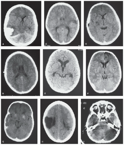
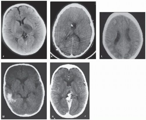
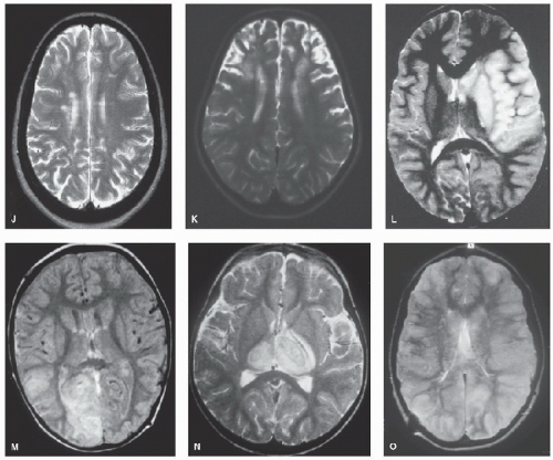
Numerous inherited conditions predispose to CVD and stroke but only 50% of children presenting with an AIS have a known predisposing condition, the commonest of which are heart disease (4,23,24) and anemias, including SCD (4,25) (Figs. 64.2J-L,P-S). Other acquired conditions such as leukemia, brain tumors (Figs. 64.2H,I), genetic disorders (including Trisomy 21 and neurofibromatosis), or single-gene disorders with a highly significant predisposition to stroke (e.g., homocystinuria, Fabry disease, and Menkes disease) are usually obvious at the time of presentation (7). Previously well children may have a recent history of trauma or infection (23,26) (e.g., with varicella—Figs. 64.2A and 64.3A), otherwise investigations may reveal a vasculopathy (23) or hereditary coagulopathy (27).

Hemorrhagic stroke and sinovenous thrombosis may also occur in the context of acquired illnesses (13,26,28), and as in adult stroke, genes that control intermediate-risk factors such as hypertension and hyperhomocysteinemia may be important.


Stroke in Children with Recognized Diagnoses


Congenital Heart Disease

Embolization of air, thrombus, or infected vegetation may occur in children with CHD, particularly during interventions such as cardiac catheterization, or surgery, or secondary to infective endocarditis. Other causes of stroke also need to be considered including sinovenous thrombosis (Figs. 64.1J,K and 64.2M-O) and primary cerebral arterial disease (Figs. 64.3 and 64.4) (4,13,23,24,29). Previously undiagnosed cardiac disease (e.g., a patent foramen ovale) may be diagnosed with contrast echocardiography (4,30,31), but there is currently no convincing evidence for treatment to prevent recurrence and investigation is therefore not essential. Asymptomatic abnormalities of the aortic valve are associated with primary CVD such as cervicocephalic dissection and moyamoya (Figs. 64.3 and 64.4) (4).


FIGURE 64.1. CT scans from children with hemorrhagic and ischemic (arterial and venous) stroke and its mimics. A: Spontaneous intracerebral hemorrhage with midline shift. B: Cortical ischemic stroke after minor head injury. C: Small infarct associated with middle cerebral artery stenosis. D: Larger infarct after recurrence of stroke in C. E: Hydrocephalus and basal ganglia infarct in tuberculous meningitis. F: Frontal infarction associated with moyamoya. G: Calcification associated with moyamoya. H: Old congenital infarct. I: Cerebellar infarction in an unconscious boy. (Continued).


FIGURE 64.1. (Continued). J: Bilateral thalamic infarction in a 4-year-old girl with severe iron deficiency anemia and sinovenous thrombosis. K: Cerebral edema and dense straight sinus thrombosis in sickle cell disease. L: Bilateral watershed ischemia secondary to rapid blood pressure reduction in severe hypertension. M: Calcification of a parieto-occipital pial angioma in Sturge-Weber syndrome. N: Vein of Galen malformation (contrast CT).


Sickle Cell Disease

The best-studied pediatric population with stroke is in children with SCD, among whom most with overt ischemic stroke (Fig. 64.2L) have intracranial, large-vessel disease (Figs. 64.3D-F) with intimal hyperplasia. Without prophylactic blood transfusion, 25% of patients will have suffered a stroke by the age of 45 years; ischemic stroke predominates in childhood, while the majority of adults have spontaneous ICH or SAH secondary to aneurysms (32), although hemorrhage secondary to hypertension and steroid use has also been well documented in children (33). Sinovenous thrombosis (Figs. 64.1K and 64.2Q), posterior leukoencephalopathy, watershed ischemia, and acute ‘silent’ infarction (Figs. 64.2R,S) have previously been under-recognized (34,35,36,37).

SCD is a complex, autosomal recessive inherited disease. The predisposition to large- and small-vessel disease, “silent” (covert) and clinical (overt) infarction, seizures, and cognitive deterioration may be, in part, related to genetic makeup (38) but is probably also linked to environmental factors, e.g., infection, poor nutrition, or hypoxemia (25,34,39,40).


Intermediate-Risk Factors for Childhood Stroke

Infection, Inflammation, and Immune Deficiency. At least one-third of cases of childhood stroke occur in the context of infection, and bacterial and tuberculous meningitis are wellrecognized associations (Figs. 64.1E, 64.2G, and 64.3B) (4,41). Overt immunodeficiency, either inherited or acquired, also appears to be an occasional association with stroke (4). Chickenpox infection is a common trigger (Figs. 64.2A and 64.3A). In SCD, high leukocyte count is a risk factor for stroke, and cerebrovascular episodes are often precipitated by infections. This group of patients is also relatively immunodeficient, secondary to either splenic autoinfarction or surgical removal.

Anemia. Stroke syndromes are well described in the hemolytic anemias, including intermediate forms of thalassemia, hereditary spherocytosis, and paroxysmal nocturnal hemoglobinuria, as well as SCD (42). Iron deficiency appears to be a risk factor for stroke in children (43).

Hyperhomocysteinemia. Classic homocystinuria (deficiency of cystathionine β-synthase) has long been recognized

as an important cause of arterial vascular disease and infarction. Homozygosity for the thermolabile variant of the methylene tetrahydrofolate reductase gene appears to be a risk factor for neonatal and childhood AIS, sinovenous thrombosis, and recurrence in childhood AIS (27). Homocysteine levels are influenced by diet; supplementation of folate, vitamin B12, and vitamin B6 may reduce plasma homocysteine levels, although few available data support efficacy in stroke prevention, and a varied, healthy diet should provide adequate intake.
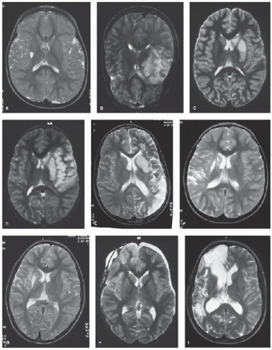


FIGURE 64.2. MRI scans from children with first and recurrent ischemic (arterial and venous) stroke and its mimics. a: Basal ganglia infarct associated with transient cerebral arteriopathy after varicella. b: Temporal infarction associated with dissection. c: Small infarct associated with middle cerebral artery stenosis. d: Larger infarct after recurrence of stroke in c. e: Recurrent infarction after dissection. F: “Silent” posterior watershed recurrent infarction after embolic occlusion. g: Deep white matter infarct in Haemophilus influenzae meningitis. h: Right frontal cortical edema after craniopharyngioma surgery. i: “Silent” recurrent infarction in the posterior watershed territory in the same patient as in h. (Continued).


Figure 64.2. (Continued). J: “Silent” infarction in the deep white matter in sickle cell anemia. k: Bilateral frontal infarction in sickle cell anemia and moyamoya. L: Large middle cerebral artery territory infarct in sickle cell anemia. M: High signal in the right occipital lobe associated with straight sinus occlusion; subacute thrombus is seen as high signal on proton density images. N: Bilateral thalamic infarction in sinovenous thrombosis (same patient as Fig. 64.1J). O: Thrombus in the straight and left transverse sinuses on coronal T1-weighted MRI.

Hypertension. Hypertension is an important risk factor for stroke in young adults and in the elderly but has largely been ignored in the pediatric literature (44). In one series, 54% of children with cryptogenic stroke and 46% of children with symptomatic stroke had systolic blood pressure above the 90th percentile and there was a significant association with cerebral arterial abnormalities (4).

Lipid Abnormalities. In a series of childhood stroke, 9% of those in whom random cholesterol was measured had high levels, while 31% had high triglyceride levels and 22% had high lipoprotein (a), a risk factor for atherosclerosis in adults (4). High lipoprotein (a) is a risk factor for stroke (27).

Disorders of Coagulation. Recognized disorders of coagulation occur acutely in a substantial proportion of children with stroke (27), but up to half resolve within 3 months of the ictus, and the prevalence of inherited coagulopathies is ˜10% in previously well patients. Polymorphisms such as factor V Leiden (which is common in Caucasian populations and is the most frequent cause of activated protein C resistance) and Prothrombin 20210 may be associated with primary and recurrent neonatal and childhood sinovenous thrombosis and AIS (7,19,27,45).

Vascular Adhesion. Adhesion of red blood cells, white blood cells, or platelets appears to be an important mechanism of endothelial damage in SCD and may play a role in stroke of other etiologies, such as moyamoya. There is evidence that different molecular mechanisms are involved in the adhesion of blood cells and platelets to the vascular endothelium in SCD. These mechanisms include adhesive ligands (e.g., von Willebrand
factor) (45) and molecules on the red blood cells and endothelial cell surfaces (e.g., vascular cell adhesion molecule).
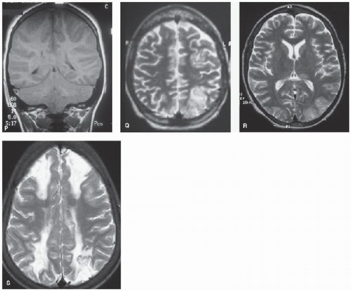


FIGURE 64.2. (Continued). P: Widespread cortical and basal ganglia hypersignal on T2-weighted axial MRI (same patient with sickle cell disease as shown in Fig. 64.1K). Q: T1-weighted coronal images showing marked swelling of the cerebral hemispheres and posterior fossa leading to tonsillar descent and brain death in same patient with sickle cell disease as shown in Figs. 64.1K and 64.2P. High signal is seen in the right transverse sinus (delta sign) due to sinovenous thrombus. R: Occipital edema in distribution compatible with reversible posterior leukoencephalopathy in sickle cell anemia and acute chest crisis. S: Bilateral watershed infarction in sickle cell anemia and facial infection.


Arterial Disease

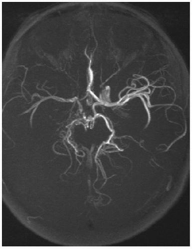
Between 30% and 80% of children with AIS have abnormal findings on cranial CT or MR angiographic studies (Figs. 64.3 and 64.4) (4,46). A less abrupt onset of stroke is characteristic of those with arteriopathy (47). Typical abnormalities include internal carotid artery (ICA) or vertebral dissection, stenosis or occlusion of the distal ICA or MCA, moyamoya syndrome (bilateral severe stenosis or occlusion of the internal carotid arteries with collateral formation), and, occasionally, patterns such as small-vessel vasculitis (4). A diagnosis of vascular disease predicts recurrent AIS (7,18) and moyamoya, and is associated with recurrent stroke and TIAs in patients with and without SCD (7). Stenoses commonly improve or stabilize [transient cerebral arteriopathy] but may progress (40). Spontaneous hemorrhage is most commonly secondary to an AVM (20), but aneurysms and pseudoaneurysms are not unusual and may occur in association with underlying conditions, including trauma (21,26).

Extracranial/Intracranial Dissection. Among children, cervicocephalic dissections are reported in 6.5%-20% of any AIS, and in up to 50% of children with posterior circulation ischemic stroke. Statistics in the US estimate an annual caseload of 56-173 children with stroke due to cervicocephalic dissections, or 1-3 cases per week (48). Risk factors include major and minor trauma (including nonaccidental inflicted injury), infection, anatomical variants of the neck, migraine, hyperhomocysteinemia, and rare disorders such as fibromuscular dysplasia, Marfan or Ehlers-Danlos syndrome, and α1-antitrypsin deficiency (48,49,50). The most frequently affected artery is the ICA, followed by the vertebral artery. In children, anterior circulation dissections are frequently intracranial (60%), affecting the intracranial ICA, MCA, and anterior cerebral artery, although circle of Willis involvement may be


difficult to distinguish from transient cerebral arteriopathy (51). Approximately 80% of posterior circulation dissections are extracranial, and more than half are located within the vertebral artery at the level C1-C2 (52). Multiple dissections are common and are seen in 8%-28% of children.
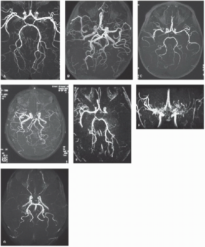


Figure 64.3. MRA from children with first and recurrent arterial ischemic stroke. a: “Transient” cerebral arteriopathy associated with basal ganglia infarct (Fig. 64.2a). b: Progressive arteriopathy associated with falloff in cognitive performance after Haemophilus influenzae meningitis and deep white matter infarction (Fig. 64.2g). c: Persistence of embolic arterial abnormality associated with “silent” recurrent infarction (Fig. 64.2F) in the posterior watershed territory. d: Unilateral arteriopathy associated with recurrent transient ischemic attacks in sickle cell anemia. e: Unilateral occlusion at the origin of the middle cerebral artery in sickle cell anemia with a large middle cerebral artery territory infarct (Fig. 64.2l). F: Bilateral moyamoya collaterals associated with severe stenosis of the distal internal carotid/proximal middle cerebral arteries. g: Bilaterally reduced flow in the middle cerebral artery without obvious moyamoya collaterals in sickle cell disease.


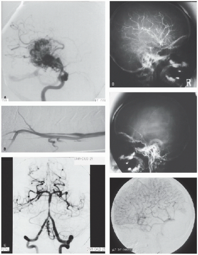
FIGURE 64.4. Conventional cerebral angiography from children with first and recurrent arterial and venous ischemic and hemorrhagic stroke. A: Arteriovenous malformation in an 11-year-old boy presenting with a spontaneous intracerebral hemorrhage. B: Typical “rat’s tail” tapering occlusion in the internal carotid artery in dissection. C: Irregularity of the midsegment of the basilar artery and the origins of both anterior inferior cerebellar arteries. D: Right-sided moyamoya collaterals. E: Paucity of vessels on the left side in the patient in D with right-sided moyamoya. F: Venous phase demonstrating absence of flow in the occluded superior sagittal and straight sinus with multiple collateral vessels draining the hemisphere toward the cavernous sinus (Fig. 64.2M).


FIGURE 64.5. Axial fat-saturated, T1-weighted MRI of the neck in a patient presenting with an acute hemiparesis shows blood in the wall characteristic of dissection.

The origin of the dissection is likely to be a small intimal tear or primary intramural hemorrhage of the vasa vasorum. Arterial dissection results in an intramural hematoma and its variable extension along the course of that artery. The magnetic resonance angiography (MRA) is not usually diagnostic (53

Only gold members can continue reading. Log In or Register to continue

Stay updated, free articles. Join our Telegram channel

Jun 4, 2016 | Posted by in CRITICAL CARE | Comments Off on Cerebrovascular Disease and Stroke

Full access? Get Clinical Tree

Get Clinical Tree app for offline access