1. Obtain the blood pressure in both arms with the patient lying supine.
2. Obtain the blood pressure with a Doppler scan at the dorsalis pedis and posterior tibial pulses on each ankle.
3. Calculate the ABI with the highest ankle pressure divided by the highest arm pressure. (Example: Right BP 140/80 mm Hg; left BP 146/88 mm Hg; right DP 136 mm Hg; right PT 124 mm Hg; left DP 128 mm Hg; left PT 132 mm Hg; right ABI = 136 mm Hg/146 mm Hg [0.93]; left ABI = 132 mm Hg/146 mm Hg [0.90].)
ABI, Ankle brachial index.
PTA may be performed on carotid, aortic, mesenteric, renal, iliac, femoral, popliteal, and tibial vessel stenosis. This procedure may be used alone or in conjunction with stent placement. Major complications after PTA or stenting include bleeding, hematoma, thrombus formation, and intimal tears (disruption of the inner lining of the vessel).3 Other complications may occur specific to the vascular bed being treated such as transient ischemic attack or stroke for carotid stenting and worsening renal failure in treatment of renal artery stenosis.3 Stents are used to compress and hold the plaque against the vessel wall and are associated with longer patency rates of the vessel.3,6 They can also be used to treat an intimal tear in the vessel wall. Stents and balloons with drug coating such as sirolimus, everolimus, and paclitaxel have been evaluated for their potential to reduce in-stent stenosis as a result of their effects on smooth muscle cell proliferation. The goal is to increase long-term patency rates. Long-term data will help to determine whether these will be an effective tool for PAD.6
Atherectomy is a technique designed for removing plaque from the vessel wall with a special rotating blade and suction apparatus. Angioplasty or stenting may follow atherectomy.3 Cryoplasty uses a freezing technique with nitrous oxide inside a balloon for the opening of occluded vessels and theoretically carries the advantage of less risk of intimal hyperplasia, vessel recoil, and dissection.5
After these procedures, patients are monitored for recovery from IV sedation and for bleeding and hematoma formation at the puncture site.3 Distal pulses are assessed bilaterally to detect any change in blood flow that may be related to formation of an embolism or thrombus for procedures that involve the abdominal vessels or extremities.3 These pulses should be compared with the baseline pulses documented before the procedure. Intake and output should be monitored closely, and adequate hydration should be maintained after any procedure with IV contrast. IV contrast can be toxic to the renal system, leading to contrast-induced nephropathy; this is characterized by an increase in creatinine of 25% from baseline within 48 hours of contrast administration. This is of greater concern in patients with preexisting diabetic nephropathy, with the incidence occurring in up to 50% of patients. Treatments such as additional IV fluids, N-acetylcysteine, sodium bicarbonate, and fenoldopam can be used to provide additional protection against contrast-induced nephropathy.7,8
Bed rest is maintained for 6 to 8 hours after the procedure with the extremity in a straight position to prevent bleeding at the puncture site. If a closure device is used at the puncture site, the patient may be allowed out of bed sooner.3,9 Any patient who undergoes an arteriogram, angioplasty, or stenting that involves the carotid or cranial circulation should undergo frequent neurologic assessment after the procedure. Special protection devices are used during angioplasty and stenting to trap any free-floating particles of plaque or thrombus that may be dislodged during the procedure. These devices serve to minimize postprocedural complications such as stroke.3
Fibrinolytic therapy is used when an embolus or thrombus has occluded a vessel. Special catheters are placed in the area of the thrombus, and agents such as alteplase, tenecteplase, or reteplase are used to lyse the clot.3 This process can be done by initial bolus and then completed via infusion and may take hours for complete lysis of the thrombus. These patients need close observation throughout the infusion for signs of bleeding, bruising, anaphylaxis, hematoma at the puncture site, and hematuria. Blood pressure should be monitored closely to decrease the risk of cerebral hemorrhage.10 Assessing for signs of cerebral bleeding is critical. If treatment is needed, aminocaproic acid (Amicar) can be used to inhibit the fibrinolytic process.4 Frequent assessment of the limb is also needed as reperfusion occurs. As the limb reperfuses, pain may actually worsen initially as microemboli break away from the thrombus and move distally to smaller vessels. As the infusion continues, pain improves as these emboli are dissolved. Frequent laboratory work includes serial monitoring of complete blood count, fibrinogen, prothrombin time and international normalized ratio (PT/INR), and partial thromboplastin time.11 Periodic assessment in radiology is done to follow the progress of the lytic agent. The infusion is discontinued when lysis is complete, fibrinogen levels drop to less than 100, bleeding occurs that necessitates transfusion, or no response to the agent is found.3
Medications Used in Vascular Surgery
Anticoagulants are among the most commonly used medications in the treatment of the patient for vascular surgery. Unfractionated heparin can be administered before, during, and after surgery. Its actions occur at multiple points in the coagulation cascade to ultimately inactivate thrombin and prevent conversion of fibrinogen to fibrin. Heparin has a short 60- to 90-minute half-life and may be administered IV or subcutaneously. The response to heparin is measured with the activated partial thromboplastin time (aPTT) and is targeted at 1.5- to 2.5-fold greater than normal to obtain a therapeutic response and prevent thromboembolism.12 Complications associated with the use of heparin include increased risk of bleeding and heparin-induced thrombocytopenia (HIT). Platelet counts should be monitored for decrease of 40% to 50% from baseline or any decrease to less than 100,000. If HIT develops, heparin must be discontinued, and alternative anticoagulants should be used.12 Protamine is the antidote for heparin; its action occurs within 5 minutes of administration. Care must be taken to avoid overly rapid administration of protamine. When administration is too rapid, side effects can include hypotension, pulmonary hypertension, shortness of breath, and flushing. The usual target dose for reversal is 1 mg of protamine for every 90 units of heparin.12
Low-molecular-weight heparins (LMWHs) such as enoxaparin (Lovenox), dalteparin (Fragmin), and tinzaparin (Innohep) may also be used in the care of the patient for vascular surgery. These drugs are administered subcutaneously and have a significantly lower molecular weight than unfractionated heparin, which gives them improved predictability in the dose response and a longer half-life. This advantage greatly reduces the need for laboratory monitoring. If testing is needed, antifactor Xa level is the test of choice for monitoring. The LMWHs are administered subcutaneously and have a significantly lower incidence rate of HIT associated with their use; they are primarily used to prevent thromboembolism after surgery but are also approved in the treatment of deep vein thrombosis (DVT) and pulmonary embolism (PE). They may also be used to bridge patients before and after surgery who require long-term anticoagulation with warfarin (Coumadin) or other anticoagulants. The decision to bridge is based on diagnosis and thromboembolic risk. Patients with renal disease may need a dose reduction depending on the severity of their disease. Complications are similar to those of unfractionated heparin. Laboratory testing should be monitored for signs of HIT, although it occurs much less frequently with the use of LMWH.3,12 New information regarding bridging indicates that less bridging may be a better option in some patients. New evidence from the BRIDGE trial indicates that bridging may do more harm than good and does not actually decrease the occurrence of thrombosis and may lead to major bleeding especially in patients with low to moderate risk of thrombosis.13 Patients with a venous thromboembolism occurring more than 3 months before surgery or with atrial fibrillation who have not had a recent stroke are usually at low to moderate risk of thrombosis and appear to be safe without bridging. Each patient should be evaluated individually.14 Patients with high risk of thrombosis and bleeding are generally bridged, and the anticoagulant is held periprocedure. If there is a low risk of thrombosis with a high bleeding risk surgery or procedure, the recommendation is to hold the anticoagulant without bridging. For patients with a high risk of thrombosis but a low risk of bleeding or low risk of thrombosis and bleeding, anticoagulants may be continued during the procedure.14
Warfarin is an oral anticoagulant that inhibits vitamin K–dependent coagulation factors and the anticoagulant proteins C and S; it has a half-life of 36 to 42 hours. Monitoring of warfarin is done with the prothrombin time (PT) and the international normalized ratio (INR).12 The PT/INR is laboratory dependent, and specific methods vary among institutions. Caregivers should be familiar with institutional methods. Warfarin is used to treat a variety of thromboembolic disorders, and it can be used to promote long-term patency of infrainguinal bypass grafts, particularly after thrombosis of previously placed grafts. Complications include increased risk of hemorrhage and skin necrosis. Patients receiving warfarin must be counseled to discontinue the drug several days before any invasive procedure to allow time for the PT/INR levels to decrease to normal. Reversal of warfarin is achieved with vitamin K or fresh-frozen plasma.12
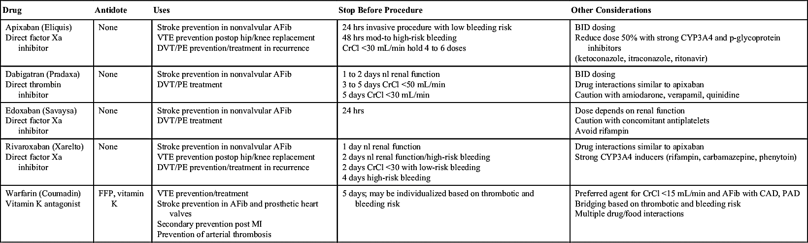
Additional oral anticoagulants are now available and are commonly referred to as direct oral anticoagulants (DOACs). These agents work at specific targets in the coagulation cascade. Table 36.1 summarizes all of the oral anticoagulants currently available including target of action, uses, antidotes, and special considerations for each drug. Rivaroxaban (Xarelto), apixaban (Eliquis), and edoxaban (Savaysa) are direct factor Xa inhibitors. Dabigatran is a direct thrombin inhibitor. They are currently approved for the prevention of stroke in nonvalvular atrial fibrillation. Rivaroxaban and apixaban are also approved for short-term thromboprophylaxis after elective hip or knee surgery. All three agents may be used in the treatment of DVT or PE. There are no reversal agents currently available for these drugs.15 Though these agents are not currently approved for treatment of arterial disease, patients may present on these medications due to comorbidities.
IV direct thrombin inhibitors act at the active site of thrombin. These drugs provide an alternative to heparin in the patient with HIT. Current drugs available in this category include argatroban (Acova) and bivalirudin (Angiomax).16 Desirudin (Iprivask) is a direct thrombin inhibitor administered subcutaneously. It may be used in patients who cannot use fondaparinux (Arixtra) or LMWH. Its only current indication is for venous thromboembolism prophylaxis in hip replacement patients.16
Parenteral factor X inhibitors currently include only fondaparinux (Arixtra). Fondaparinux activates antithrombin III, leading to inactivation of factor X. It is administered subcutaneously and requires no laboratory monitoring. Its primary use is prevention of DVT and treatment of acute coronary syndromes. It may be used in patients unable to use LMWH or unfractionated heparin due to heparin-induced thrombocytopenia. There is no reversal agent for fondaparinux.3,16
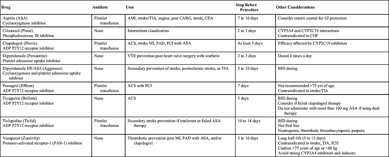
Antiplatelet agents include aspirin, cilostazol (Pletal), clopidogrel (Plavix), dipyridamole (Persantine), dipyridamole ER/aspirin (Aggrenox), prasugrel (Effient), ticagrelor (Brilinta), ticlopidine (Ticlid), and vorapaxar (Zontivity). These agents are summarized in Table 36.2. They may be used in the patient with vascular disease as a preventative measure for myocardial infarction and stroke or as part of medical management for patients after placement of infrainguinal bypass grafts, carotid endarterectomy, and peripheral and carotid stenting.3,12,17 These drugs exhibit an irreversible permanent effect on the platelet for its lifespan and produce a qualitative effect on the platelet measured with the bleeding time. Platelet counts are not affected by these agents. Cilostazol also has some vasodilatory effects and is contraindicated in patients with heart failure. Patients should be counseled regarding the discontinuation of these drugs 5 to 10 days before invasive procedures to decrease the risk of bleeding.3,12,17 Patients at moderate to high risk for cardiovascular events may continue their antiplatelet agents to the time of surgery.17
Glycoprotein IIb/IIIa inhibitors are parenteral agents that also interfere with platelet aggregation and include abciximab (Reopro), eptifibatide (Integrilin), and tirofiban (Aggrastat). These agents are administered by IV infusion. Patients who have had drug-eluting coronary stent placement within the previous 12 months of a surgical procedure may need to be bridged with a GP IIb/IIIa inhibitor to discontinue their antiplatelet drugs for surgery or may be instructed to continue their aspirin and clopidogrel (Plavix) up to surgery.12,17
Preprocedural Assessment
The PAD patient often presents with multiple comorbidities and/or risk factors including advanced age, tobacco use with or without chronic pulmonary disease, diabetes mellitus, impaired renal function, coronary artery disease, hypertension, and congestive heart failure.17 Optimizing the patient’s status before interventional and surgical procedures is vital to the outcome of the postoperative course. Cardiac risk, fluid management, pharmacotherapy, renal evaluation, and medical management of other comorbidities before surgery is vital to patient safety.18
The Vascular Study Group of New England Cardiac Risk Index (VSG-CRI) was developed as a means of cardiac risk prediction for patients undergoing noncardiac vascular surgery. The index includes nine variables that comprehensively evaluate cardiac risk in the vascular patient population. These variables are listed in Box 36.2. The index has been used to predict the cardiac risk in carotid endarterectomy, lower extremity bypass, endovascular repair of abdominal aortic aneurysm repair, and open infrarenal abdominal aortic aneurysm repair. Even though endovascular procedures may carry a lower risk of a cardiac event, patients should be stratified in the event that an endovascular procedure needs to be converted to an open procedure or additional procedures are required.18 The American Heart Association currently recommends that patients with intermediate or high cardiac risk undergo a cardiac stress test with exercise or pharmacologic agents and an electrocardiogram (ECG). Stress echocardiograms have been shown to be twice as accurate in predicting the postoperative occurrence of cardiac events and may be the best tool for evaluating cardiac risk. Some patients may require cardiac intervention before vascular surgery. Patients considered low risk for a postoperative event will require no further intervention.18 Surgery in patients who have undergone coronary stent placement should be delayed 4 to 6 weeks due to increased risk of stent thrombosis. Dual antiplatelet therapy should be continued before the surgery unless the bleeding risk outweighs the risk of thrombosis. For patients who have undergone placement of drug-eluting stents, surgery should be delayed for at least 6 months if possible.18
Perioperative use of beta-blockers has been associated with decreased morbidity and mortality in the high-risk vascular surgery patient, particularly those undergoing lower extremity bypass or open abdominal procedures.3,18,19 There is evidence that, although cardiac events are decreased, there may be an increased risk of stroke.18,19,20 Incidence of perioperative myocardial infarction ranges from 14% to 17.8% for open abdominal procedures and 5% to 15% for lower extremity bypass procedures.20 Women are at particular risk for complications after vascular surgery and also have lower survival rates. Their responses to beta blockade may also be less beneficial than in men.21 Current literature supports the use of beta-blockers in patients stratified as high risk for elective vascular procedures while using caution with these agents in low-risk patients.18 The benefits of beta-blocker usage in the first 2 years after myocardial infarction include reduction in the incidence of sudden death and the risk of reinfarction.22
Fluid management before surgery is important due to the risk of blood loss that may lead to hemodynamic instability. Impaired fluid balance may lead to cardiac and pulmonary complications as a result of increased vascular permeability and edema. Some patients have been shown to fare better postoperatively with tighter fluid management to reduce the risk of hypervolemia, which can lead to pneumonia, pulmonary edema and longer hospital stays postoperatively.18
Preprocedure evaluation should include medication history to include particularly anticoagulants, antiplatelets, and dietary supplements that may increase risk of bleeding or thrombosis. Patients may be taking a wide variety of medications due to multiple comorbidities. Preoperative control and management of medications for hypertension, diabetes, and chronic pulmonary disease is vital to positive outcomes after surgery. Uncontrolled hypertension can increase the risk of perioperative stroke. Uncontrolled diabetes increases the risk of postoperative infection and contributes to an increase in postoperative morbidity and mortality. Elevations in blood sugar may also lead to osmotic diuresis resulting in volume depletion.22 Patients with chronic pulmonary disease may also be oxygen dependent and need further assessment with pulmonary function testing before surgery as well as pretreatment to maximize their pulmonary status.23 Assess tobacco use and counsel the patient to stop smoking 8 weeks before surgery. Tobacco use increases the risk of postoperative infection and delays healing. Stopping the use of tobacco can also benefit the patient with chronic lung disease.22
Chronic kidney disease (CKD) is another common comorbidity in the vascular disease patient. This may occur because of various pathologies including age-related changes, hypertension, diabetes, or primary renal disease and is defined as glomerular filtration rate less than 60.22 These patients may have anemia, thrombocytopenia, and electrolyte imbalance as well as acid-base disturbances. CKD can contribute to major postoperative complications. Due to the use of contrast for radiologic studies and endovascular procedures, patients with CKD are at high risk for further impairment of renal function and the development of contrast-induced nephropathy. Contrast using gadolinium may be an alternative to decrease this risk. Though very rare, gadolinium can contribute to the development of systemic fibrosis if used in patients with nephrogenic systemic fibrosis.18
Patient assessment includes verification of the surgical procedure and the patient’s understanding of the procedure as well as the type of anesthesia scheduled. Assess circulation in the extremities to establish the baseline for lower extremity and abdominal procedures. Assess and establish the baseline cranial nerve function for cerebrovascular procedures. Instructions in the preprocedure assessment should also include any skin preparation ordered such as special showers with antibacterial soap and withholding oral intake as directed before surgery.22
Evidence-Based Practice
Wiseman and colleagues examined the rates of surgical site infections (SSI) in 49,817 patients who underwent vascular surgery for aneurysm and lower extremity occlusive disease from 2005 to 2012 to determine the occurrence of no SSI, in-hospital SSI, or SSI after discharge. SSI is one of the most common complications occurring after vascular surgery. Factors evaluated that contribute to SSI include female gender, obesity, diabetes, smoking, hypertension, coronary artery disease, chronic obstructive pulmonary disease, dyspnea, neurologic disease, prolonged operative time over 4 hours, American Society of Anesthesiologists class 4 or 5, lower extremity revascularization of aortoiliac procedure, and groin anastomosis. Results indicated that comorbidities were the leading contributor to postdischarge SSI. In-hospital factors predictive of in-hospital SSI were length of operative time and emergency case status.
Related posts:


Stay updated, free articles. Join our Telegram channel

Full access? Get Clinical Tree


