| Test | Purpose |
| FVC (forced vital capacity): Records maximum amount of air that can be exhaled as quickly as possible after maximum inspiration. | Provides an indication of respiratory muscle strength and ventilatory reserve. Often reduced in obstructive disease (because of air trapping) and in restrictive disease. |
| FEV1 (forced expiratory volume in 1 sec): Records maximum amount of air that can be exhaled in first second of respiration. | Effort dependent and declines with age. Reduced in certain obstructive and restrictive disorders. |
| FEV1/FVC: Ratio of expiratory volume in 1 sec to FVC | Provides a more sensitive indicator of obstruction to airflow. Ratio is normal or increased in restrictive disease. |
| FEF25%–75%: Records forced expiratory flow over 25%–75% volume (middle half) of FVC. | This measure provides a more sensitive index of obstruction in smaller airways. |
| FRC (functional residual capacity): Amount of air remaining in lungs after normal expiration. | Increased FRC indicates hyperinflation or air trapping, which can result from obstructive disease. |
| TLC (total lung capacity): Amount of air remaining in lungs at end of maximum inhalation | Increased TLC indicates air trapping associated with obstructive pulmonary disease. Decreased TLC indicates restrictive disease. |
| RV (residual volume): Amount of air remaining in lungs at end of a full, forced exhalation | RV is increased in obstructive pulmonary disease, such as emphysema. |
| DLco (diffusion capacity of carbon monoxide): Reflects surface area of alveolocapillary membrane | DLco is reduced when alveolocapillary membrane is diminished, such as in emphysema, pulmonary hypertension, and pulmonary fibrosis. |
Surgical Procedures
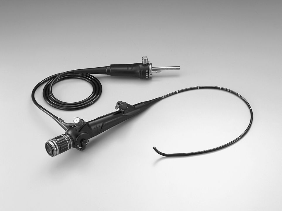
Surgical procedures can be diagnostic or therapeutic in nature. Diagnostic procedures can include bronchoscopy, mediastinoscopy, laryngoscopy, and thoracoscopy. Bronchoscopies are performed to visualize the airway or remove abnormal tissue, mucous plugs, or foreign bodies (Fig. 34.1). They also aid in evaluating lung lesions and staging of lung cancer. Complications can include airway obstruction, hypoxemia, pneumothorax, hemorrhage, or cardiovascular problems such as dysrhythmias or hypotension.
Mediastinoscopy is performed for direct visualization of lymph nodes or tumors at the tracheobronchial junction, subcarinal, or upper lobe bronchi via a lighted scope. The potential for hemorrhage is present because of the close proximity of the innominate vessels and aortic arch to the mediastinoscope. Other complications can include venous air embolism, vagally mediated reflex bradycardia from compression of the trachea or great vessels, airway or esophageal injury including subcutaneous emphysema, chest pain, or pneumothorax. Recurrent laryngeal nerve injury can occur and manifest symptoms such as hoarseness or vocal cord paralysis.7 A laryngoscopy is performed to visualize or biopsy the oropharynx, laryngopharynx, larynx, or proximal trachea. Complications include trauma to the lips, mucous membranes, teeth, or eyes; rupture of the esophagus; hypoxemia; or laryngospasm. Endobronchial ultrasound (EBUS) is a minimally invasive technique that allows the proceduralist to see beyond the lumen of the airway. There are two EBUS systems currently available—the radial probe EBUS allows for evaluation of central airways and accurate definition of airway invasion and facilitates the diagnosis of peripheral lung lesions; and the linear EBUS guides transbronchial needle aspiration of hilar and mediastinal lymph nodes.8
Thoracoscopy is the insertion of an endoscope, a narrow-diameter tube with a camera attachment, through a small incision in the chest wall for examination of the lungs or other structures in the chest cavity without the use of a large incision. It is performed for basic diagnostic (undiagnosed pleural fluid or pleural thickening) and therapeutic procedures (pleurodesis). Complications can include bleeding, infection of the pleural space, and injury to intrathoracic organs, atelectasis, and respiratory failure.9 This procedure is different from video-assisted thoracoscopic surgery (VATS), an invasive procedure that uses a high-level access platform and multiple ports for separate viewing and working instruments to access pleural space.9 VATS can be diagnostic or therapeutic and is used often for biopsy of mediastinal masses, to perform wedge resections, to obtain hemostasis, or to evacuate blood clots. A variety of procedures can be performed via thoracoscopy, from lung volume reduction to a biopsy and excision of mediastinal lesions. Robotic-assisted thoracic procedures can enhance the speed and safety of VATS. Smaller incisions are used for robotic surgery, which may contribute to less postoperative pain and morbidity.10 In one randomized study, VATS was associated with less postoperative pain and better quality of life than anterolateral thoracotomy for 1 year postsurgery.11
A significant advantage of thoracoscopy is that it is minimally invasive and results in less incisional pain. It can also decrease recovery time and length of hospital stay. In some facilities, patients come to the PACU with a small chest tube that is removed if chest radiograph results are clear; the patient is then allowed to go home in a few hours. VATS may be converted to an open surgery if there is an inability to achieve one-lung ventilation, extensive pleural adhesions, uncontrolled or significant intraoperative bleeding, an inability to identify target lesion for biopsy, or technical difficulties with or, rarely, primary failure of video equipment and/or endoscopic instruments.12
Therapeutic thoracic surgeries may include pectus excavatum, chest wall reconstruction, wedge resection of a lung lesion, segmentectomy, lobectomy, or pneumonectomy. Excision of the right lung is less tolerated than removal of the left lung because of the larger vascular bed and breathing capacity. Other thoracic surgeries include lung volume reduction for removal of emphysematous lung tissue or lung transplant.
Perianesthesia Nursing Care after Thoracic Procedures
Admission assessment in the PACU is the same as for any other surgical patient (see Chapters 27 and 28). Common problems that lead to delayed discharge from the hospital for the patient who has undergone a thoracic procedure include inadequate pain control, prolonged air leak, severe nausea, fever, debility, and arrhythmias.6 Postoperative care should target prevention or speedy treatment of these complications. Some specific issues for the patient after thoracic surgery are discussed later in this chapter.
Positioning
Positioning after thoracic procedures varies; therefore, medical orders must be checked. The patient may be kept in a side-lying position until awake and then the head of the bed is elevated 30 to 45 degrees to facilitate ventilation. This position allows the diaphragm to drop into normal position, thus enhancing lung expansion and, if present, facilitating chest tube drainage. After lobectomy, segmentectomy, and wedge resection, the patient can be turned freely from side to side to allow full expansion of lung tissue on both the operative and nonoperative sides. After pneumonectomy, the patient may be placed on the back or on the operative side.13 The patient is not positioned side-lying on the nonoperative side because the mediastinum is no longer confined by lung tissue and may move freely, thereby compressing the remaining lung or creating traction or torsion of the vena cava. In addition, if the bronchial stump ruptures and bleeds profusely, the unaffected lung is compressed by secretions from the pneumonectomy site.
Position changes are important after thoracic surgery. If the patient undergoes an outpatient procedure such as bronchoscopy, position changes are made independently. If the patient has a chest tube in place, the perianesthesia nurse needs to assist with position changes to ensure system patency and patient comfort. Position changes also include early return to ambulation with the goal of promoting patient comfort, drainage of secretions, and prevention of venous stasis and atelectasis.
Respiratory Assessment and Care
On arrival, the patient is placed on oxygen via the delivery system required per the extent of the patient’s surgery, preexisting medical conditions, and need for continued assistance. Continued assistance may include a nasal cannula after bronchoscopy, face mask or face tent, or mechanical ventilation. Delivered oxygen should be given with humidification to help thin tracheobronchial secretions and thus permit the ciliary mechanism and coughing to clear the airway.
The perianesthesia nurse should assess respiratory function on arrival, beginning with inspection of the patient’s respiratory effort and ease of effort. Respiratory rate is noted; a rate of 10 to 20 breaths per minute is considered normal. A rate of greater than 20 is considered to be tachypnea and may be caused by pain, hypoxemia, hypoventilation, or secretions. The use of pulse oximetry helps in the quick assessment for hypoxemia. A respiratory rate of less than 10 is considered bradypnea, which may occur as a result of anesthetic and opioid administration. The patient should also be assessed for the quality of respirations. The patient may have a respiratory rate within normal limits but not deep enough to blow off the carbon dioxide of normal respirations. Some PACUs have the capability to monitor end-tidal CO2 with a capnograph. In situations (e.g., thoracic surgery) where it is possible that the patient’s respiratory status has been compromised and the patient is at higher risk for hypoventilation, capnography monitoring and assessment may offer improved patient safety.14,15 The nurse should auscultate the patient’s lungs to assure that respirations are of good quality. Appropriate pain management can promote effective ventilation. The patient may also arrive in the PACU intubated with either a T piece, if respiratory effort is sufficiently present but loss of airway patency is a concern, or mechanical ventilation, if airway and ventilation are concerns.
Breath sounds should be assessed for depth, clarity, and the presence of adventitious sounds including crackles, rhonchi, or a pleural friction rub. The use of accessory muscles should be noted. Accessory muscle actions include nasal flaring, suprasternal retractions, diaphragmatic breathing, and intercostal retractions.
The regularity of breathing is assessed as regular, irregular, or ventilated. Ventilator settings are confirmed. If arterial blood gases are drawn, adjustments are made, if necessary, after assessment of results. Ongoing pulse oximetry monitoring is necessary for any patient who has undergone a thoracic surgical procedure with capnography available if needed.
Intubation might have been used to protect the airway, to assist ventilation, or to provide a means for management of secretions through suctioning. Tracheal suctioning of the patient after thoracic surgery may be necessary to assist in removal of accumulated secretions.
Respiratory Management
The modified stir-up regimen, including positioning, mobilization, sustained maximal inspiration (SMI), cascade coughing, and pain relief, is especially important for a patient recovering from a thoracic surgical procedure. Positioning and mobilization have already been discussed. The SMI and cascade coughing exercises are the easiest ways to maintain a patent airway after the patient is reactive to verbal commands. Preoperative teaching is extremely important; the patient who has been well educated and knows what is expected after surgery can cooperate by taking a deep breath, holding it for 3 seconds, exhaling (the SMI), taking a deep breath, and coughing throughout exhalation (the cascade cough). Effective preoperative teaching enhances the effectiveness of the modified stir-up regimen even if the patient is not fully reactive.
When the patient is fully conscious, rigorous SMIs and cascade coughing are continued every hour. This regimen is most effective with the patient sitting to allow full lung expansion. If the patient cannot sit, raise the head of the bed and have the patient bend the knees to relax the abdominal muscles. The patient is instructed to inspire deeply and hold the breath for 3 seconds to expand the lungs and relax the abdominal muscles so that the belly pouches out. Four to five SMIs are taken, and the patient is instructed to perform the cascade cough to clear the tracheobronchial tree of accumulated secretions. After the patient performs about three cascade coughs, a forceful cough is then usually produced spontaneously, thus clearing the airways of secretions. Endotracheal secretions are usually excessive after thoracic surgery because of manipulation and irritation of the tracheobronchial tree during the operation and intubation, decreased lung ventilation, and a decreased cough reflex. Pain, fear, or both may interfere with the patient’s ability to perform the SMI and cascade cough.
Pain Management
Although pain after bronchoscopy is usually limited to a sore throat, the patient undergoing thoracic surgery should be told before surgery to expect a fair amount of postoperative incisional pain. The patient should also be told that pain relief measures are available and may include epidural analgesia, patient-controlled analgesia, and nurse-administered opioids. The thoracotomy incision is an extremely painful incision because of irritation from respiratory effort and any upper body movement (Fig. 34.2).1 Because acute pain after thoracic surgery has been linked to chronic thoracic pain months later, appropriate pain relief must occur. In one study of the incidence of chronic postsurgical pain (CPSP), 37.6% of patients who had undergone a thoracotomy were diagnosed with CPSP at 4.4 months. The pain persisted after 1 year in 19.1% of thoracotomy patients and for 2 years in 13.2% of these patients.16 In another study, multiple predictors were identified for CPSP following thoracic surgery including younger age (<60 years old), female gender, duration of postoperative chest tube drainage of more than 4 days, postoperative pain management techniques, and preexisting hypertension.17 Patients who developed CPSP after thoracic surgery were found to have significantly decreased physical function and worse quality of life.17
Related posts:


Stay updated, free articles. Join our Telegram channel

Full access? Get Clinical Tree


