FIG. 54.1 Trauma bay at the NATO Role 3 Multinational Medical Unit.
Prehospital Phase
During the prehospital phase, vital information regarding the trauma patient’s condition at the scene and MOI reveals important clues in the clinical finding of how the patient presents in the resuscitation area or later in the postanesthesia care unit (PACU). If the patient had a prolonged extrication period at the scene, the airway may have been compromised; the patient may have active or uncontrolled bleeding; the patient may have been exposed to environmental elements (e.g., decreasing core temperature) and/or if the patient was resuscitated at the scene or during transport.
Other conditions at the scene that can influence the trauma patient’s outcome include such considerations as (1) whether restraint devices were used; (2) whether combat protection gear was worn; (3) whether airbags were deployed during impact; (4) whether the passenger was ejected from the vehicle; (5) in what position the patient was found; (6) whether the vehicle rolled over; (7) whether the windshield was broken; (8) the speed at which the vehicle was traveling; (9) where the impact was on the vehicle; (10) whether the patient sustained an impaled object; (11) whether the patient wore a motorcycle helmet; (12) whether other fatalities occurred at the scene; and (13) what, if anything, bystanders did to assist the victim. All these observations by the first providers help to piece each part of the trauma puzzle together to ensure a comprehensive approach to the management of the trauma patient.
Mechanism of Injury
The pattern or MOI simply refers to the manner in which the trauma patient was injured.8 For accurate assessment of the trauma patient in the PACU, the nurse needs a basic understanding of the different types of MOIs. Patterns of injury are related to the categories of the injuring force and the subsequent tissue response. A thorough understanding of these aspects of injury helps in determining the extent and nature of the potential injuries. Damage occurs when the force deforms tissues beyond failure limits.8 Injuries result from different kinds of energy (kinetic forces, such as motor vehicle crashes [MVCs], falls, or bullets) or acute exposure (thermal, chemical, electrical, radiation, or high-yield explosives) to the tissues and underlying structures. Some of the major factors that influence the severity of the injury are the velocity of the objects and the force in terms of physical motion to moving or stationary bodies. The force is the mass of an object multiplied by the acceleration. Numerous studies conclude that the MOI helps identify common injury combinations, predict eventual outcomes, and explain the type of injury sustained.8 Although a certain pattern of injury may be predictable for specific injuries, trauma patients may sustain other injuries. A thorough assessment for identification of all actual and potential injuries is needed.8,9
Various forms of traumatic injuries are blunt force (high-velocity); penetrating such as those that cut or pierce; falls from great heights; firearms; and chemical, electric, radiant, and thermal burns. MVCs create impressive forces that can fracture extremities, crush organs, and lead to massive blood loss and soft tissue damage. At the time of a crash, three impacts occur: (1) vehicle to object, (2) body to vehicle, and (3) organs within body. Forces are exerted in relation to acceleration, deceleration, shearing, and compression.8,9 Acceleration-deceleration injuries occur when the head is thrown rapidly forward or backward, resulting in sudden alterations. The semisolid brain tissue moves more slowly than the solid skull and collides with the skull, causing injury. The injury where the brain makes contact with the skull is called a coup. The brain injury can also occur as the brain tissue is thrown in the opposite direction, causing damage in the contralateral skull surface, which is known as contrecoup injury. Ever-changing MOIs also create the need for new nursing educational programs and competencies for postanesthesia nurses to stay up-to-date and to advance practice.
Blunt Trauma
Blunt trauma is one of the major types of trauma injuries that is best described as nonpenetrating trauma. Blunt force refers to physical trauma to a body part, either by impact, injury, or physical force. Blunt forces produce crushing, shearing, or tearing of the tissues, both internally and externally.8–11 High-velocity MVCs and falls from great heights cause blunt trauma injuries associated with direct impact, deceleration, continuous pressure, and shearing and rotary forces.8–11 These blunt trauma injuries are usually more serious and life-threatening than other types of trauma because the extent of the injuries is less obvious and diagnosis is more difficult. Because blunt trauma injuries can leave little outward evidence of the extent of internal damage, the nurse must be extremely vigilant and astute in making observations and ongoing assessments.
When the body decelerates, the organs continue to move forward at the original speed. As the body’s organs move in the forward direction, they are torn from their attachments by rotary and shearing forces.8–11 Furthermore, blunt forces disrupt blood vessels and nerves. This MOI to the microcirculation causes widespread epithelial and endothelial damage and thus stimulates cells to release their constituents and further activates the complement, the arachidonic acid, and the coagulation cascade that activated the systemic inflammatory response syndrome. This unique inflammatory response is covered later in this chapter. Finally, blunt trauma may mask more serious complications related to the pathophysiology of the injury.
Penetrating Trauma
The increased rate of interpersonal violence as well as mass shootings in the United States and penetrating trauma is contributing to increased morbidity and mortality.12 Penetrating trauma refers to an injury produced by a foreign object such as stab wounds and firearms. The severity of the injury produced by a foreign body is related to the underlying structures that are damaged. The MOI causes the penetration and crushing of underlying tissues and the depth and the diameter of the wound that results from penetrating trauma. Tissue damage inflicted by bullets depends on the bullet’s size, velocity, range, mass, and trajectory. Knives often cause stab wounds, but other impaling objects can cause damage. Tissue injury depends on length of the object, the force applied, and the angle of entry. These penetrating wounds cause disruption of tissues and cellular function and thus result in the introduction of debris and foreign bodies into the wound.8,11 Impaled objects are left in place until definitive surgical extraction is available because of the tamponade effect of vascular injuries. Finally, the insult to the body may occur as local ischemia or extend to a fulminant hemorrhage from these penetrating injuries.10
Contusion of Tissues
When blunt trauma is significant enough to produce capillary injury and destruction, contusion of tissues occurs. Consequently, the extravasation of blood causes discoloration, pain, and swelling.8–13 If a large vessel ruptures, a hematoma may produce a distinct palpable lesion. With a massive contusion or hematoma, an increase in myofascial pressures often results in a sequela known as compartment syndrome.9–13 A compartment is a section of muscle enclosed in a confined supportive membrane called fascia; compartment syndrome is a condition in which increased pressure inside an osteofascial compartment impedes circulation and impairs capillary blood flow and cellular ischemia, resulting in an alteration in neurovascular function.9–13 This syndrome occurs more frequently in the lower leg or forearm but can occur in any fascial compartment. Damaged vessels in the ischemic muscle dilate in response to histamine and other vasoactive chemical substances such as the arachidonic cascade and oxygen-free radicals. This dilation, with resultant leakage of fluid from capillary membrane permeability, results in increased edema and tissue pressure.9 The increased edema and pressure compress capillaries distal to the injury, impeding microvascular perfusion. These pathologic changes cause a repetitive cycle within the confined tissues, which increases swelling and leads to increased compartment pressures. Fascial compartment syndrome can be measured if indicated. Normal pressure is more than 10 mm Hg, but a reading of more than 35 mm Hg suggests possible anoxia.11,13 A fasciotomy may be indicated to prevent muscle or neurovascular damage.
Stabilization Phase
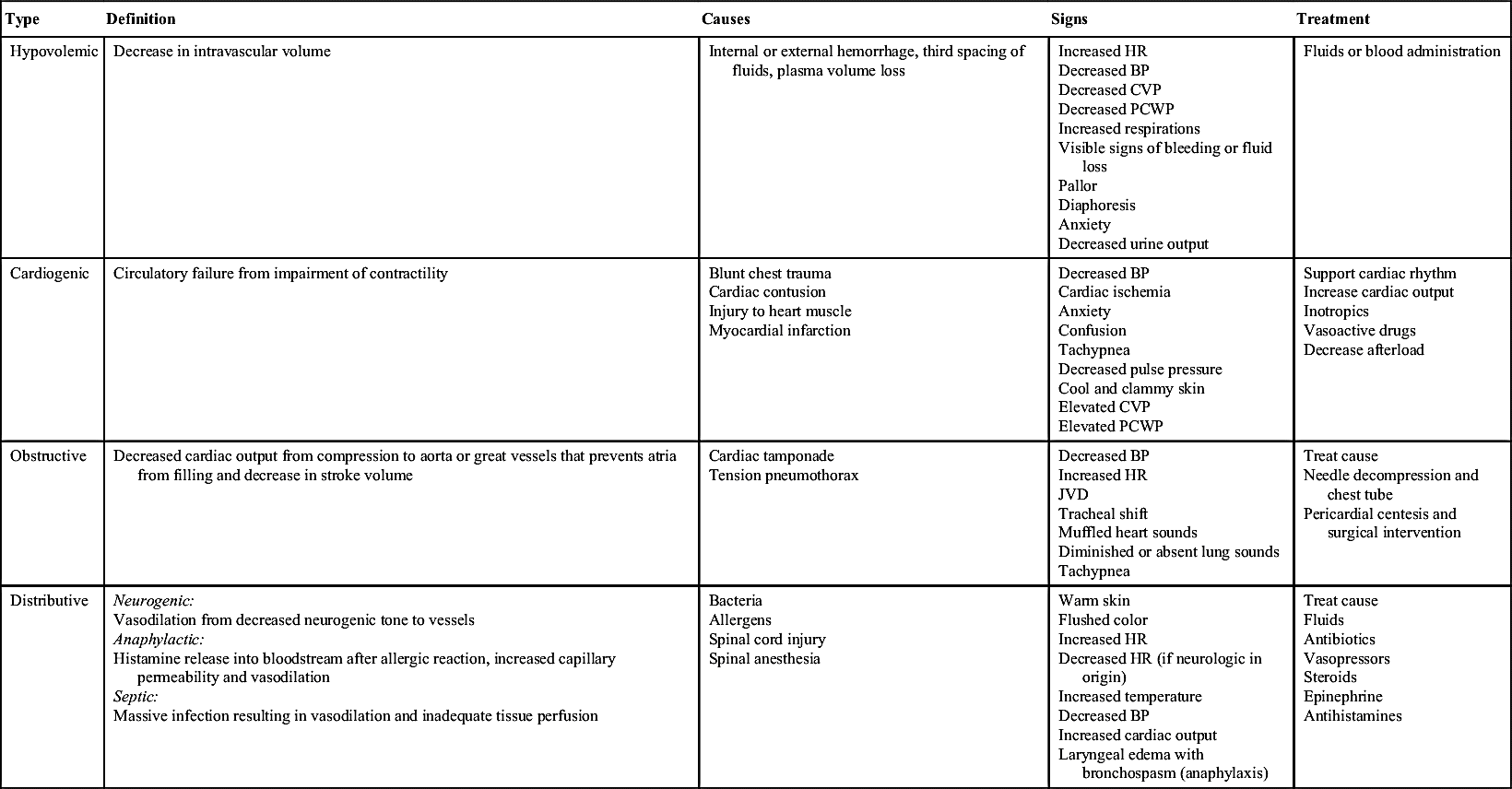
The initial assessment, resuscitation, and stabilization processes initiated in the emergency department and trauma center extend into the operating room (OR), the PACU, and the critical care unit. Temperature of the trauma rooms may be increased to prevent hypothermia during resuscitation. Because the most common cause of shock (Table 54.1) in the trauma patient is hypovolemia from acute blood loss, the ultimate goal in fluid resuscitation is prompt restoration of circulatory blood volume through replacement of fluids so that tissue perfusion and delivery of oxygen and nutrients to the tissues should be maintained.10–15 Rapid identification and ensuing implementation of correct aggressive treatment are vital for the trauma patient’s survival. Although hypovolemia is the most common form of shock in the trauma patient, cardiogenic shock, obstructive shock (tension pneumothorax, cardiac tamponade), and distributive shock (neurogenic shock, burn shock, anaphylactic shock, and septic shock) can occur. Rapid-volume infusers deliver warmed intravenous fluids at a rate of 950 mL/min with large-bore intravenous catheters.15 Many trauma centers initially infuse 2 to 3 L of lactated Ringer or normal saline solutions and then consider blood products. The fluids should be warmed to prevent or minimize hypothermia. Crystalloids, colloids, or blood products can be used for effective reversal of hypovolemia.
Crystalloids are electrolyte solutions that diffuse through the capillary endothelium and can be distributed evenly throughout the extracellular compartment. Examples of crystalloid solutions are lactated Ringer solution, Plasma-Lyte, and normal saline solution. Although controversy exists regarding crystalloid versus colloid fluid resuscitation in multiple trauma, the American College of Surgeons Committee on Trauma recommends that isotonic crystalloid solutions of lactated Ringer or normal saline solution be used for that purpose.15 Furthermore, crystalloids are much cheaper than colloids. Administration of crystalloids should be threefold to fourfold the blood loss.15
Colloid solutions contain protein or starch molecules or aggregates of molecules that remain uniformly distributed in fluid and fail to form a true solution.13–16 When colloid solutions are administered, the molecules remain in the intravascular space, thereby increasing the osmotic pressure gradient within the vascular compartment. Volume for volume, the half-life of colloids is much longer than that of crystalloids. Colloid solutions commonly used are plasma protein fraction, dextran, normal human serum albumin, and hetastarch.
Tranexamic acid (TXA) is a synthetic version of amino acid lysine. TXA is an antifibrinolytic that inhibits activation of plasminogen, a substance responsible for dissolving clots, and should be administered early to be effective in reducing the risk of bleeding and death from exsanguination.17
Researchers in several randomized controlled trials have used hypertonic solutions to resuscitate patients in shock.13–21 According to Beekley,22 hypertonic saline solution (3% sodium chloride) can be used in resuscitation of the child with a severe head injury because it maintains blood pressure and cerebral oxygen delivery, decreases overall fluid requirements, and results in improved overall survival rates.19 In addition, patients with low Glasgow Coma Scale (GCS) scores from head injuries have improved survival rates in the hospital.19
Although crystalloid and colloid solutions serve as primary resuscitation fluids for volume depletion, blood transfusions are necessary to restore the capacity of the blood to carry adequate amounts of oxygen. Furthermore, blood component therapy is considered after the trauma patient’s response to the initial resuscitative fluids has been evaluated.11,13 In an emergency, universal donor blood (type O negative for women in childbearing years) packed red blood cells can be administered for patients with exsanguinating hemorrhage. Untyped O negative whole blood can also be given to patients with an exsanguinating hemorrhage. Other blood products, such as platelets and fresh frozen plasma, may need to be given to the trauma patient because of a consumption coagulopathy. Most notable are the leukemic trauma patients with low platelet counts. With fluid resuscitation of these patients with immunosuppression, colloids are contraindicated because of the antiplatelet activity that exacerbates hemorrhaging.17 Type-specific blood oftentimes is available within 10 minutes and is preferred over universal donor blood. Fully cross-matched blood is preferred in situations that can warrant awaiting type and cross match, which often takes up to 1 hour.11,13–15 Finally, the therapeutic goal of all blood component therapy is to restore the circulating blood volume and to give back other needed blood with red blood cells and clotting factors to correct coagulation deficiencies.21–22
New evidence recognizes “permissive hypotension” by keeping the patient’s systolic blood pressure at approximately 90 mm Hg correlates with better outcomes owing to conservation of important clotting factors.22,23 In addition, there also seems to be a protective mechanism of myocardial suppressive factors that conserve homeostasis of fluid shifts. The intent of this protective response is to prevent further hemorrhaging or bleeding out of the red blood cells and clotting factors.24 By sustaining the hypotension, the blood pressure supports basic perfusion until the patient is in the OR and surgically resuscitated.19–23 Damage control resuscitation and warm, fresh, whole blood are now associated with better survival rates in combat-related massive hemorrhage injuries. Restoration of blood volume before homeostasis is achieved may have adverse complications of exacerbation of blood loss from increase in blood pressure.18–26
In summary, fluid resuscitation of the trauma patient is essential to ensure that adequate circulating volume and vital oxygen and nutrients are delivered to the tissues. However, new studies recommend permissive hypotension with the use of damage control resuscitation to decrease mortality and morbidity and optimize the patient’s survival.18–26 These combat-related studies are now influencing the management of civilian resuscitation of massive hemorrhage injuries in trauma centers throughout the United States.3
Diagnostic Studies and Protocols
Diagnostic tests and laboratory studies have a vital role in establishing the patient’s baseline and current status. The results of these tests predicate the treatment protocols that are initiated. Comprehensive diagnostic studies are required to establish an accurate diagnosis and to plan effective treatment of the patient with multiple injuries. The initial routine studies may be arterial blood gas determinations, urinalysis (myoglobinuria), complete blood count, electrolyte levels, lactate, prothrombin and partial thromboplastin times, and type and cross match. Other diagnostic studies that can be ordered for suspected injuries include lateral cervical spine, upright anteroposterior chest, and anteroposterior pelvic radiographs; computed tomographic scan; 12-lead electrocardiogram; ultrasound scan; and toxicology laboratory studies. Diagnostic peritoneal lavage is performed only on the severely injured patient with hypotension, especially if the result of an abdominal examination is suggestive of injury or is unreliable. Pregnancy tests should be performed on all women of childbearing age but should not delay treatment of life-threatening injuries.
Collaborative Approach
Poor and inaccurate communication, including communication gaps, may lead to uncertainty and, frequently, to inappropriate decisions about patient care and ultimately result in patient harm either through inefficiency or suboptimal management.27 Collaborative practice is essential in the care of the trauma patient. During the prehospital phase, vital communication with the trauma team is initiated at the trauma center. Subsequently, when the patient is first admitted to the PACU, the approach to patient care is comprehensive. Together, the postanesthesia nurse, anesthesia provider, respiratory therapist, surgeon, and radiology technician create a collaborative environment so that care becomes focused and directed. This collaborative practice continues through the intraoperative and postanesthesia periods.
Having a standard evidence-based approach to care delivery decreases the number of communication errors and provides a more reliable approach to patient care.27,28 The anesthesia provider and surgical team communicate a comprehensive system report to the perianesthesia nurse and verbally review any definitive findings of the computed tomographic scan including whether generalized edema or lesions are found. Vital nursing information is communicated to the receiving postanesthesia nurse caring for the trauma patient. The anesthesia provider and trauma surgeon should communicate the significant findings during the intraoperative period that may be problematic during the recovery phase, what the PACU nurse should look for and report promptly to these physicians, and what the PACU nurse should do to prevent harm during the postanesthesia phase of care.27 In many ways, it is even more important that accurate information and anticipatory guidance be effectively transmitted in the PACU hand-off.27
Postanesthesia Phase I
Before the shock trauma patient is admitted to the PACU, the postanesthesia nurse begins preparing for the PACU admission. When the OR nurse calls the PACU to notify the admitting nurse that the trauma surgeon is beginning to close the patient’s surgical site, important prehospital and intraoperative information is communicated. The transfer of care or hand-off communication from the OR nurse to PACU nurse consists of a detailed yet succinct report of all pertinent findings such as surgical operation, type of anesthesia including opioids, isolation status, vital signs, oxygen saturation, ventilation settings, hemodynamic monitoring, drains, vasoactive drugs, intravenous sites with types of solutions, and other pertinent findings so that the PACU nurse can begin to prepare for the patient’s needs.
Primary Assessment
Foremost, the trauma nursing assessment that occurs in the combat trauma resuscitation unit or the shock trauma resuscitation bay should always focus first on uncontrolled hemorrhage or massive bleeding to control life-threatening bleeding and even prevent death.11 The PACU nursing assessment of the shock trauma patient in the PACU begins with evaluation of the ABCDs (airway, breathing, circulation, and disability). Most important, this vital primary assessment should focus first on the patency of the airway and effective breathing due to the effects of anesthesia and surgery. This airway assessment begins with proper positioning of the patient’s head, with cervical spine protection always maintained if injury is suspected. Cervical collars should not be removed unless specifically directed by the trauma surgeon or neurosurgeon after confirmation of the absence of spinal cord injury. The patient may need to have the airway cleared with suctioning and removal of secretions or blood. In addition, airway adjuncts may be needed, such as oropharyngeal and nasopharyngeal airways. If the patient is intubated via endotracheal tube or nasotracheal tube, ventilatory support should be provided with the proper settings to achieve optimal oxygenation and ventilation.
Next, the postanesthesia nurse evaluates the patient’s work of breathing. While recalling the MOI, such as blunt or penetrating trauma to the chest, the nurse should be highly suspicious of pulmonary contusions, fractured ribs, or injuries from shearing forces. The nurse assesses spontaneous respirations, respiratory excursion, chest wall integrity, symmetry, depth, respiratory rate, use of accessory muscles, and the work of breathing. With palpation, the nurse should evaluate for the presence of subcutaneous emphysema, hyperresonance or dullness over the lung fields, and tracheal deviation. With auscultation, the nurse assesses the lungs for bilateral breath sounds and evaluates for adventitious breath sounds. In addition, pulse oximetry and end-tidal CO2 monitoring augment the complete respiratory assessment of the trauma patient.
After a thorough evaluation of the airway and breathing, the nurse begins the circulatory assessment. With the use of palpation, the nurse evaluates the circulation; checks the quality, location, and rate of the pulses; and compares the right with the left and the upper extremities with the lower. If the nurse can palpate a radial pulse, the arterial pressure is at least 80 mm Hg. If no radial pulse is palpable, the nurse then palpates the femoral pulse (a situation that indicates a pressure of 70 mm Hg). If only a carotid pulse is palpable, the arterial pressure is approximately 60 mm Hg. The patient’s blood pressure and pulse (rate and rhythm) should be monitored via the cardiac monitor. Any changes in the patient’s appearance should be investigated and prompt the nurse to reassess the patient. Pulseless electric activity may show as electric impulses on the cardiac monitor without the presence of a palpable pulse. Pulseless electric activity may be seen in the trauma patient related to a variety of causes such as pneumothorax, cardiac tamponade, hypovolemia, or hypothermia.
Simultaneously during the palpation of pulses, the nurse assesses the patient’s skin temperature, color, and capillary refill. Capillary refill is a good indicator of tissue perfusion, especially in children. Another aspect of circulatory assessment is observation of the patient for any significant or uncontrollable bleeding from the operative site. The nurse should inspect the peripheral, central, and arterial lines to ensure the patency of the lines and integrity of the sites. Each line should be identified and labeled with the date and time to distinguish the type of parenteral fluid and medication administration in use.
The final component of the primary survey is the disability or neurologic examination. The patient’s mental status should be assessed with AVPU (Alert, Voice, Pain, Unresponsive) scale or the GCS. The AVPU is described as follows: A for awake and responding to nurse’s questions, V for verbal response to nurse’s questions, P for responding to pain, and U for unresponsive.10,11 The GCS is used in many PACUs for evaluating neurologic status and predicting outcomes of severe trauma. This neurologic scoring system allows for constant evaluation from field to emergency room to PACU. One must remember that anesthesia blunts the neurologic response; therefore, the response is not as useful in the immediate postanesthesia period. Next, bilateral pupil response is evaluated for equality, roundness, and reactivity: brisk, slow, sluggish, or no response to light and accommodation. Before the primary assessment is complete, the PACU nurse quickly reassesses the ABCDs for stability and then is ready to receive a report from the anesthesia provider.
Anesthesia Report
The anesthesia provider’s report provides valuable information concerning the trauma patient’s presenting status to the perianesthesia nurse.27 This report includes significant facts that pertain to the MOI, prehospital phase, admitting and stabilization period, operative report, intubation, anesthetic agents, estimated blood loss, fluid resuscitation, cardiopulmonary status, and treatment abnormalities.
In the review of the anesthesia report, the nurse should note any difficulty in intubation of the patient. The nurse should take note if the oral secretions are tinged pink or bloody, which can indicate an infectious process, pulmonary edema, trauma, or uncontrolled hemorrhage.
Another important aspect of the anesthesia report is estimated blood loss and fluid replacement, which are carefully monitored through the hemodynamic status of the trauma patient. With the use of arterial lines and pulmonary artery catheters, the anesthesiologist can closely monitor the patient’s hemodynamic status. In severe chest injuries, closed chest drainage units and autotransfusions or Cell Saver blood recovery systems can be used to conserve the vital life-sustaining resource blood. Because the goal of treatment is to keep the patient in a hemostatic state, the intraoperative trending should reveal that the patient is volume supported because the body responds hypermetabolically to trauma and achieving that state ensures the delivery of oxygen and other nutrients to essential tissues in the body. End organ perfusion is monitored and measured with the urinary output and hemodynamic monitoring. Urinary catheters are essential in management of fluids in the trauma patient and assessment of kidney function. Hemodynamic monitoring reflects the body’s hydration status and reveals the work of the heart. A detailed operative report reveals the surgical insult to the patient. It also presents a comprehensive review of all anesthetic agents that reflects a rapid sequence of induction, balanced analgesia, and heavy use of opioids including the time these agents were given and the amounts and types of muscle relaxants and reversal agents used. The anesthesia report should reveal any untoward events that occurred during surgery, such as hypothermic or hypotensive events, and significant dysrhythmias including ischemic changes. Chapter 26 also has information on the patient hand-off.
Secondary Assessment
After the anesthesia report is received, a brief initial primary survey is completed, and the postanesthesia nurse begins the secondary comprehensive survey with a high degree of suspicion concerning the trauma patient’s MOI for specific perianesthesia problems. The initial surgery often is directed at repair of the major life-threatening injuries such as a ruptured aorta. Consequently, additional injuries may manifest themselves during this later time in the PACU after swelling and bruising are allowed to develop. During the comprehensive secondary survey, head-to-toe assessment is performed as the trauma patient is emerging from anesthesia. The perianesthesia nurse may discover other injuries such as pulmonary, cardiac, or renal contusions and compartment syndrome of different extremities.
The head-to-toe assessment begins with a neurologic assessment including the patient’s level of consciousness; the appropriateness of verbal response; pupillary reactivity, size, and shape; equality of pupillary response to light and accommodation; movement; sensation; and pain response in the extremities. Each aspect is carefully evaluated and documented. Next, the head and face are inspected for abrasions, lacerations, puncture wounds, ecchymosis, and edema. These structures are palpated for subcutaneous emphysema or tenderness. The eyes are assessed for gross vision by asking the patient to identify the number of fingers the nurse is holding up. Furthermore, the eyes are evaluated for ecchymosis, “raccoon eyes,” and possible conjunctival hemorrhage. Extraocular movements are also evaluated by asking the patient to follow the nurse’s finger in six directions. The presence of maxillofacial injuries can be of great concern because of the potential to compromise the patient’s airway.
The ears are inspected for Battle sign (ecchymosis behind the ears). The nose is examined for drainage of blood or clear fluid. Clear fluid draining from the nose or ears should be checked for the presence of cerebrospinal fluid (CSF). A draining nose or ears should never be packed. If CSF is suspected or the Battle sign is seen, a nasogastric tube should never be inserted through the patient’s nose. An orogastric tube is the placement of choice. Finally, the nurse should immediately report a positive CSF finding to the trauma surgeons.
The neck should be evaluated for edema, ecchymosis, tracheal deviation, pulsating or distended neck veins, and subcutaneous emphysema. As the perianesthesia nurse continues the assessment of the chest, the anterior and the lateral thorax and axilla are inspected for lacerations, abrasions, contusions, puncture wounds, ecchymosis, and edema. The nurse carefully palpates the chest for tenderness and subcutaneous emphysema. The chest wall is observed for symmetry, depth, and equality of expansion and excursion. If the trauma patient has a flail chest from the injury sustained, astute monitoring for effective oxygenation and ventilation is essential. Breathing is observed for rate, degree of effort, use of accessory muscles, or paradoxical chest wall movements. Breath and heart sounds are auscultated, noting adventitious lung sounds (e.g., wheezing, rales, friction rubs) or murmurs, bruits, and muffled heart sounds. The perianesthesia nurse carefully notes facial expressions or body reactions that may suggest possible cardiac contusions or rib fractures. The operative site and all dressings and drains should be assessed and described. Furthermore, all drains should be labeled, drainage of fluid is measured, and color and consistency are described. Precise documentation of all fluid output is essential for accurate fluid replacement.
The next areas to be inspected are the abdomen, pelvis, and genitalia. All abrasions, contusions, edema, and ecchymosis are noted. The abdomen is auscultated for bowel sounds before palpation for tenderness and rigidity. The nasogastric tube, jejunostomy, or tube drainage is examined for color, consistency, and amount of fluid. In suspected internal abdominal or retroperitoneal hemorrhage, abdominal compartment measurements should be assessed. Some common causes of abdominal compartment syndrome are pelvic fractures, hemorrhagic pancreatitis, ruptured abdominal aortic aneurysm, blunt and penetrating abdominal trauma, bowel edema from injury, septic shock, and perihepatic or retroperitoneal packing for diffuse nonsurgical bleeding. If intraabdominal hypertension or abdominal compartment syndrome are suspected or considered, the standard of care is measurement of bladder pressures.29 A catheter or similar device is connected to a pressure transducer connected to the urinary catheter. The urinary catheter is clamped near the connection, 50 mL of saline solution is instilled to the bladder, the transducer is leveled at the symphysis pubis, and the pressure is measured at end expiration. If the pressure is elevated, a decompression laparotomy should be performed to release the pressure that develops from bowel edema.11,13 The abdomen is then left open and covered with a sterile wound vacuum until the swelling has resolved and the abdomen can be closed.
The pelvis is palpated for stability and tenderness, especially over the crests and the pubis. Priapism, which is persistent abnormal erection, may be noted.11,13 In addition, preexisting genital herpes may also be present. The urinary catheter is inspected for color and amount of drainage. Urinary output should be at least 0.5 to 1.0 mL/kg/h in adults and 1 to 2 mL/kg/h in children.11 Hematuria can indicate kidney or bladder trauma.11 Furthermore, urinary output must be vigilantly monitored to ensure a minimum of 30 mL/h in adults so that the patient does not develop acute renal failure from rhabdomyolysis, which can occur after traumatic injuries. The vagina and rectum are checked carefully for neurologic function and bloody drainage.
All extremities are examined for circulatory, sensory, and motor functions with range of motion. Because the trauma patient is rushed to the OR to correct life-threatening injuries, minor soft tissue injuries often may be missed. Later in the PACU, these soft tissue and musculoskeletal injuries can develop into compartment syndrome. Each extremity must be thoroughly examined for abrasions, contusions, puncture wounds, ecchymosis, and edema. If neurovascular compromise is found, an arteriogram or venogram can be performed as a conclusive diagnostic study.
The patient is then log-rolled onto the side with maintenance of cervical spine integrity for assessment of the back, flanks, and buttocks for abrasions, contusions, and tenderness. Rectal tone is noted if spinal cord injury is suspected. Posterior chest assessment is completed as a last step in the head-to-toe assessment. Finally, a detailed yet succinct description of the primary and secondary assessment is documented.
Pain
Assessment and management of pain are important parts of the scope of care provided to the trauma patient in the PACU. The trauma patient may have musculoskeletal injuries or ruptured organs that cause severe pain, which may include more than the surgical site. The perianesthesia nurse needs to recognize that because pain is subjective, verbal, nonverbal, and hemodynamic changes that indicate the patient may be exhibiting signs of pain should be noted. Pain can manifest itself with increased heart rate, increased blood pressure, pallor, tachypnea, guarding or splinting, and nausea and vomiting. Pain scales should be used to augment the nursing assessment of pain.30,31
Optimal management of acute pain may use the following techniques: (1) patient-controlled analgesia with intravenous or epidural infusions, (2) intravenous intermittent doses for pain or sedation, and (3) major plexus blocks. Guidance for medications should be based on choosing drugs that minimize cardiovascular depression and intracranial hypertension. A higher incidence rate of substance abuse, both alcohol and recreational or addictive mind-altering drugs, in the trauma patient population may require higher doses of opioids or analgesics.30,31 Other complementary techniques, such as music therapy and guided imagery, may be initiated and used as adjuncts when the trauma patient returns for subsequent surgical procedures or wound debridement. Finally, continual pain assessment is vital to the patient’s optimal care.
Nausea
Nausea can be a concern for the trauma patient. Trauma patients rarely fast before the traumatic event; therefore, patients enter anesthesia with a potentially full stomach. Vomiting can lead to aspiration and a host of issues. Extubation of trauma patients should be delayed until the gag reflex returns to avoid aspiration. Many times, nasogastric tubes are present, but if the patient has consumed food before the event, particles may be too large to be evacuated. Nausea needs to be treated after surgery and is seen with a higher incidence rate in the traumatized patient.
Related posts:


Stay updated, free articles. Join our Telegram channel

Full access? Get Clinical Tree


