Cardiopulmonary Arrest


Cardiopulmonary Arrest


By necessity, most recommendations for treating cardiopulmonary arrest are not derived from high quality randomized human studies but rather come from retrospective series, animal experiments, and expert opinion. Treatment recommendations traditionally have been intricate and most applicable to patients who sustained cardiac death, especially outside the hospital. Since the focus of this book is on the hospitalized critically ill patient, some of the discussion that follows will naturally differ from widely disseminated generic recommendations. Outside the hospital, and in the coronary care unit and cardiac catheterization lab, most arrests are due to ventricular tachycardia (VT) and ventricular fibrillation (VF) in patients with ischemic heart disease. As a corollary, because VT or VF is so likely to be the cause of death in cardiovascular ICU, such patients should almost always be treated immediately with unsynchronized cardioversion. This fact underscores the utility of installing automated external defibrillators (AEDs) in public places. By contrast, a respiratory event (aspiration, excessive sedation, pulmonary embolism, airway obstruction) is much more likely to occur in a hospitalized patient. It follows that arrests on a general ward or noncardiac ICU are more likely to respond to a nonarrhythmia directed intervention, often one involving the lungs.


▪ PRIMARY PULMONARY EVENTS (RESPIRATORY AND PULMOCARDIAC ARREST)

Patients found unresponsive without respirations but with an effective pulse have suffered a respiratory arrest. Failure to rapidly restore oxygenation results in hypoxemia and progressive acidosis that culminate in reduced contractility, hypotension, and eventual circulatory collapse. Although the etiology of many respiratory arrests remains uncertain even after thorough investigation, the cause often can be traced to respiratory center depression (e.g., sedation, coma, stroke, high intracranial pressure) or to failure of the respiratory muscle pump (e.g., excessive workload, impaired mechanical efficiency, small or large airway obstruction, or muscle weakness). Tachypnea usually is the first response to stress, but as overloading continues, the respiratory rhythm disorganizes, slows, and eventually ceases. Initially, mild hypoxemia enhances the peripheral chemical drive to breathe and stimulates heart rate. Profound hypoxemia, however, depresses neural function and produces bradycardia refractory to sympathetic and parasympatholytic influences. At this point, cardiovascular function usually is severely disordered, because cardiac and vascular smooth muscle function poorly under conditions of hypoxia and acidosis and cardiac output falls as heart rate declines. The observation that nearly one half of hospitalized arrest victims exhibit an initial bradycardic rhythm, underscores the role of respiratory causes of circulatory arrest.


FIGURE 20-1 Change in arterial partial pressure of oxygen and carbon dioxide after respiratory arrest (normal lungs). Oxygen concentrations fall precipitously to dangerously low levels within minutes. By contrast, the rise in carbon dioxide tensions is much slower, requiring 15 to 20 min to reach levels sufficient to produce life-threatening acidosis.

In hospitalized, especially critically ill patients, the partial pressure of arterial oxygen (PaO2) plummets shortly after ventilation ceases because limited O2 stores are rapidly consumed. Reserves are diminished by diseases which reduce baseline saturation (e.g., chronic obstructive pulmonary disease [COPD], pulmonary embolism), lower functional residual capacity (e.g., morbid obesity, pregnancy), or both (e.g., pulmonary fibrosis, congestive heart failure). Ambulatory patients who suffer sudden cardiac arrest have substantially greater O2 reserves because they typically do not have diseases causing significant desaturation or thoracic restriction. For this reason, attention to oxygenation is much more important in the hospitalized arrest victim, whereas establishing artificial circulation and prompt rhythm correction are priorities for the “cardiac” death patient. Unlike O2, CO2 has a huge storage pool and an efficient buffering system. Therefore, PaCO2 initially builds rather slowly, at a rate of 6 to 9 mm Hg in the first apneic minute and 3 to 6 mm Hg/min thereafter (Fig. 20-1). However, as the apneic patient develops metabolic acidosis from tissue hypoxia, H+ combines with HCOimage 
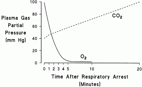
to dramatically increase the rate of CO2 production. The net effect of these events is that life-threatening hypoxemia occurs long before significant respiratory acidosis.


▪ PRIMARY CARDIOVASCULAR EVENTS (CARDIOPULMONARY ARREST)

The heart may abruptly fail to produce an effective output because of an arrhythmia or suddenly impaired pump function resulting from diminished preload, excessive afterload, or decreased contractility. The normal heart compensates for changes in heart rate over a wide range through
the Starling mechanism. Thus, cardiac output usually is maintained by compensatory chamber dilation and increased stroke volume despite significant slowing of rate. Children and adults with dilated or stiff hearts lose this reserve and are highly sensitive to bradycardia.

Decreases in left ventricular preload sufficient to cause cardiovascular collapse usually are the result of venodilation, hemorrhage, pericardial tamponade, or tension pneumothorax. In contrast to the left ventricle, which is constantly adapting to a widely changing afterload, the right ventricle is not adept at adjusting quickly to increased impedance. Therefore, abrupt increases in right ventricular afterload (e.g., air or thromboembolism) are much more likely to cause catastrophic cardiovascular collapse. Acute dysfunction of the cardiac muscle fiber can result from tissue hypoxia, severe sepsis, acidosis, electrolyte disturbance (e.g., hypokalemia), or drug intoxication (e.g., β-blockers). Regardless of the precipitating event, patients with narrowed coronary arteries are particularly susceptible to the adverse effects of a reduced perfusion pressure.

Neural tissue is disproportionately sensitive to reduced blood flow. Circulatory arrest always produces unconsciousness within seconds, and respiratory rhythm ceases rapidly thereafter. Thus, ongoing respiratory efforts indicate very recent collapse or the continuation of effective blood flow below the palpable pulse threshold. (In a person of normal body habitus, a systolic pressure of approximately 80, 70, or 60 mm Hg must be present for a pulse to be detected at radial, femoral, or carotid sites, respectively.)


▪ CARDIOPULMONARY RESUSCITATION

Cardiopulmonary resuscitation (CPR) was conceived as a temporary circulatory support procedure for otherwise healthy patients suffering sudden cardiac death. In most cases, coronary ischemia or primary arrhythmia was the inciting event. Since its inception however, CPR use has been expanded to nearly all types of patients who suffer an arrest. Understandably, with wider application, its success rate has declined. Currently, less than one half of all patients undergoing CPR will be resuscitated initially, and well less than one half of these initial survivors live to hospital discharge. Even more discouraging, at least one half of the discharged patients suffer neurological damage severe enough to prohibit independent living. Despite the success portrayed on television, as few as 5% of all CPR recipients enjoy even a near-normal postarrest life. In addition, pharmacoeconomic analyses suggest that in hospital, resuscitation may be the least cost effective treatment delivered with any regularity. The likelihood of successful CPR (discharge without neurological damage) depends on the population to whom the procedure is applied and the time until circulation is restored. Brief periods of promptly instituted CPR are highly successful when applied to patients with sudden cardiac death, but when CPR is used as a “last rite” for progressive multiple organ failure, the likelihood of benefit approaches zero.


Principles of Resuscitation

This chapter emphasizes general principles of resuscitation which are enduring, intentionally omitting details that are not evidence based or are likely to change. Current expert recommendations for resuscitation are much simpler than those in the past and stress the importance of effective circulatory support and prompt shock of VT and VF de-emphasizing respiratory support. While that advice makes sense for most out of hospital events, in the hospital the resuscitation team must quickly consider the specific circumstances of each arrest to determine the best course of action (Table 20-1). For example, a mechanically ventilated patient found in VF will not be saved by a formulaic approach to arrhythmia treatment if it is not recognized that the cause of the event is a tension pneumothorax or airway obstruction. Because survival declines exponentially with time after arrest (Fig. 20-2), most successfully resuscitated patients
are revived in less than 10 min. To this end, first responders should summon help and begin effective chest compression. If the cardiac rhythm can be monitored and is VT or VF, maximal energy unsynchronized direct current (DC) cardioversion should be delivered as quickly as possible. If these initial actions are unsuccessful, institution of more prolonged, “advanced” resuscitation measures may be indicated.


FIGURE 20-2 Probability of successful initial resuscitation after cardiopulmonary arrest. Exponential declines in survival result in low success rates after 6 to 10 min of full arrest conditions.


TABLE 20-1 COMMON CLINICAL SCENARIOS OF CARDIOPULMONARY ARREST


SETTING


LIKELY ETIOLOGY


APPROPRIATE INTERVENTION


Early during mechanical ventilation


Misplaced ET tube


Confirm proper location by visualization and auscultation, CO2 detector


Tension pneumothorax


Physical examination, chest tube placement


Hypovolemia


Fluid bolus


Auto-PEEP


Reduce VE, increase expiratory time, bronchodilator, suction airway


Hypoxemia


Check ET placement, oximeter saturation, administer 100% O2


During chronic mechanical ventilation


ET tube displacement


Confirm proper ET placement by auscultation and chest radiograph


Hypoxemia


Confirm oxygenation with oximeter or ABG, increase FiO2


Tension pneumothorax


Physical examination, chest tube placement


Auto-PEEP


Reduce VE, increase expiratory time, bronchodilator


Mucus plugging


Suction airway


Post-central line placement/attempt


Tension pneumothorax


Physical examination, chest tube placement


Tachyarrhythmia


Withdraw intracardiac wires or catheters, try cardioversion/antiarrhythmic


Bradycardia/heart block


Withdraw intracardiac wires or catheters, try chronotropic drugs, temporary pacing


During dialysis or plasmapheresis


Hypovolemia Transfusion reaction


IgA deficiency: allergic reaction Hyperkalemia


Fluid therapy Stop transfusion; treat anaphylaxis


Stop transfusion, treat anaphylaxis


Check K+, treat empirically if ECG suggests hyperkalemia


During transport


Displaced ET tube Interruption of vasoactive drugs


Early identification using end-tidal CO2 Restart IV access


Acute head injury


Increased intracranial pressure (especially with bradycardia)


Diabetes insipidus: hypovolemia (especially with tachycardia)


Lower intracranial pressure (ICP): hyperventilation, mannitol, 3% NaCl


Administer fluid


Pancreatitis


Hypovolemia


Fluid administration


Hypocalcemia


Calcium supplementation


After starting a new medicine


Anaphylaxis (antibiotics)


Stop drug, administer fluid, epinephrine, corticosteroids


Angioedema (ACE inhibitors)


Hypotension/volume depletion (ACE inhibitors)


Volume expansion


Methemoglobinemia


Methylene blue


Toxin/drug overdose Cyclic antidepressants


Seizures/tachyarrhythmias


Sodium bicarbonate


β-Blocker/Ca2+ blocker


Severe bradycardia


Chronotropes, pacing, glucagon, insulin + glucose


Organophosphates carbamates


Severe bradycardia


Decontamination, atropine, pralidoxime


MAO inhibitor CO, cyanide


Hypertension Hypoxia


Drug removal Oxygen, sodium nitrite + sodium thiocyanate


After myocardial infarction


Tachyarrhythmia/VF Torsade de pointes


DC countershock, lidocaine Cardioversion, Mg, pacing, isoproterenol, stop potential drug causes


Tamponade, cardiac rupture


Pericardiocentesis, fluid, surgical repair


Bradycardia, AV block


Chronotropic drugs, temporary pacing


After trauma


Exsanguination


Fluid/blood administration, consider laparotomy-thoracotomy


Tension pneumothorax


Physical examination, chest tube placement


Tamponade


Pericardiocentesis/thoracotomy


Abdominal compartment syndrome


Measure bladder pressure, decompress abdomen


Burns


Airway obstruction


Intubate, reintubate


Hypovolemia


Fluid administration


Carbon monoxide


100% O2


Cyanide


Sodium nitrite-thiosulfate


ABG, arterial blood gases; ACE, angiotensin-converting enzyme; AV, atrioventricular; DC, direct current; ECG, electrocardiogram; ET, endotracheal; PEEP, positive end-expiratory pressure; VF, ventricular fibrillation.


The primary activities of resuscitation include (i) team direction, (ii) circulatory support, (iii) cardioversion/defibrillation, (iv) airway management and ventilation, (v) establishing intravenous access and administering drugs, and (vi) performance of specialized procedures (e.g., pacemaker and chest tube placement). Managing a cardiopulmonary arrest usually requires five persons. Additional personnel may be needed for particular tasks such as documentation, chart review, and communication with the laboratory or other physicians, but limiting the number of people involved to the minimum required avoids pandemonium.

Jul 17, 2016 | Posted by in CRITICAL CARE | Comments Off on Cardiopulmonary Arrest

Full access? Get Clinical Tree

Get Clinical Tree app for offline access